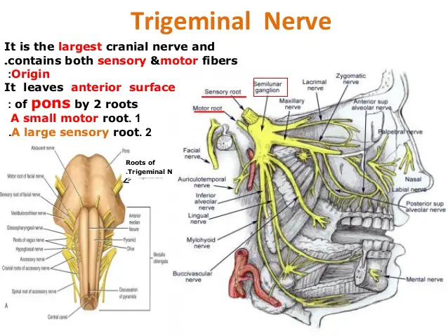
Grip Muscles – Anatomy and Exercise
| | |

Grip Muscles – Anatomy and Exercise

Introduction The muscles that are involved in the act of gripping are known as Grip Muscles, the Grip Muscles are flexor digitorum profundus, flexor digitorum superficialis, flexor digiti minimi brevis, flexor pollicis longus, extensor digitorum, lumbricals, interossei, adductor pollicis. Anatomy of Grip Muscles 1. Flexor Digitorum Profundus Origin It originates from the upper 3/4 of…

| |

Scapular muscles- Anatomy and Exercise

Introduction The scapula provides attachment for several groups of muscles. scapular muscles include the supraspinatus, subscapularis, teres minor, infraspinatus, deltoid, and teres major. These muscles attach to the scapular surface and assist with abduction and external and internal rotation of the glenohumeral joint. Anatomy of scapular muscles 1. Supraspinatus Origin It originates from the medial…

Abdominal Muscle – Anatomy and Exercise
| | |

Abdominal Muscle – Anatomy and Exercise

Introduction The abdominal muscles form the anterior and lateral abdominal wall and consist of the external abdominal obliques, the internal abdominal obliques, the rectus abdominis, and the transversus abdominis. Acting together these muscles form a firm wall that protects the viscera and they help to maintain erect posture. In addition the contraction of these muscles…

Gluteal Muscle Anatomy and Exercise
| | |

Gluteal Muscle Anatomy and Exercise

Anatomy of Gluteal Muscle Gluteus muscle, any of the large, fleshy muscles of the buttocks, stretching from the back portion of the pelvic girdle (hipbone) down to the greater trochanter, the bony protuberance at the top of the femur (thighbone). These include: 1) gluteus maximus2) gluteus medius3) gluteus minimus. 1) GLUTEUS MAXIMUS: This is a…

Calf Muscle Anatomy: Anatomy and Exercise
| | |

Calf Muscle Anatomy: Anatomy and Exercise

Anatomy of the Calf muscle The calf muscle is located on the back of the lower leg and is actually made up of two muscles: The medial and lateral heads of the gastrocnemius muscle, which is the most superficial, are present. The gastrocnemius muscle belly is formed by the convergence of its two heads. The…

Hamstring Muscle: Anatomy And Exercise
| | |

Hamstring Muscle: Anatomy And Exercise

Hamstring Muscle Anatomy The hamstrings are a group of muscles and their tendons at the rear of the upper leg. They include the biceps femoris, semitendinosus, and semimembranosus. The hamstrings flex the knee joint and extend the thigh to the backside of the body. They are used in walking, running, and many other physical activities….

ATRIAL SEPTAL DEFECT
| | |

ATRIAL SEPTAL DEFECT

OVERVIEW An atrial septal defect (ASD) is a hole in the wall between the two upper chambers of your heart (atria). The condition is present at birth (congenital). Small defects may never cause a problem and may be found incidentally. It’s also possible that small atrial septal defects may close on their own during infancy…

Quadriceps Muscle
| | | |

Quadriceps Muscle

Quadriceps femoris muscle Anatomy The quadriceps Muscle also called simply the quadricep Femoris or Knee extensor is a large muscle group that Covers the Front Of the Thigh And the four muscles Group That Makes One Strongest Muscle Of The Lower Limb ( on the front of the thigh). It is the super extensor muscle…

Iliopsoas Muscle Anatomy: Know Your Muscles
| | |

Iliopsoas Muscle Anatomy: Know Your Muscles

The term Iliopsoas means the combination of the psoas major muscle & the iliacus muscle Joining at their inferior ends. These muscles are distinct in the abdomen, but usually indistinguishable in the thigh. As such, they are usually given the common name “iliopsoas” and are referred to as the “dorsal hip muscles”.The psoas minor Also…