List of Veins of the Human Body
Introduction
Veins are a vital component of the circulatory system, working alongside arteries and capillaries to facilitate the circulation of blood throughout the body. In contrast to arteries, which transport oxygen-rich blood from the heart to the body’s tissues, veins carry deoxygenated blood back to the heart for reoxygenation. However, there are special cases such as the pulmonary veins, which transport oxygenated blood from the lungs to the heart
Anatomy of Veins: The three main layers that are made up of a vein are the tunica adventitia, medium, and intima. The deepest layer, or tunica intima, is made up of endothelial cells that smooth the blood’s surface. The middle layer, known as the tunica media, is composed mainly of smooth muscle fibers and elastic tissue. However, it is thinner in veins compared to arteries. The tunica adventitia, or externa, is the outer layer composed of connective tissue, providing support and structure to the vein.
List of veins of the human body
An inventory of the human body’s veins:
Veins of the heart
The veins of the heart, also known as the cardiac veins, play a crucial role in returning deoxygenated blood from the myocardium (heart muscle) back to the right atrium of the heart. These veins are essential for maintaining adequate blood supply to the heart muscle itself, ensuring its proper function. Here’s an overview of the main cardiac veins:
Coronary sinus: The coronary sinus, also referred to as the cardiac veins, plays a critical role in returning deoxygenated blood from the heart muscle (myocardium) back to the right atrium of the heart. These veins are essential for maintaining adequate blood supply to the heart muscle itself, ensuring its proper function. Here’s an overview of the main cardiac veins:
- Great cardiac vein – It Is Also known as the anterior interventricular vein, the great cardiac vein accompanies the anterior interventricular artery (left anterior descending artery) within the anterior interventricular sulcus. It removes blood from the interventricular septum and the front (anterior) part of the left ventricle.
- Oblique vein of the left atrium – The oblique vein of the left atrium is also known as the oblique vein of Marshall (OVM). Located on the posterior surface of the left atrium, the coronary sinus is a small vein that is a remnant of the left superior vena cava developed during embryonic development. It is a left superior vena cava remnant that developed during embryonic development. The Oblique vein of marshall is often associated with atrial arrhythmias like atrial fibrillation and can serve as a pathway for ablation procedures in certain cases. Its significance lies in its potential role in cardiac electrophysiology and interventions for certain heart rhythm disorders.
- Middle cardiac vein – The middle cardiac vein runs alongside the posterior interventricular artery (posterior descending artery) within the posterior interventricular sulcus. It drains the posterior part of the left ventricle and interventricular septum.
- Small cardiac vein – Also known as the right marginal vein, it runs along the right side of the heart’s margin alongside the right marginal artery. The blood is removed from the right atrium and ventricle.
Venous Drainage into the Right Atrium: Besides the coronary sinus, smaller veins directly drain into the right atrium, including:
- Anterior Cardiac Veins: These veins drain the anterior surface of the right ventricle and may directly empty into the right atrium or join other veins before doing so.
- Thebesian Veins: These are small veins that drain the myocardium directly into the chambers of the heart. They are particularly numerous in the right atrium but are also found in the other chambers.
Veins of Superior vena cava – The brachiocephalic veins, also called innominate veins, are directly responsible for creating the superior vena cava (SVC). The superior vena cava is formed by joining the left and right brachiocephalic veins. Here’s a bit more detail:
Brachiocephalic vein
Right Brachiocephalic Vein (Right Innominate Vein):
- It is formed by the confluence of the right subclavian vein (which drains the right arm) and the right internal jugular vein (which drains the right side of the head and neck).
- The right brachiocephalic vein is typically shorter than the left one.
· Left Brachiocephalic Vein (Left Innominate Vein):
- It was formed by the union of the left subclavian vein (which drains the left arm) and the left internal jugular vein (which drains the left side of the head and neck).
- Establishing the Superior vena cava, this crosses the midline anterior to the trachea and integrates with the right brachiocephalic vein.
Pericardial veins – The pericardial veins are a network of veins that drain blood from the pericardium, the membranous sac surrounding the heart. These veins are relatively small and variable in their anatomy. They play a role in returning deoxygenated blood from the pericardium to the systemic venous circulation. However, the pericardial veins are not as well-defined or extensively studied as other venous systems in the body.
- The surrounding venous structures, such as the cardiac, pulmonary, and bronchial veins, may receive the pericardial veins’ drainage.
- Bronchial Veins: Pericardial veins might additionally empty into the bronchial veins, which frequently empty into the superior vena cava or the azygos vein.
Veins of Lungs
The Oxygenated blood is transported from the lungs to the left atrium of the heart by the pulmonary veins. Here’s a detailed overview:
- Right Superior Pulmonary Vein (RSPV):
- The right lung’s upper lobe is drained.
- It typically enters the posterior aspect of the left atrium, slightly above and to the right of the opening of the inferior vena cava.
- Right Inferior Pulmonary Vein (RIPV):
- Drains the middle and lower lobes of the right lung.
- It enters the left atrium slightly inferior to the RSPV.
- Left Superior Pulmonary Vein (LSPV):
- The left lung’s upper lobe is drained.
- It enters the left atrium directly.
- Left Inferior Pulmonary Vein (LIPV):
- The lower lobe of the left lung is drained.
- It enters the left atrium directly.
These veins are essential for oxygenating blood. After receiving oxygen from the alveoli (tiny air sacs) in the lungs, the blood is collected by the pulmonary veins and transported back to the heart’s left atrium. From there, it’s pumped into the left ventricle and then circulated throughout the body.
Vein of Thyroid
The thyroid gland is mainly supplied by three groups of veins.
- Superior Thyroid Veins:
- The upper pole of the thyroid gland is where the superior thyroid veins usually originate.
- They drain blood from the superior portion of the thyroid gland and usually run upward to join the internal jugular vein.
- Middle Thyroid Veins:
- The veins originating in the middle thyroid come from the central part of the thyroid gland.
- These veins collect blood from the middle section of the thyroid gland and typically empty into the internal jugular vein or the superior thyroid vein.
- Inferior Thyroid Veins:
- The inferior thyroid veins arise from the lower pole of the thyroid gland.
- These veins collect blood from the lower part of the thyroid gland and usually empty into the brachiocephalic vein, which then connects to the superior vena cava.
Veins of Laryngeal
The veins of the larynx drain blood from this anatomical region and contribute to the overall venous drainage of the head and neck. The laryngeal veins can be categorized into multiple groups.
- Superior Laryngeal Vein:
- Arises from the superior laryngeal artery.
- Drains the supraglottic region (above the vocal folds) of the larynx.
- Generally empties into the internal jugular vein.
- Inferior Laryngeal Vein (Recurrent Laryngeal Vein)
- Arises from the inferior thyroid veins.
- Drains the area below the vocal folds in the larynx, known as the subglottic region.
- The usual direction of drainage is into the inferior thyroid veins or the brachiocephalic vein.
Veins of head and neck
The veins of the head and neck form an extensive network that drains blood from various structures in this region and ultimately returns it to the heart. Here’s an overview:
- Internal Jugular Vein:
- The internal jugular vein is one of the major veins of the neck, running alongside the internal carotid artery.
- Blood is collected from the brain, face, and neck.
- External Jugular Vein: The external jugular vein lies closer to the surface of the neck. It drains blood from the scalp and the superficial structures of the neck.
- Facial Vein: The facial vein drains blood from the superficial structures of the face. It starts at the inner corner of the eye as the angular vein and collects blood from the nasal veins. also includes Angular vein, Supratrochlear veins, Supra-orbital vein, External nasal veins, Deep facial vein, External palatine vein, Submental vein
- Superficial Temporal Vein: Drains blood from the temporal region of the scalp. The retromandibular vein is usually formed by the joining of the maxillary vein with it.
- Retromandibular Vein: The retromandibular vein is created when the superficial temporal vein and the maxillary vein come together. It often divides into anterior and posterior branches, which can join with the facial vein and the common facial vein, respectively.
Veins of brain
The veins of the brain, also known as cerebral veins, are responsible for draining blood from the brain and carrying it back to the heart. They form a complex network that helps regulate blood flow within the brain. These veins are crucial for maintaining proper circulation and oxygenation to support brain function.
The major veins of the brain include:
- Superior Sagittal Sinus: Located in the midline of the brain, running along the superior surface of the brain in the longitudinal fissure between the two cerebral hemispheres. The superficial veins in the brain are where blood is gathered before flowing into the confluence of sinuses.
- Inferior Sagittal Sinus: The inferior sagittal sinus can be found in the lower region of the falx cerebri and gathers blood from the inner surface of the cerebral hemispheres. The connection of sinuses is formed by the joining of the straight sinus.
- Straight Sinus: Lies in the tentorium cerebelli, draining blood from the deep structures of the brain and cerebellum. The confluence of sinuses is typically the point where it connects with the inferior sagittal sinus.
- Cerebral veins
- Superficial cerebral veins
- The basal vein and great cerebral vein are types of internal cerebral veins.
- Transverse Sinuses: Paired sinuses located in the posterior part of the brain, within the tentorium cerebelli. Blood is received from the point where sinuses come together, and then it flows as sigmoid sinuses.
- Sigmoid Sinuses: Curved continuation of the transverse sinuses that drain into the jugular veins. They are named for their sigmoid (S-shaped) course within the skull.
- Cavernous Sinuses: The cavernous sinuses are found in pairs on either side of the sella turcica, a hollow in the sphenoid bone. blood is received from various sources, such as the superior and inferior ophthalmic veins, before being drained into the petrosal sinuses and the internal jugular veins. These veins play a crucial role in maintaining proper cerebral circulation, and any obstruction or damage to them can lead to serious neurological issues. Top of Form
Veins of brainstem
The brainstem is a vital structure that connects the brain to the spinal cord and plays a critical role in many basic functions such as breathing, heart rate regulation, and consciousness. Like other parts of the brain, the brainstem has its own network of veins responsible for draining blood from its tissues. Here are some key points about the veins of the brainstem:
- Basilar Vein: This vein is one of the main drainage pathways for the brainstem. It typically runs along the ventral (front) surface of the brainstem, following the course of the basilar artery. The basilar vein receives blood from various tributaries draining the brainstem.
- Pontine Veins: These veins drain the pons, which is a part of the brainstem involved in functions such as sleep, respiration, swallowing, bladder control, and equilibrium. The pontine veins typically drain into the basilar vein or other nearby veins.
- Medullary Veins: These veins drain the medulla oblongata, which is the lowermost part of the brainstem. The medulla is involved in controlling vital autonomic functions such as heart rate, blood pressure, and reflexes. Medullary veins drain into various venous structures surrounding the brainstem.
Veins of the Cerebellum
While not part of the brainstem itself, the cerebellum is closely associated with it and plays a crucial role in motor coordination and balance. The brainstem also receives assistance from veins draining the cerebellum when it comes to venous drainage.
Orbital veins
The orbital veins are veins located within the orbit, the bony cavity in the skull that houses the eyeball and associated structures. These veins play a crucial role in draining blood from the orbit and surrounding structures, ultimately returning it to systemic circulation. Here are some key points about the orbital veins:
- Superior Ophthalmic Vein: This vein is one of the main venous channels in the orbit. It typically originates near the medial angle of the eye and courses superiorly within the orbit, running along the superior orbital fissure. It receives blood from various veins within the orbit and often communicates with the cavernous sinus.
- Superior ophthalmic vein:
- Nasofrontal vein
- Ethmoidal veins
- Lacrimal vein
- Vorticose veins
- Ciliary veins
- Central retinal vein
- Episcleral vein
- Inferior Ophthalmic Vein: This vein is smaller than the superior ophthalmic vein and typically accompanies the inferior ophthalmic artery. The blood is drained from the lower areas of the orbit, and it can occasionally connect with the pterygoid plexus.
- Superficial Veins: These veins are located within the eyelids and drain blood from the superficial tissues of the orbit, including the skin and conjunctiva. They ultimately contribute to the venous drainage of the orbit.
Veins of thorax
The thoracic region of the body contains several important veins that play crucial roles in the circulation of blood. Here are some of the key veins in the thorax:
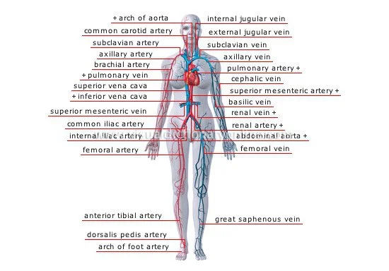
Superior Vena Cava (SVC): The superioe vena cava is a large vein that carries deoxygenated blood from the upper half of the body (head, neck, arms, and upper torso) to the right atrium of the heart.
- Superior vena cava are as follows:
- Internal jugular vein
- Lingual vein
- Dorsal lingual veins
- Sublingual vein
- Deep lingual vein
- Superior thyroid vein
- Middle thyroid veins
- Sternocleidomastoid vein
- Superior laryngeal vein
- Facial vein
- Angular vein
- Supratrochlear veins
- Supra-orbital vein
- External nasal veins
- Deep facial vein
- External palatine vein
- Submental vein
- Retromandibular vein
- Superficial temporal veins
- Middle temporal vein
- Transverse facial vein
- Maxillary veins
- Pterygoid plexus
- External jugular vein
- Posterior auricular vein
- Anterior jugular vein
- Suprascapular vein
- Transverse cervical veins
- Dural venous sinuses
- Transverse sinus
- Confluence of sinuses
- Marginal sinus
- Occipital sinus
- Petrosquamous sinus
- Sigmoid sinus
- Superior sagittal sinus
- Inferior sagittal sinus
- Straight sinus
- Inferior petrosal sinus
- Superior petrosal sinus
- Cavernous sinus
- Sphenoparietal sinus
- Diploic veins
- Emissary’s veins
- Cerebral veins
- Superficial cerebral veins
- Internal cerebral veins
- Basal vein
- Great cerebral vein
Inferior Vena Cava (IVC): This is the largest vein in the human body, and it carries deoxygenated blood from the lower half of the body (abdomen, pelvis, and legs) to the right atrium of the heart.
Inferior vena cava :
- Inferior phrenic veins
- Lumbar veins
- Ascending lumbar vein
- Hepatic veins
- Renal veins
- Right suprarenal vein
- Right ovarian vein
- Right testicular vein – Pampiniform plexus
- Common iliac vein
- Median sacral vein
- Iliolumbar vein
- Internal iliac vein
- Superior gluteal veins
- Inferior gluteal veins
- Obturator veins
- Lateral sacral veins
- Vesical veins
- Middle rectal veins
- Internal pudendal vein – It further includes three more veins, Deep veins of the clitoris, Deep veins of the penis, Inferior rectal veins
- Posterior labial veins
- Posterior scrotal veins
- External iliac vein
- Inferior epigastric vein
- Deep circumflex iliac vein
Brachiocephalic vein :
- Inferior thyroid vein
- Inferior laryngeal vein
- Pericardial veins
- Pericardiophrenic veins
- Bronchial veins
- The vein of the spine – Vein at the base of the skull and Vein in front of the spine are Occipital vein and Anterior vertebral vein, Deep cervical vein
- Internal thoracic veins: Superior epigastric veins, Musculophrenic veins, and Anterior intercostal veins are the internal thoracic veins.Supreme intercostal vein
- Azygos Vein: The azygos vein is a major vein on the right side of the body that drains blood from the posterior thoracic wall and empties into the Superior vena cava. It also plays a role in providing collateral circulation in case of superior vena cava obstruction.
- Hemiazygos Vein: This vein is located on the left side of the vertebral column and drains blood from the left side of the posterior thoracic wall. The usual connection is with the azygos vein or the accessory hemiazygos vein, and it eventually empties into the superior vena cava.
- Accessory Hemiazygos Vein: Similar to the hemiazygos vein, this vein is located on the left side of the vertebral column and drains blood from the left side of the posterior thoracic wall. It typically connects with the azygos vein or the hemiazygos vein and eventually drains into the superior vena cava.
Veins of abdomen
The abdominal region contains a complex network of veins that play essential roles in draining blood from various organs and tissues and returning it to the heart. Here are some of the key veins in the abdomen:
- Inferior Vena Cava (IVC): The Inferior vena cava, as mentioned earlier, is the largest vein in the body and receives blood from the lower half of the body, including the abdomen, pelvis, and lower limbs. It transports blood without oxygen back to the right atrium of the heart.
- Hepatic Veins: These veins drain blood from the liver and ultimately empty into the Inferior vena cava The hepatic veins are critical for carrying nutrient-rich blood away from the liver after it has been processed.
- Renal Veins: The renal veins remove the contents of blood from the organ of the human body named the kidneys. The left renal vein typically receives blood from the left kidney and then joins the Inferior vena cava, while the right renal vein directly empties into the Inferior vena cava.
Renal veins:
- Gonadal Veins: These veins drain blood from the gonads (testicles in males and ovaries in females). The Inferior vena cava frequently receives drainage directly from the right gonadal vein, whereas the left renal vein is usually the recipient of drainage from the left gonadal vein.
- Superior Mesenteric Vein (SMV): The superior mesenteric vein drains blood from the small intestine, cecum, ascending colon, and part of the transverse colon. Forming the portal vein, it combines with the splenic vein. Superior mesenteric vein:
- Right gastro-omental vein
- Ileocolic vein
- Appendicular vein
- Right colic vein
- Middle colic vein
- Inferior Mesenteric Vein (IMV): Blood from the descending colon, sigmoid colon, and rectum is drained by the Inferior Mesenteric Vein (IMV). It typically joins the splenic vein.
- Inferior mesenteric vein
- Left colic vein
- Sigmoid veins
- Superior rectal vein
- Inferior mesenteric vein
Portal Vein: The portal vein, although not an abdominal vein in a strict sense, should be noted as it gathers blood from the superior mesenteric vein and inferior mesenteric vein and transports it to the liver for processing. Later on, it combines with the hepatic veins and after a period of time it drains into the inferior vena cava.
Hepatic portal vein
The hepatic portal vein is a crucial blood vessel in the abdomen that plays a central role in the circulatory system, particularly in the liver’s function.
- Cystic vein
- Para-umbilical veins
- Left gastric vein
- Right gastric vein
- Splenic vein – Left gastro-omental vein
Veins of the upper limb
The veins of the upper limb are crucial for returning deoxygenated blood from the arm, forearm, and hand back to the heart. Here’s an overview of the major veins of the upper limb:
- Cephalic Vein: The Cephalic Vein, a prominent superficial vein in the upper limb, runs alongside the brachial artery and converges to form the axillary vein. It begins in the hand, typically from the dorsal venous network of the hand, and runs along the lateral aspect of the forearm and arm. The cephalic vein often courses proximally along the lateral border of the biceps brachii muscle and pierces the clavipectoral fascia to drain into the axillary vein near the deltopectoral triangle.
- Basilic Vein: Another prominent superficial vein, the basilic vein, begins in the hand from the dorsal venous network and ascends along the medial aspect of the forearm and arm. It typically courses proximally along the medial border of the biceps brachii muscle and penetrates the deep fascia to join the brachial veins or the axillary vein.
- Brachial Veins: These veins are formed by the union of the basilic and cephalic veins in the arm. The brachial artery is where they travel, and they come together to create the axillary vein.
- Subclavian Vein: Beyond the outer edge of the first rib, the axillary vein extends and is referred to as the subclavian vein. After passing beneath the clavicle, it combines with the internal jugular vein to create the brachiocephalic vein.
- Axillary Vein: The axillary vein begins at the lower border of the teres major muscle and extends to the outer border of the first rib, where it becomes the subclavian vein. It receives blood from the brachial veins and the basilic vein and is a major conduit for blood flow from the upper limb to the heart.
Axillary vein :
- Subscapular vein
- Circumflex scapular vein – The vein that surrounds the shoulder blade is known as the circumflex scapular vein, and it connects with the thoracodorsal vein. The posterior circumflex humeral vein and/or the anterior circumflex humeral vein are also joined with it.
- Lateral thoracic vein
Deep veins of upper limbs
- Radial Vein: Accompanying the radial artery along the lateral aspect of the forearm is the radial vein. It receives blood from the deep palmar venous arch, dorsal venous network, and radial collateral veins. The radial vein typically merges with the ulnar vein to form the brachial veins in the forearm.
- Ulnar Vein:The ulnar vein travels alongside the ulnar artery on the inner aspect of the forearm. It receives blood from the deep palmar venous arch, dorsal venous network, and ulnar collateral veins. The radial vein typically combines with the ulnar vein to create the brachial veins in the forearm.
Veins of lower limbs
The veins of the lower limbs play a crucial role in returning deoxygenated blood from the legs and feet back to the heart. Here’s an overview of the major veins of the lower limbs:
- Great Saphenous Vein: The great saphenous vein is one of the longest veins in the body. It originates from the dorsal venous arch and ascends along the medial aspect of the leg and thigh. It receives blood from tributaries such as the anterior and posterior accessory saphenous veins. The great saphenous vein typically joins the femoral vein in the groin region. It also includes external pudendal veins.
- Small Saphenous Vein: The small saphenous vein begins on the lateral aspect of the foot and runs upward along the back of the leg. It typically courses along the posterior aspect of the leg, receiving blood from tributaries such as the sural veins. The small saphenous vein typically connects with the popliteal vein at the back of the knee.
Deep veins of lower limbs
- Femoral Vein: In the thigh, there is a deep vein called the femoral vein. The popliteal vein runs alongside the femoral artery and collects blood from the great saphenous vein, deep femoral vein, and other smaller veins. As it goes under the inguinal ligament, the femoral vein substitute into the external iliac vein.
- Profundus femoris vein – A significant vein situated in the thigh is known as the “profundus femoris vein,” also referred to as the “deep femoral vein” or “profunda femoris vein.”. It receives blood from various tributaries in the thigh, including branches from the muscular compartments of the thigh, and it contributes to the venous drainage of the lower limb.
- Popliteal Vein: The popliteal vein continues from the femoral vein as it traverses the adductor hiatus of the adductor magnus muscle. It receives blood from the small saphenous vein, deep veins of the leg, and knee-joint veins. The popliteal vein becomes the femoral vein as it passes above the knee.
Popliteal vein :
- Sural veins
- Anterior tibial veins
- Posterior tibial veins
- Fibular veins
Veins of the vertebral column
- Anterior internal vertebral venous plexus
- Basivertebral veins
- Anterior spinal veins
- Posterior spinal veins
Types of Veins: The size, location, and function of veins are used to categorize them. veins include those that are large in diameter and play significant roles in the systemic circulation, such as the superior and inferior vena cava, which return blood from the upper and lower body to the heart, respectively. Major veins often have accompanying arteries of the same name and serve specific regions of the body.
Functions of Veins: The primary function of veins is to return deoxygenated blood from the body’s tissues back to the heart and lungs for reoxygenation. This process is essential for maintaining adequate oxygen supply to tissues and organs. Additionally, veins contain valves that prevent the backflow of blood and facilitate unidirectional flow toward the heart, especially in regions where gravitational forces may impede blood return, such as the lower extremities.
Clinical Significance: Veins play a crucial role in various medical procedures and diagnostic techniques. They are commonly used for venipuncture to obtain blood samples for laboratory testing, intravenous (IV) therapy for medication administration, and surgical procedures such as vein grafting for coronary artery bypass surgery. Vein abnormalities or diseases, such as deep vein thrombosis (DVT) or varicose veins, can have significant implications for cardiovascular health and may require medical intervention.
Disclaimer: The information provided about veins in the human body is intended for general informational purposes only. It is not meant to serve as a replacement for expert medical diagnosis, guidance, or care. Never be embarrassed to get advice from your doctor or another qualified healthcare provider when you have questions about a medical issue.
We strive to provide accurate and up-to-date information. However, the content may not always reflect the most current medical research or developments. Therefore, we make no representation or warranty of any kind, express or implied, regarding the completeness, accuracy, reliability, suitability, or availability of the information provided. You accept all risk when you make use of any information in this content. We shall not be liable for any loss, injury, or damage arising from or related to the use of the information provided.
FAQs
How many veins are in the human body?
The vena cava is the primary vein in your body and is located in the upper right part of your chest, known as the superior vena cava. Its function is to transport blood from your head, neck, arms, and chest back to your heart.
What are the 4 major veins?
The major vessels connected to the heart are the aorta, pulmonary trunk, pulmonary veins, and vena cava (both superior and inferior).
What are the 4 cardiac veins?
The coronary sinus receives drainage from the following tributaries: Great cardiac vein.
Oblique vein of the left atrium.
Posterior vein of the left ventricle.
Middle cardiac vein.
Small cardiac vein.
Which vein is most important?
The jugular veins play a crucial role as the head, particularly the brain, has a significant oxygen demand. Despite the average human brain weighing around 3 lbs., it receives approximately 15% to 20% of the blood pumped out by the heart.
Which are the thinnest veins in the body?
Capillaries – Tiny, extremely thin-walled vessels known as capillaries serve as a connection between arteries (which carry blood away from the heart) and veins (which carry blood back to the heart).
What are the six large veins?
The large blood vessels connected to the heart are known as the great vessels. These include the inferior vena cava, superior vena cava, pulmonary arteries, pulmonary veins, and the root of the aorta.
What color are veins?
The hemoglobin in deoxygenated blood traveling through your veins behaves in the same way. Although your veins reflect a dark red color, your skin appears more blue than red, creating an optical illusion that makes our veins look blue, green, or purple despite the actual redness of our blood!
Which vein is closest to the heart?
The blood from your lower legs is carried by your saphenous veins to the veins in your upper legs. From there, the blood continues its journey upward to your inferior vena cava, which is a large vein that directly empties blood into your heart.
What are the three common veins?
The main superficial veins include the basilic, cephalic, and median cubital veins. The basilic and cephalic veins originate around the wrist and extend towards the upper part of the forearm.
What is the most popular vein?
The superficial veins of the upper limb, particularly the dorsal metacarpal veins and median cubital vein, are common sites for venous access. While dorsal metacarpal veins are like better for venous cannulation, there is bounded information available regarding their anatomical variation.
What is the complete number of veins in the human body?
The human body contains veins and arteries that distribute blood to all areas of the body, comprising at least 34 major veins and numerous smaller veins that link with the capillaries.