Vastus intermedius muscle: Anatomy, Function, Exercise
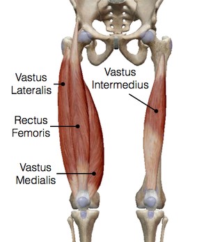
The vastus intermedius muscle is a muscle in the thigh. It is located in the quadriceps region, between the other three quadriceps muscles: the rectus femoris, the vastus lateralis, and the vastus medialis. Like the other quadriceps muscles, the vastus intermedius functions as an extensor of the knee. The muscle also plays a role in helping to stabilize the Knee joint.
Origin of Vastus imtermedius
Originating from the upper two-thirds of the anterior and lateral surfaces of the femur and the intermuscular septum
The origin of the vastus intermedius is along the lateral side of the femur.
Insertion
The fibers of the vastus intermedius fan out and insert along the lateral side of the knee, form the deep part of the tendon and
then inserts into the lateral side of the patella.
Blood supply
The descending branch of the lateral circumference femoral artery.
Innervation
The Vastus Intermedius muscle nerve supply from a branch of the Femoral nerve, originating from lumbar nerve roots are 2, 3, and 4 nerves.
How to Assess of Vastus intermedius muscle?
Assessment of the muscle cannot be done separately, therefore quadriceps muscles examination and test for knee extensor is used for its power.
How to do Palpation of Muscle?
The muscle alone is difficult to palpate. It is located under the rectus femoris muscle, the least superficial muscle in the thigh’s muscles; therefore, it cannot be isolated for exercise and/or massage.
How to stretch the Vastus intermedius muscle?
This muscle is difficult to stretch separately, After maximum knee flexion is achieved, extra internal rotation is helpful, However, it cannot be proved that it separately stretches the muscle. It is not a two-joint muscle, therefore it cannot be further stretched by hip extension as the rectus femoris can. It is also not manipulated with massage therapy to stretch the fibres sideways as the vastus lateralis and vastus medialis muscles are.
What is the main function of the vastus intermedius?
The main function of the Vastus intermedius is the Knee extension in the Middle range of Knee movement.
What muscle is under the vastus intermedius?
Vastus Intermedius is located in the middle of thigh, Below the Rectus femoris in the anterior compartment of the thigh and while medial side located Vastus medialis, and lateral side located Vastus Lateralis.
Is vastus intermedius a flexor?
How do I strengthen my vastus intermedius?
Vastus intermedius cannot be strengthened separately, Quadriceps muscle strengthening exercises also strengthen the Vastus intermedius muscle.
How do you train vastus intermedius?
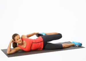
If you want to train your vastus intermedius, you should do regular exercise of the quadriceps muscles mainly stretching exercises and strengthening exercises.
Why is it called vastus intermedius?
The muscle is located in between or in the middle of the thigh, underneath the Rectus femoris and medial side located Vastus medialis, and the lateral side located Vastus Lateralis.
Clinical Importance
The vastus intermedius tear is mainly a sports injury, mainly overuse sports injury, in which pain and spasms in the front of the high with also knee pain and painful walking are the symptoms associated.
Vastus intermedius pain treatment mainly uses of RICE Principle (Rest, Elevation, Ice, Compression) for 2 to 3 weeks depending upon the level of Injury. After the rest period is over, regular stretching and strengthening exercises are advised for you.