Layers of Heart
What is the Heart?
The pump that circulates blood throughout your body is the heart, a fist-sized organ. It serves as the main component of your circulatory system.
The heart is composed of four main chambers that are driven by electrical impulses and are composed of muscle. Your heart’s operation is controlled by your brain and nerve system.
Function
The primary job of the heart is to pump blood throughout your body. Additionally, your heart
- Regulates the heart rate’s rhythm and tempo.
- keeps your blood pressure stable.
How do other organs and your heart function together?
Your heart regulates your heart rate and other bodily processes in concert with other systems. The principal frameworks are:
Nervous system:
Your heart rate is regulated in part by your neurological system. Your heart beats more quickly during activity and more slowly during rest thanks to the messages it delivers.
Endocrine system:
Hormones are released by the endocrine system in humans. Your blood pressure is influenced by these hormones, which instruct your blood vessels to dilate or widen. Your thyroid gland’s hormones can alter the rate at which your heart beats.
Structure of Heart
The human heart is a muscular organ with four chambers that is roughly the size and shape of a closed fist, with two-thirds of its mass located to the left of the midline.
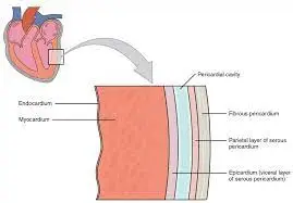
The parietal layers of a serous membrane line the pericardial sac, which encloses the heart. The epicardium is the outermost layer of the heart wall, the myocardium is the intermediate layer, and the endocardium is the innermost layer.
Layers of the Heart Wall
3 layers of tissue form the heart wall. The heart walls have three layers the epicardium, which is the outermost, the myocardium, which is the intermediate layer, and the endocardium, which is the innermost.
Chambers of the Heart
There are four chambers within the heart’s interior cavity:
- Right atrium
- Right ventricle
- Left atrium
- Left ventricle
The blood flows into the two thin-walled chambers called atria. The heart’s two ventricles are powerful chambers with thick walls that yank blood out of the organ. Variations in the quantity of myocardium present, which represents the amount of force each chamber must provide, cause differences in the thickness of the heart chamber walls.
Systemic veins supply deoxygenated blood to the right atrium, while pulmonary veins supply oxygenated blood to the left atrium.
Valves of the Heart
The heart, like pumps, requires a series of valves to maintain the fluid flow in a single direction. Two different kinds of valves in the heart maintain the blood’s proper flow.
Semilunar valves are found at the bases of the major veins that exit the ventricles, whereas atrioventricular valves, also known as cuspid valves, are found between the atria and ventricles.
The tricuspid valve is the right atrioventricular valve. The bicuspid, or mitral, valve is part of the left atrioventricular system. The pulmonary semilunar valve is located between the pulmonary trunk and the right ventricle. The left ventricle and the aorta are separated by the aortic semilunar valve.
Atrioventricular valves seal during ventricle contraction to stop blood from returning to the atria. Semilunar valves close to stop blood from returning to the ventricles when the ventricles relax.
Pathway of Blood through the Heart
The atria and ventricles contract simultaneously, despite the convenience of first describing the blood flow through the right side of the heart before moving on to the left. The heart functions as a pair of simultaneous pumps, one on the left and one on the right. After passing through the right ventricle and the right atrium, blood is pushed to the lungs where oxygen is taken in. Blood travels from the lungs to the left atrium and subsequently to the left ventricle. It is pumped to the systemic circulation from there.
Blood Supply to the heart
As a working muscle, the heart wall’s myocardium requires a steady flow of nutrients and oxygen to keep it functioning properly. Because of this, the heart muscle contains a vast network of blood capillaries that carry oxygen to the contracting cells and eliminate waste.
Blood is supplied to the walls of the myocardium by the ascending aorta’s left and right coronary arteries. Blood travels via the myocardium’s capillaries before entering the cardiac (coronary) vein system. The coronary sinus, which opens into the right atrium, receives the majority of the cardiac veins’ drainage.
The Layers of the Heart Wall
The heart wall is made up of connective tissue, endothelium, and cardiac muscle. It is the cardiac muscle that allows the heart to contract and enables the synchronization of the heartbeat. Three layers consist of the heart wall: the epicardium, myocardium, and endocardium.
Epicardium: the heart’s outermost layer of protection.
Myocardium: the heart’s muscular middle layer wall.
Endocardium: the inner layer of the heart.
Epicardium–The 1st layer
The outer layer of the heart wall is called the epicardium, or epicardium. Since it makes up the pericardium’s interior layer, it is also referred to as the visceral pericardium. Adipose tissue and elastic fibers make up the majority of the loose connective tissue that makes up the epicardium.
The epicardium helps to produce pericardial fluid and protects the inner layers of the heart. This fluid lowers friction between the pericardial membranes by filling the pericardial space. The coronary blood arteries, which carry blood to the heart wall, are also located in this layer of the heart. The epicardium’s inner layer and the myocardium are near one another.
What is the function of the epicardium?
Beyond functioning as the heart’s outermost layer of protection, the epicardium has a variety of other purposes. It has a protective purpose and is the deepest layer of the pericardium, the membrane that surrounds the heart. In addition, the epicardium secretes substances critical to the survival and proliferation of cardiomyocytes, or the heart muscle cells, and provides signals for appropriate heart development and maturation inside the embryo.
Additionally, the cells that originate from the epicardium can serve as producers of cardiac cells, or cells that can differentiate into a wide variety of other cell types. The heart’s damage response to heart illness, including myocardial infarctions (heart attacks), and the subsequent regeneration of the heart, are influenced by epicardial signals.
What distinguishes the epicardium from the pericardium?
A component of the pericardium is the epicardium. It is known as the visceral layer of the serous pericardium and is the innermost layer. The terminology and the particular cardiac layers that they are discussing are the primary differences. The parietal layer of the serous pericardium, the visceral layer of the serous pericardium, and the outer fibrous layer, often known as the fibrous pericardium, are the three distinct layers that surround the heart in the pericardium.
What are the key facts that one should know about the epicardium?
The outermost layer of the heart is called the epicardium, and it is the innermost layer of the pericardium. It is made up of connective tissue, fat, and mesothelial cells. It is next to the middle layer of muscle tissue called the myocardium. The term “endocardium” refers to the innermost layer.
The visceral section of the serous layer is formed by the epicardium, which is partially comprised of the pericardium. The epicardium performs a variety of tasks, including guarding the heart, generating substances that aid in the healthy development of cardiac cells, and guaranteeing a suitable reaction to damage to cardiac cells.
Myocardium-The 2nd layer
In the heart wall, the middle layer is called the myocardium (myocardium). It is made up of the cardiac muscle fibers that allow the heart to contract. The heart wall’s thickest layer, the myocardium, varies in thickness depending on the location of the heart. Since the left ventricle produces the energy required to pump oxygenated blood from the heart to the rest of the body, its myocardium is the thickest. The peripheral nervous system regulates involuntary activities such as heart rate and cardiac muscle contractions.
Specialized myocardial muscle fibers enable cardiac conduction. These fiber bundles, which are made up of Purkinje fibers and the atrioventricular bundle, transport electrical impulses from the heart’s core to the ventricles.
Location: It is the middle layer situated between the epicardium and the endocardium.
Myocardium Structure
According to histology, the heart muscle cells (cardiomyocytes), which are grouped into a group known as myofibers, plus a small number of fibroblast cells make up the myocardium. The intercalated discs serve as a link between the cardiomyocytes.
It is the heart wall’s thickest layer anatomically. Specifically in the left ventricle, which has the thickest layer, it is thicker in the ventricular wall than in the auricular wall. Three sub-layers comprise the left ventricular myocardium: the subepicardial, mid-myocardial, and subendocardial layers.
It has an abundance of cardiac lymphatic channels, coronary veins, and coronary arteries. Additionally, a cardiac conduction system made up of specific cardiomyocytes innervates it.
Myocardium-associated Diseases
The myocardium is affected by a wide range of diseases collectively known as myocardial diseases. Some of the typical myocardial diseases are given below:
| Disease | Introduction |
| Myocardial Infraction | Blockage of the blood flow in the myocardium leads to a heart attack. |
| Myocarditis | Inflammation of the myocardium |
| Myocardial fibrosis | A condition of the formation of excessive fibrous tissue in the myocardium. |
| Myocardial ischemia | Reduction in the oxygen supply to the myocardium due to restriction in blood flow. |
Endocardium
The endocardium is the smooth, thin tissue that lines the heart’s chambers and valves. The deepest layer of the heart’s walls has all of the blood arteries that are required and acts as a barrier between the bloodstream and the cardiac muscles. The conduction system of the heart, which controls the contraction of the cardiac muscles, is also housed there.
Because the endocardium plays such an important function in controlling the heartbeat and pumping blood through the heart, issues with it can have a significant negative impact on health. The most prominent of them is endocarditis, which is an infection and inflammation of these tissues that mostly affects the valves.
Let’s quickly review the endocardium’s structure, functions, and relationship to various health issues.
Function of endocardium
The four chambers of the heart, which are responsible for pumping blood throughout the body, are covered with endocardium. It performs two crucial roles as the heart’s innermost layer of walls:
Anatomic function: The endocardium, a tissue lining the inside of the heart, maintains blood flow through the organ independent of the myocardium, or cardiac muscles. It also covers the valves that open and close to control how much blood flows through the heart’s chambers.
Conduction system: Electrical signals that pass through endocardial-embedded nerves control heart rhythm and activity. Because of the connections between these neurons and the myocardium, the muscle pumps blood throughout the body by contracting and relaxing.
Anatomy
It’s crucial to understand the endocardium’s location and structure in addition to what it accomplishes.
Location
The endocardium, which is the heart’s inner lining, is located along the walls of the left and right ventricles as well as the left and right atria. Furthermore, the outer layer of the tricuspid, pulmonary, mitral, and aortic valves—which act as portals between the chambers—represents this tissue. The walls of the heart are composed of three layers, one of which is the endocardium. It connects to the myocardium, the thickest layer made up of the heart muscles, as the innermost of these. The epicardium, a tissue that envelops the myocardial, houses the principal arteries, veins, and nerves that supply the heart.
Structure endocardium
The three sublayers that make up the endocardium determine its purpose. These are the following:
The innermost layer called the endothelium, regulates all material exchange between the heart muscles and the circulation. It is made up of specialized endothelial cells, the same kind that lines the veins and arteries.
The smooth muscle in the elastic tissue layer presses against the veins that go through it, and the connective tissue.
The outermost layer of the endocardium that connects to the heart muscle is called the subendocardial layer. It has Purkinje fibers, which carry electrical signals to the myocardium, and fibrous collagen cells, which give structure and stability, in addition to nerves and arteries.
Endocardial associate disease
| Diasease | Disease |
| Infective Endocarditis | Inflammation of the endocardium due to infection. |
| Endocardial Fibroelastosis | A condition where the elastic and fibrous layer of the endocardium is thickened. |
| Nonbacterial Thrombotic Endocarditis (NBTE) | A condition characterized by the formation of a blood clot on the endocardium, leading to inflammation. |
| Libman-Sacks Endocarditis | A condition is characterized by the formation of a blood clot on the endocardium, leading to inflammation. |
| Carcinoid Heart Disease (CHD) | A condition characterized by forming small sterile vegetations, primarily on the valves. |
Endocarditis
An infection and inflammation of the endocardium is called endocarditis. It is the most important and prevalent endocardial ailment. In summary:
Symptoms: A variety of symptoms, such as fever, chills, exhaustion, chest pains, edema in the belly or limbs, night sweats, and aches and pains in the muscles and joints, are associated with endocarditis. There are wider health repercussions if left untreated.
Causes: Although it can have other causes, this disease is typically brought on by an infection with bacteria known as infective endocarditis.
Diagnosis: Blood tests and cardiac imaging, including magnetic resonance imaging (MRI), cardiac angiography, and echocardiograms, are used by clinicians to diagnose this illness if it is suspected.
Treatment: The primary line of treatment is intravenous (IV) antibiotics; however, surgery may be used if these don’t yield results.
Prognosis: Endocarditis has a bad prognosis, with 10 to 26% of patients having in-hospital mortality and 60 to 70% projected to live at five years. The ailment typically occurs in people who have had previous heart problems or who have artificial valves. The patient’s age and past medical history have a significant influence on this illness.
MVP, or mitral valve prolapse
An unusually thick tissue on the mitral valve (between the left ventricle and atrium), including the endocardium, is the defining feature of this congenital disease. Blood spills back into the left atrium as a result of MVP causing the valve to “flop” back into place.
It is mostly asymptomatic, although symptoms include weariness, chest discomfort, and occasional episodes of fast heartbeat. Imaging methods, such as echocardiograms (echo), are used to detect it. Generally, no treatment is needed; however, if the condition progresses to the point where valve leakage occurs, intervention is required to stop a heart attack or stroke.
Heart Cancer
Hedinger syndrome, which is often called “carcinoid syndrome,” is a group of illnesses caused by the overproduction of hormones. Therefore, among the symptoms are facial flushing, persistent diarrhea, hypotension, and so forth.
This may eventually result in right heart failure, which can cause weariness, leg edema, fast and irregular heartbeats, and sharp swings in weight. Standard cardiac imaging methods, including computer tomography (CT) scanning and electrocardiograms (EKG), are used to diagnose it. Treatment options include medication or surgical excision of the troublesome tissues.
FAQ
Which four components of the heart are named?
The heart’s four chambers
A little collecting chamber called an “atrium” and a big pumping chamber called a “ventricle” are located on either side of the wall. There are two upper chambers, the left and right atriums, and two bottom chambers, the left and right ventricles.
Which three layers make up the pericardium?
Pericardium: Structure and Purpose
membranes pericardiales
The outer fibrous sac covering the heart is called the fibrous pericardium.
The layer that lies between the visceral and fibrous pericardia is called the parietal pericardium.
The inner layer of the pericardium and the outer layer of the heart wall are together referred to as the visceral pericardium.
What is epicardium known by another name?
Epicardium: Definition, Uses, and Additional Information Osmotic
A component of the pericardium is the epicardium. It is known as the visceral layer of the serous pericardium and is the innermost layer.
What does endocardium mean?
The endocardium is the smooth, thin tissue that lines the heart’s chambers and valves. The deepest layer of the heart’s walls has all the blood arteries that are required and act as a barrier between the lifeblood and the cardiac muscles.
The myocardium is placed where?
Cardiomyocytes make up the myocardium, the layer of muscle that makes up the heart. The heart’s myocardium, which is thinner in the atria and thicker in the ventricles, is present in the walls of all four chambers.
References
- Bailey, Regina. “The 3 Layers of the Heart Wall.” ThoughtCo, Apr. 5, 2023, thoughtco.com/the-heart-wall-4022792.
- Professional, C. C. M. (n.d.). Heart. Cleveland Clinic. https://my.clevelandclinic.org/health/body/21704-heart
- Structure of the heart | SEER training. (n.d.). https://training.seer.cancer.gov/anatomy/cardiovascular/heart/structure.html
- Gurarie, M. (2023, August 11). Definition and function of endocardium. Verywell Health. https://www.verywellhealth.com/endocardium-definition-5088789
- Osmosis – epicardium: what is it, functions, and more. (n.d.). Osmosis. https://www.osmosis.org/answers/epicardium
- Dahal, P. (2023, August 6). Layers of the heart: epicardium, myocardium, endocardium. Microbe Notes. https://microbenotes.com/layers-of-the-heart/#2-myocardium-middle-layer-of-heart