Development of the nervous system
Introduction
The brain is a compound organ comprised of gray parts and white matter, which can be hard to differentiate Starting from an embryologic position allows us to understand more easily how the parts relate or connect to each other. The embryonic nervous system initiates as a very simple structure—fundamentally just a straight line, which then gets increasingly compound.
Many structures that are seen to be adjoining in the adult brain are not connected, and the connections that exist may seem random But there is an underlying order to the system that derives from how different parts develop. By following the developmental pattern or figure, it is possible to learn what the major regions or areas of the nervous system are.
Neural Tube
To initiate a sperm cell and an ovum fuse to become a fertilized egg. The fertilized ovum, or zygote, starts dividing or splitting to generate (produce) the cells that make up a whole organism. Sixteen days after fertilization, the developing (mature)embryo’s cells belong to one of three germ layers that give rise (generate) to the various tissues in the body. The endoblast, or internal tissue, is responsible for generating (producing) the lining tissues of different spaces within (inside) the body, similarly the mucosae of the digestive and respiratory systems. The mesoderm, or middle tissue, gives rise (generates) to nearly all of the muscle and connective tissues. eventually, the ectoderm, or outer tissue, get mature into the integumentary system (the skin) and the nervous system. It is likely not difficult or hard to see that the outer tissue of the embryo becomes the outer envelope of the body. But how is it answerable for the nervous system?
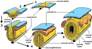
As the embryo get develops (matures) or is enlarged, a portion of the ectoderm differentiates into a specialized region or area of neuroectoderm, which is the precursor (forerunner)for the tissue of the nervous system. Molecular signals impact cells in this area to differentiate into the neuroepithelium, forming a neural plate. The cells then begin or initiate a change in shape, causing the tissue to clasp and fold inward (inside). A neural groove forms, noticeable as a line along with the dorsal surface of the embryo. The ridge or elevation-like border on either side of the neural groove is referred to or named the neural fold. As the neural folds come together and converge (mixed), the underlying structure forms into a tube just under the ectoderm called the neural tube.
Cells from the neural folds then individual(independent) from the ectoderm to form a cluster of cells referred to as the neural crest, which runs lateral side to the neural tube. The neural crest runs apart from the mature or embryonic, central nervous system (CNS) that will form along with the neural groove and develops into some parts of the peripheral nervous system (PNS), comprised of the enteric nervous tissue. Many tissues that are not part or element of the nervous system also get arising from the neural crest, similar to craniofacial cartilage and bone, and melanocytes (pigmentation cells).
At this point or margin, the early nervous system is a simple, hollow tube. It course from the anterior end (front side) of the embryo to the posterior end. initiate at 25 days, the anterior(front) end develops into the brain, and the posterior portion (region )becomes the spinal cord. This is the most general arrangement alignment of tissue in the nervous system, and it gives origin to more compound structures by the 4th week of development or growth.
Primary Vesicles
As the anterior end of the neural tube starts to develop into the brain, it undergoes a couple of enlargements (expansion) the result is the production (making) of sac-like vesicles. same as the child’s balloon animal, the long, straight neural tube starts to take on a new shape. 3 vesicles construct at the first stage, which is called primary vesicles.
The prosencephalon – is the forward-most vesicle, and the name can be loosely translated to mean forebrain. The mesencephalon(middle) is the next vesicle, which can be termed the midbrain. The 3rd vesicle at this stage is the rhombencephalon. The first part of this word is also the root of the word rhombus, which is a geometrical figure with 4 sides of equal length (a square is a rhombus with 90° angles). Whereas prosencephalon and mesencephalon translate into the English words forebrain and midbrain, there is no word for 4 sided-figure brains.” However, the third vesicle can be called the hindbrain. One way of analysis about how the brain is scheduled is to use these three areas—forebrain, midbrain, and hindbrain which are based on the primary vesicle stage of maturity or stage of the growth
Secondary Vesicles
The brain continues to develop, and the vesicles differentiate again. The 3 primary vesicles become (set off) 5 secondary vesicles. The prosencephalon develops into two new vesicles called the telencephalon and the diencephalon. The telencephalon will become the cerebrum. The diencephalon gives origin to different adult structures; 2 that will be very important are the thalamus and the hypothalamus. In the embryonic diencephalon, a structure known as the eye cup growth will in the end become the retina, the nervous tissue of the eye called the retina. This is an uncommon example of nervous tissue developing as part of the central nervous system structures in the embryo, but becoming a peripheral structure in the fully formed nervous system.
The mesencephalon does not modify into any finer divisions. The midbrain is a settled region of the brain at the primary vesicle stage of growth and residue that way. The rest of the brain develops around it and comprises a large percentage of the mass of the brain. splitting the brain into the forebrain (front), midbrain (middle), and hindbrain(back) is useful in comprising its developmental pattern, but the midbrain is a small portion of the whole brain, relatively speaking.
The rhombencephalon gets matured into the metencephalon and myelencephalon. The metencephalon counterpoise to the adult structure known as the pons and also gives origin to the cerebellum. The cerebellum is accountable for about 10 percent of the mass of the brain and is a very important structure in itself. The most remarkable connection between the cerebellum and the leftover brain is at the pons because the pons and cerebellum develop out of the same vesicle. The myelencephalon corresponds to the adult structure named the medulla oblongata. The structures that come from the mesencephalon and rhombencephalon, except for the cerebellum, collectively comprise the brainstem, which remarkably comprises the midbrain, pons, and medulla.
Spinal Cord Development
during the brain developing from the anterior neural tube, and The spinal cord is developing from the posterior neural tube. However, its structure does not vary from the basic arrangement of the neural tube. It is a long, straight cord with a small, hollow (empty) space down the center. The neural tube is defined in terms of its anterior versus posterior parts, but it also has an anterior–posterior measurement. As the neural tube splits from the rest of the ectoderm, the side nearest to the surface is dorsal, and the deeper side is ventral.
As the spinal cord develops, the cells comprising the wall of the neural tube proliferate(multiply)and are distinguished into the neurons and glia of the spinal cord. The dorsal tissues will be associated with sensory functions, and the ventral tissues will be connected with motor functions.
Relating Embryonic Development to the Adult Brain
- Embryonic development can assist in understanding the anatomy of the adult brain because it establishes a groundwork on which more compound structures can be built. First, the neural tube establishes the anterior–posterior measurement of the nervous system, which is termed the neuraxis. The embryonic nervous system in mammals can be said to have a standard positioning. Humans (and other primates, to some degree) make this elaborate by standing up and walking on two legs. The anterior–posterior measurement of the neuraxis overlays the superior–inferior measurement of the body. However, there is a major curve between the brainstem and forebrain, which is termed the cephalic flexure. Because of this, the neuraxis begins in an inferior position the end of the spinal cord, and ends in an anterior position, the forepart of the cerebrum.
- the primary vesicles help to fix the basic areas of the nervous system: forebrain, midbrain, and hindbrain. These divisions are useful in definite situations, but they are not equal regions. The midbrain is small compared with the hindbrain and especially the forebrain. The secondary vesicles go on to settle the major areas of the adult nervous system. The telencephalon is the cerebrum, which is the major part of the human brain. The diencephalon continues to be suggested by this Greek name because there is no better term for it (dia- = “through”). The diencephalon is between the cerebrum and the rest of the nervous system and can be reported as the region via which all predictions have to pass between the cerebrum and everything else. The brainstem comprises the midbrain, pons, and medulla oblongata, which communicate to the mesencephalon, metencephalon, and myelencephalon. The cerebellum, being a large part of the brain, is contemplated as a separate region.
- One other benefit of contemplating embryonic growth is that definite connections are more evident because of how these adult anatomies are related. The retina, which begins as part of the diencephalon, is primarily connected to the diencephalon. The eyes are just below the anterior-most part of the cerebrum, but the optic nerve expands back to the thalamus as the optic tract, with branches into an area of the hypothalamus. There is also a connection between the optic tract to the midbrain, but the mesencephalon is near the diencephalon, so that is not difficult to visualize. The cerebellum get originates out of the metencephalon, and its largest white matter connection is to the pons, also from the metencephalon. There are connections between the cerebellum and both the medulla and midbrain, which are the nearest structures in the secondary vesicle stage of growth. In the adult brain, the cerebellum seems close to the cerebrum, but there is an indirect connection between them.
- Another aspect of the adult central nervous system(CNS) structures that are related to embryonic growth is the ventricles—open spaces within the central nervous system (CNS)where cerebrospinal fluid (CSF) circulates. They are the remains of the empty center of the neural tube. The 4 ventricles and the tubular spaces related to them can be linked back to the empty center of the embryonic brain
Disorder of the …
Development of the Nervous System: Spina Bifida
- The early formation of the nervous system depends on the formation of the neural tube. A groove forms along the dorsal surface of the embryo, which becomes deeper until its border meets and closes off to form the tube. If this fails to occur, especially in the posterior aspect where the spinal cord forms, a developmental deficit called spina bifida occurs. The closing of the neural tube is very important for more than just the proper development of the nervous system. The surrounding tissues are dependent on the proper growth of the tube. The connective tissues surrounding the central nervous system can be involved as well.
- There are three classes of this disease; occult, meningocele, and myelomeningocele. The first type, spina bifida occulta, is the mildest because the vertebral bones do not fully nearby the spinal cord, but the spinal cord itself is not affected. No functional dissimilarity may be observed which is what the word occult means; it is hidden or secret spina bifida. The other two types both include the formation of a cyst—a fluid-filled sac of the connective tissues that wrapped the spinal cord called the meninges. “Meningocele” means that the meninges protrude via the spinal column but nerves may not be included and few symptoms are present, though complications may begin later in life. “Myelomeningocele” means that the meninges protrude and spinal nerves are included and therefore severe neurological symptoms can occur.
- frequently surgery to close the opening or to eliminate the cyst is important. The earlier that surgery can be performed, the better the chances of controlling or limiting forward damage or infection at the opening. For many children with meningocele, surgery will reduce the pain, although they may feel some functional loss. Because the myelomeningocele form of spina bifida includes more extensive damage to the nervous tissue, neurological damage may continue but symptoms can frequently be handled. Complications of the spinal cord may present at later stages in life, but overall life expectancy is not decreased.
FAQ
What happens when your nervous system is off?
a person may feel the sudden onset of one or more symptoms, similar to tingling, Numbness, weakness, or inability to move a part or all of one side of the body (paralysis). Dimness, blurring, loss of vision, or double vision in one or both eyes. Loss of speech, trouble in understanding the speech.
What is the development of the nervous system?
The nervous system develops from the ectoderm following an inducible signal from the mesoderm. The initial mesodermal cells precipitate to form the notochord, which elongates under the primitive band along the with the anterior—posterior axis of the developing embryo.
What are 5 amazing facts about the nervous system?
There are two portion of the nervous system.
Neurons are made of three parts.
Neurons may look different from one another.
There are two types of nervous systems.
The body has billions of nerve cells.
Neurons are programmed to do different things.
What are 3 common complication of the nervous system?
Disorders of the nervous system
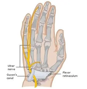
Structural disorders, similar as brain or spinal cord injury, cervical spondylosis , Bells palsy,carpal tunnel syndrome,peripheral neuropathy, and Guillain-Barré syndrome,. brain or spinal cord tumors,
Infections, similar as meningitis, encephalitis, polio, and epidural abscess.
Can we live without the nervous system?
Our Circulatory System and Nervous System We could not live without nervous system Our circulatory and nervous system are connected via organ and bodily functions. Our brains regulate these functions via our nervous system.
Which food is good for nerves weakness?
Six considerable Plant-Based Foods to Fight Nerve Pain
Green and leafy vegetables. spinach,broccoli and asparagus all comprises fibers, vitamin B, a nutrient important for nerve regeneration or nerve regrowth and nerve function.
Fruits. Eat more than one fruit daily to help heal damaged nerves.
Zucchini.
Sweet potato.
Quinoa.
Avocado.
At what age does the nervous system develop?
At just six weeks, the embryo’s brain and nervous system start to develop, although the compound parts of the brain continue to grow and develop via the end of pregnancy, with growth ending around the age of 25.
At what age is the nervous system gets fully developed?
about 25 years old
In reality, most of the building blocks of the nervous system are in place when we’re born. But some very important anatomy and pathways aren’t well mature until we’re about 25 years old. Even after that, we carry on with learning, grow, and changing.
Is the nervous system necessary?
The nervous system plays an important role in nearly every aspect of our health and well-being. It mentors everyday activities similar to waking up; automatic activities similar to breathing; and complex processes like thinking, reading, remembering, and feeling emotions.
How do you look after the nervous system?
Steps to keep the central nervous system healthy
do Exercise on a daily basis.
Get enough sleep.
Expose the body to sunlight.
Add meditation to the daily routine for good health.
Walk barefoot.
Drink green tea.
The food you eat matters.
Which fruit is best for nerves?
Antioxidants
This can be influential to optimize the myelin sheath and prevent any damage to the nerve. Good sources of antioxidants are berries similar to blueberries, blackberries, or raspberries, dark leafy greens, fatty fish, and walnuts. Try to intake rainbow food every day!
Which drink is good for nerves?
rather than coffee or energy drinks, try green tea. It has less than half the caffeine of coffee and includes healthy antioxidants, as well as theanine, an amino acid that has a soothing effect on the nervous system.
What helps improve nerves?
pomegranates, bananas, oranges, and prunes, which are very good sources of potassium, Vitamin B — Vitamins B1, B2, and B6 help the nerves to send impulses from the brain to the body.,while milk, leafy greens, and eggs are rich sources of calcium.
Which brain function develops first?
even if the brainstem is the first part of the brain to develop or mature, the higher parts are evolving at the same time but at different rates.
Which is the largest nervous system in the human body?
The central nervous system (CNS) is the largest portion or largest part of the nervous system and it comprises the brain and spinal cord.