Cubital Fossa
Introduction
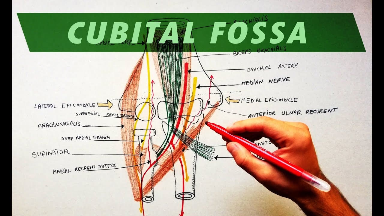
The cubital fossa, also known as the antecubital fossa, is a triangular depression or hollow in the anterior (front) aspect of the elbow joint.
It is located on the inner side of the elbow, and its boundaries are formed by various anatomical structures, including muscles, tendons, and bones.
The cubital fossa is an important area in clinical medicine and anatomy due to its accessibility and the presence of several significant structures within it.
Due to its triangular shape, the cubital fossa has three edges and an inferiorly directed apex. It also has a floor and a roof, and the structures that make up its contents can be found throughout.
Borders
The cubital fossa has three borders, a roof, and a floor, and is triangular in shape:
- Lateral border – Brachioradialis muscle.
- Medial border – Pronator teres muscle.
- Superior border – the epicondyles of the humerus.
- Roof: fascia, subcutaneous fat, skin, and bicipital aponeurosis.
- Floor: Proximally Brachialis muscle while supinator located distally on the floor.
The medial nerve, brachial artery, and ulnar artery are partially protected by the bicipital aponeurosis. The median cubital vein, which can be accessible for venipuncture, passes through the roof.
Structure
From lateral to medial, the cubital fossa has four primary vertical structures.
Even though the radial nerve passes close by the brachioradialis muscle, it is not generally technically regarded as being a member of the cubital fossa. The radial nerve splits into its deep and superficial branches as it moves along.
Biceps Tendon
The biceps tendon enters the cubital fossa before connecting to the radial tuberosity immediately distal to the radius neck.
Brachial Artery
The forearm receives oxygenated blood from the brachial artery. At the top of the cubital fossa, it divides into the radial and ulnar arteries.
Between the two heads of the pronator teres, the medial nerve exits the cubital. The majority of the flexor muscles in the forearm are supplied by it.
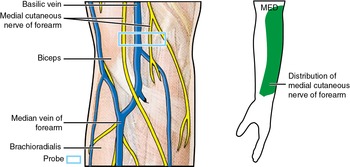
A number of superficial veins can also be found on the cubital fossa’s roof. A typical location for venepuncture is the median cubital vein, which joins the basilic and cephalic veins and is easily accessible.
Physiologic Variants
According to the presence of the median cubital vein (MCV) or median antebrachial vein, the superficial veins of the cubital fossa are anatomically divided into four categories.
- Type I: The cephalic vein (CV) and basilic vein (BV) are joined in the cubital region by the prominent median antebrachial vein. N type is another name for this.
- Type II: In the cubital region, the basilic and cephalic veins are connected by the median cubital vein. This kind is also referred to as type M type.
- Type III: The brachial cephalic vein has inadequate or no development in the cubital area.
- Type IV: The cephalic vein and basilic vein do not communicate.
The most typical type is type II, followed by type I, which has the cephalic and basilic veins joined by the median cubital vein. Despite the fact that type I and type II were the most common kinds of male and female, respectively, there is no statistical difference between the two. Additionally, there was no difference in the frequency of the kinds in the right and left upper limbs.
Because these superficial veins come in such a variety, mispuncture has been linked to unfavourable outcomes like bruising, hematomas, and sensory changes in a number of healthcare systems. There are two common venous return patterns in the cubital fossa, which are known to most medical professionals. This version emphasises the value of venipuncture using an intravenous illuminator.
Surgical Considerations
Pulsatile hematomas known as brachial artery pseudoaneurysms arise bleeding into soft tissues. Despite being uncommon, brachial artery pseudoaneurysms are more common after interventional operations than after diagnostic ones. Pseudoaneurysmal complications might pose a major risk to the patient’s life and the injured limb.
Clinical Importance
Brachial Pulse & Blood Pressure
The brachial artery in the cubital fossa is covered by the stethoscope when taking blood pressure. The artery connects the biceps tendon medially. In the cubital fossa, just medial to the tendon, the brachial pulse can be felt.
The brachial artery and median nerve, which are the more significant contents of the cubital fossa, have been protected by the bicipital aponeurosis (the cubital fossa’s ceiling), which was historically known as the “grace of God” tendon.
Venipuncture
For venous access (phlebotomy), the region just superficial to the cubital fossa is frequently employed. The superficial veins in the cubital fossa of the upper limbs, which include the cephalic, basilic, median cubital, and antebrachial veins and their tributaries, are one of the most often used locations for venipuncture. This area is traversed by numerous superficial veins.
The accessible medial cubital vein joins the basilic and cephalic veins. It can therefore be used frequently for venipuncture. Additionally, a peripherally inserted central catheter may be implanted using it.
The antecubital fossa is statistically the least painful area for peripheral intravenous access while having a higher risk of venous thrombosis.
Supracondylar Fracture
This fracture frequently affects young patients and typically happens when someone falls onto a hyperextended elbow. The fracture runs transversely between the two epicondyles. Although less than 5% of instances involve it, it can also occur when someone falls onto an elbow that is bent.
The cubital fossa’s contents may be impacted by the displaced fracture pieces, resulting in injury. Interference with the brachial artery’s ability to supply the forearm with blood might result from direct injury or post-fracture edoema. Volkmann’s ischemic contracture may be a result of the following ischemia.
The second most frequent nerve compression syndrome in peripheral nerve compression disease is cubital tunnel syndrome. The cubital tunnel is the most frequent location of entrapment for the ulnar nerve, though it can happen at other points along its course as well, such as the deep flexor-pronator aponeurosis, the arcade of Struthers, the medial intermuscular septum, and the medial epicondyle. the uncontrollable flexion of the hand, caused by fibrotic and short flexor muscles.
Cubital Tunnel Syndrome
A disorder when the ulnar nerve is compressed or stretched, resulting in tingling or numbness in the ring and little fingers, pain in the forearm, and/or weakness in the hand. When the elbow is bent for a lengthy period of time, such as while holding a phone or sleeping, these symptoms are frequently experienced.
In order to determine how much the nerve and muscle are being impacted, nerve testing (EMG/NCS) may occasionally be required. The initial course of action is to abstain from symptoms’ causes. To prevent the elbow from bending at night, you can wear a splint or loosely wrap a pillow or towel around it. Keeping the “funny bone” free of stress is also beneficial.
FAQs
What are the three contents of the cubital fossa?
The brachial artery, biceps tendon, radial nerve, and median nerve are all located within the cubital fossa.
What structure is in the cubital fossa?
The biceps tendon, which passes through and connects to the radial tuberosity of the radius, is probably the most noticeable structure in the cubital fossa.
The radial and ulnar arteries branch out of the brachial artery near its apex as it passes through the fossa just medial to the biceps tendon.
What are the major veins in the cubital fossa?
Cephalic, basilic, median cubital, and median antebrachial are the cubital fossa’s primary superficial veins. The cephalic vein, which typically arises at the lateral end of the dorsal venous network of the hand, is the longest vein in the upper limb.
Why it is called cubital fossa?
The Cubital Fossa is a triangular depression on the anterior surface of the elbow that is situated between the forearm and the arm, with the triangle’s tip pointing distally. Due to its location anterior to the elbow, it is also referred to as the “antecubital”.
What is the function of the cubital fossa?
A fat-filled, triangular region called the cubital fossa is located anterior to the elbow joint. The pathway it provides for vascular and neurological processes traveling between the upper arm and forearm makes this tiny region anatomically significant.
References
- Bains, K. N. S. (2023, July 17). Anatomy, shoulder and upper limb, elbow cubital fossa. StatPearls – NCBI Bookshelf. https://www.ncbi.nlm.nih.gov/books/NBK459250/
- TeachMeAnatomy. (2020, November 8). The Cubital Fossa – Borders – Contents – TeachMeAnatomy. https://teachmeanatomy.info/upper-limb/areas/cubital-fossa/
- Cubital fossa. (2023, May 17). In Wikipedia. https://en.wikipedia.org/wiki/Cubital_fossa


