Ethmoid Bone
Introduction
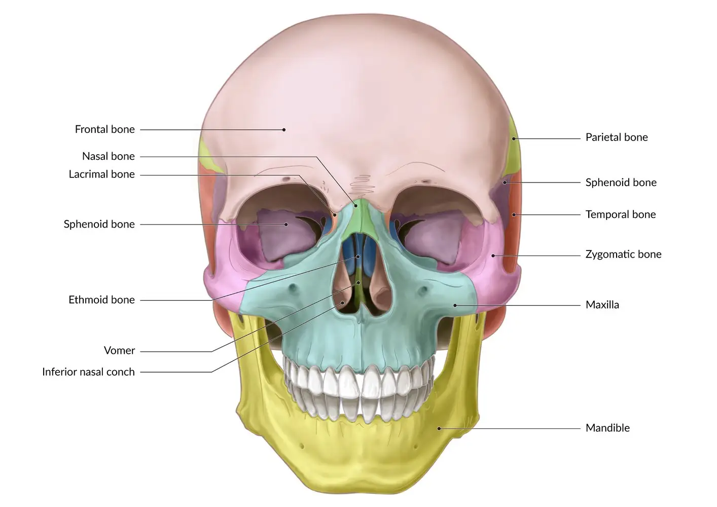
The ethmoid bone is an unpaired cranial bone located in the central region of the face, between the two orbits. It forms the roof of the nasal cavity and the floor of the anterior cranial fossa in the skull. The ethmoid bone plays a vital role in providing structural support and housing the olfactory nerve endings responsible for the sense of smell.
It is one of the most complex bones in the human skull, featuring several thin bony plates and cellular cavities. The highly porous nature of the ethmoid bone makes it very light and fragile. In this article, we will explore the anatomy and physiology of this multi-functional cranial bone.
Anatomy
Ethmoid Bone Parts
The ethmoid bone has several parts that enable it to carry out its complex functions:
Perpendicular Plate
- Thin, quadrilaterals-shaped bony plate projecting downward from the cribriform plate
- Forms the superior part of the nasal septum which divides the left and right air passages
- Articulates with vomer bone to complete the nasal septum
- Has a small opening for passage of the nasociliary nerve
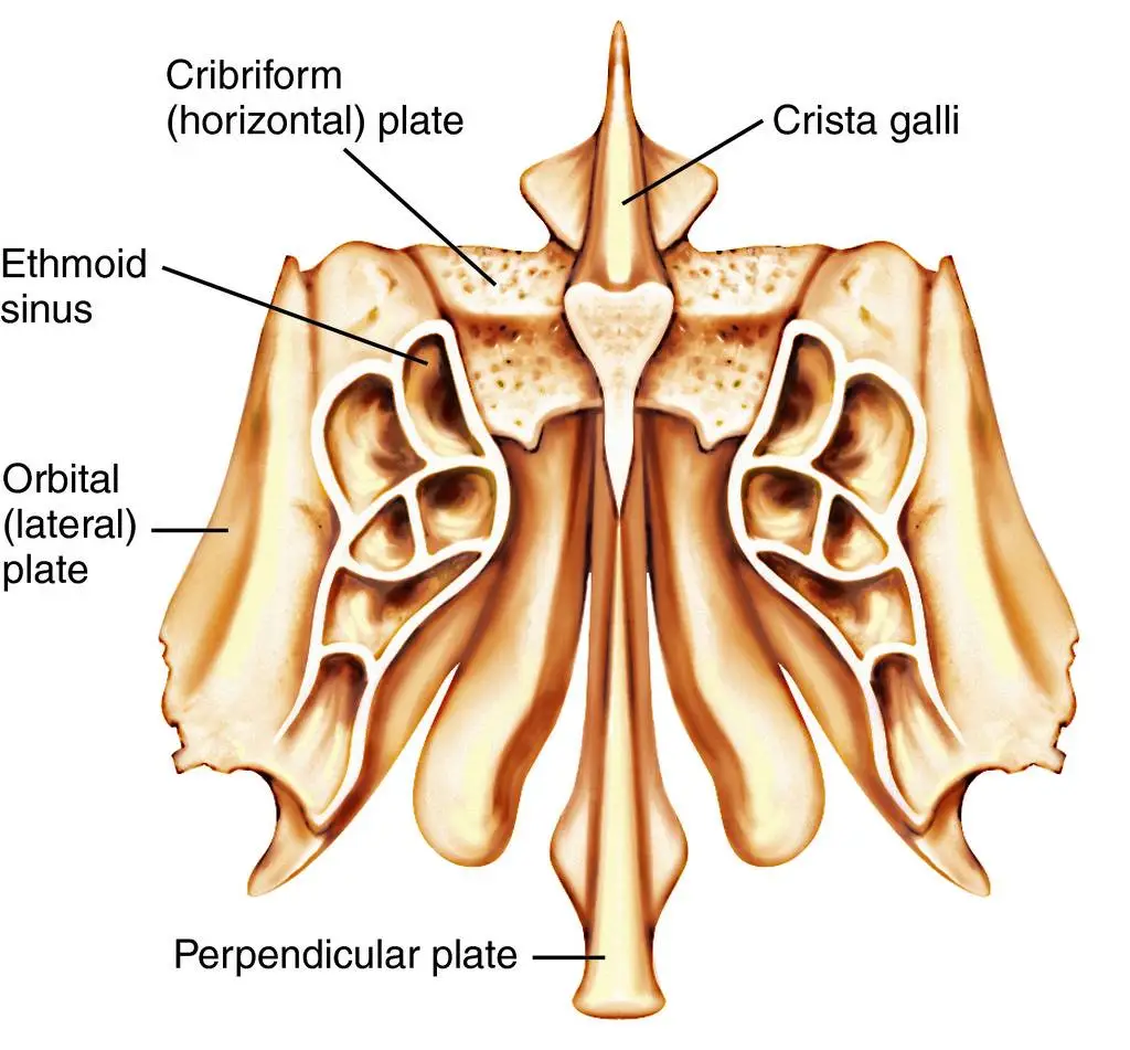
Labyrinth (Lateral Masses)
- Consist of extremely thin-walled bony plates bordering upper parts of nasal cavities
- Contain numerous small air-filled cavities called ethmoidal cells or sinuses
- Cells are arranged in anterior, middle, and posterior groups based on location
- Anterior cells open into the middle meatus of the nasal cavity
- Posterior cells open into the superior meatus
- Sinuses make bone lightweight, resonate sound, humidify air, produce mucus
Orbital Plates
- Irregularly shaped bony plates projecting upward to form part of the medial walls of orbits
- Separate ethmoidal cells from the orbital cavities
- Have small openings for passage of nerve branches and blood vessels
Cribriform Plate
- Horizontal bony plate forming the roof of the nasal cavity
- Contain multiple small foramina for passage of olfactory nerve fibers
- Forms part of the base of the anterior cranial fossa
- Has lateral grooves that become ethmoidal canals carrying nerves and vessels
The complex internal structure of the ethmoid bone facilitates its vital neurosensory and respiratory functions. The delicate arrangements of plates and cells make it vulnerable to fractures and infections.
Borders and Articulations
The ethmoid bone articulates with several other facial and cranial bones:
Frontal Bone
- The ethmoid articulates anteriorly with the vertical portion of the frontal bone
- This occurs at the chorionic junction between the nasal bones and frontal bone
- The cribriform plates meet to form the crista galli structure
Sphenoid Bone
- Posteriorly, the sphenoid bone forms the primary articulation
- Specifically where the sphenoid sinus meets the posterior ethmoid sinuses
- It forms a key part of the central skull base
Lacrimal Bone
- The medial walls of the orbits contain the lacrimal bone
- This is the main lateral articulation of the ethmoid bone
- Separates the ethmoid from the eye sockets
Nasal Bone
- Inferior nasal septum articulates with the nasal crest of the maxillary bone
- Septal cartilage also attaches to the nasal bones here
- Allows ethmoid to take part in nose infrastructure
Foramina and Openings
Cribriform Plate
- Contains multiple openings transmitting olfactory nerves
- Over 20 foramina in honeycomb pattern pierced by filaments
- Allows passage of olfactory nerves through bone to the nasal mucosa
Additional Openings
- Grooves for ethmoidal nerves, arteries, and veins
- Small canals transmit vessels and nerves to the nasal cavity
- Connect anatomy between the cranial vault and nasal region
Embryology
The ethmoid bone begins as cartilage during fetal development and undergoes endochondral ossification, unlike other cranial bones.
- Formed from the anterior cartilaginous part of the base of the embryonic cranium
- Ossification centers appear in the perpendicular plate during 4th fetal month
- Additional centers occur in labyrinths, orbital, and cribriform plates by 5th month
- Sinuses are visible by 5th month and expand considerably after birth
- Bone ossifies fully before puberty but enlarges outward as sinus cells grow
Nerves
The ethmoid bone transmits olfactory nerves and contains branches of the ophthalmic and maxillary nerves:
- Olfactory nerves – I cranial nerve fibers pass through holes in the cribriform plate to reach nasal mucosa
- Nasociliary nerve – a branch of the ophthalmic nerve that runs in a groove on labyrinths
- Anterior and posterior ethmoidal nerves – branches supply mucosa of sinuses
Blood Supply
Arterial Supply:
- The ocular artery branches into the anterior and posterior ethmoidal arteries.
- Sphenopalatine artery
- a branch of the maxillary artery
- Septal branches of the superior labial artery
Venous Drainage
- Through facial veins ultimately to the internal jugular vein
Lymphatic Drainage
Lymph vessels present near the ethmoid bone drain to deep cervical lymph nodes. Specific nodes include:
- Superficial parotid nodes
- Upper deep cervical nodes
- Retropharyngeal nodes
- Submandibular nodes
The rich nerve supply, blood vessels, and lymphatic channels allow the metabolic needs of the ethmoid bone to be met and linked to the rest of the head.
Location & Functions
The ethmoid bone sits in an anteromedial position within the central skull base. It articulates laterally with the orbital plate of the frontal and sphenoid bones. The ethmoid bone interacts with several other adjacent facial bones:
- Frontal bone – anterior articulation
- Sphenoid bone – posterior articulation
- Lacrimal bone – lateral articulation
- Nasal bone – inferior articulation
This central position allows the ethmoid bone to connect the nasal cavity to the orbits and cranial cavity. The integral functions of the ethmoid bone include:
- Forming part of the medial walls of orbits – The orbital plates contribute to the inner orbital walls, providing muscular attachments around the eyes.
- Forming the roof of the nasal cavity – The horizontally oriented cribriform plate creates the ceiling of the nasal cavity, just beneath the cranial base.
- Housing the paranasal sinuses – The ethmoid bone contains numerous small air-filled spaces known as ethmoidal sinuses, divided into anterior, middle, and posterior groups.
The complex anatomy of the ethmoid bone enables it to provide both structural and functional support in the delicate central areas of the skull base and nasal cavity. The bone plays a vital role in olfaction, respiration, orbital integrity, and anterior cranial fossa stability.
Common Disorders of the Ethmoid Bone
Ethmoid Sinusitis
Causes and Symptoms
- Caused by nasal inflammation, allergies, infections
- Swelling blocks sinus drainage leading to mucus buildup
- Symptoms include nasal congestion, facial pain, headaches
- May also cause fever, sinus pressure, loss of smell
Diagnostic Procedures
- Physical exam of nose, sinuses, throat, ears, etc.
- Nasal endoscopy to visualize sinus openings
- CT scan of sinuses to see blockages
- Allergy testing and microbial cultures
Treatment Options
- Nasal corticosteroid sprays and antihistamines
- Saline irrigation for clearing mucus
- Antibiotics if bacterial infection is present
- Surgery if medications fail to improve drainage
Ethmoid Bone Fractures
Types of Fractures
- Linear fractures of thin orbital or cribriform plates
- Comminuted fractures with shattered bony pieces
- Common in trauma from vehicle accidents
Signs and Symptoms
- Periorbital bruising, pain, and swelling
- CSF rhinorrhea or bleeding from the nose
- Altered vision, ocular misalignment
- May see CNS issues from trauma
Management and Healing
- Conservative treatment for non-displaced fractures
- Surgery for displaced bones or cerebral fluid leaks
- Monitoring for meningitis risk and vision loss
- Healing aided by bone grafting if needed
Nasal Polyps and Ethmoidal Tumors
Understanding Nasal Polyps
- Abnormal mucous membrane growths in the nose/sinuses
- Arise commonly from ethmoidal mucosa
- Allergic rhinitis and chronic inflammation involved
Ethmoidal Tumors
- Inverted papilloma – benign tumor but locally aggressive
- Squamous cell carcinoma – malignant tumor
- Esthesioneuroblastoma – a rare cancer of olfactory nerves
Screening, Diagnosis, and Treatment
- Nasal endoscopy and CT scans detect lesions
- Biopsy analyses tumor type
- Polypectomy or surgical resection
- Chemoradiation therapy for cancers
Summary
The ethmoid bone is an unpaired cranial bone located centrally in the skull between the orbital cavities and the nasal cavity. It has several vital functions including forming the roof of the nose, the medial orbital walls, and housing the paranasal sinuses.
The complex internal structure of the ethmoid bone features thin perforated plates and an intricate labyrinth of air cells. The main parts include the perpendicular plate, forming the nasal septum; the labyrinth holding the anterior, middle, and posterior ethmoidal sinuses; the orbital plates constituting the medial orbital walls; and the horizontal cribriform plate with multiple openings for olfactory nerves.
The central position of the ethmoid bone allows it to articulate with the adjacent frontal, sphenoid, lacrimal, and nasal bones. It also contains various openings and grooves that allow the passage of important nerves and blood vessels. The rich nerve supply provides innervation to the sinuses and nasal cavity.
Due to the fragile construction and vital location, the ethmoid bone is susceptible to fractures and common disorders like sinusitis. Chronic inflammation can impair sinus drainage leading to fluid buildup, pain, and infections. Trauma may cause linear cracks or fragmentation of the thin ethmoid walls. Tumors and polyps may also arise within the sinus mucosa.
Treatment often relies on endoscopic surgery to access the internal anatomy for drainage, bone repair, or lesion removal. A comprehensive understanding of ethmoid bone anatomy is critical for managing associated disorders.
FAQs
Where is the ethmoid bone located?
The ethmoid bone is located at the midline of the central skull base, between the nasal cavity and orbital sockets, behind the bridge of the nose.
What structures articulate with the ethmoid bone?
The ethmoid bone articulates with the frontal, sphenoid, lacrimal, and nasal bones.
What is the function of the cribriform plate?
The cribriform plate forms the roof of the nasal cavity and has multiple small openings that allow passage of olfactory nerve fibers carrying the sense of smell.
How many ethmoidal sinuses are there?
There are several ethmoidal air cell sinuses surrounding the nasal cavity, usually divided into anterior, middle, and posterior groupings.
What are some common disorders of the ethmoid bone?
Chronic inflammatory conditions like ethmoid sinusitis, nasal polyps, tumors, and fractural cracks or breaks are frequently associated with disorders.
Why is the ethmoid bone important?
The ethmoid bone is important because it supports the medial walls of the eye sockets, houses the sense of smell nerves, allows sinus resonance, and anchors central skull base stability.
References
- Ethmoid bone. (2023, October 30). Kenhub. https://www.kenhub.com/en/library/anatomy/the-ethmoid-bone
- Yu, M. (2023, July 24). Anatomy, Head and Neck, Ethmoid Bone. StatPearls – NCBI Bookshelf. https://www.ncbi.nlm.nih.gov/books/NBK544328/
- Hayes, K. (2023, June 6). The Anatomy of the Ethmoid Bone. Verywell Health. https://www.verywellhealth.com/ethmoid-bone-anatomy-4689011
- Ethmoid Bone | Complete Anatomy. (n.d.). www.elsevier.com. https://www.elsevier.com/resources/anatomy/skeletal-system/axial-skeleton/ethmoid-bone/23626#section-list-of-clinical-correlates-4
- https://th.bing.com/th/id/R.47da8a9885943dd0dbb3dff5bd26bf62?rik=CqrX4btQCHx8UA&riu=http%3a%2f%2fimg.tfd.com%2fMosbyMD%2fethmoid_bone.jpg&ehk=L421ODVbMPpMBgJ9TTn0IXLhwfAex9U%2bGRAA02lO0Rs%3d&risl=&pid=ImgRaw&r=0
- https://th.bing.com/th/id/OIP.CflUiWHtObme1YzHyUjyGAHaFP?rs=1&pid=ImgDetMain