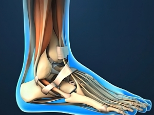
Foot and Ankle Tendons: Your Guide to Anatomy, Function, and Common Issues
The foot and ankle have a lot of tendons that run from the lower leg to the foot. These contain the peroneus tendons as well as the extensor tendons. Tendons are groups of tissue connecting muscles to bones.
The tendons that intersect that ankle and foot control the movement of the foot and ankle.
Damage to the foot and ankle tendons is a common reason for foot pain, commonly induced by overuse, overstretching, or an injury.
Introduction
Tendons are thick bands of connective tissue that attach muscles to bone. When a muscle contracts, the tendon stretches on the bone causing the joint to move. There are multiple tendons situated in the foot and ankle all responsible for different ankle, foot, and toe activities. Tendons also provide stability around the foot and ankle
We are going to look at the positions of the various tendons in the foot and ankle:
Lateral Ankle Tendons
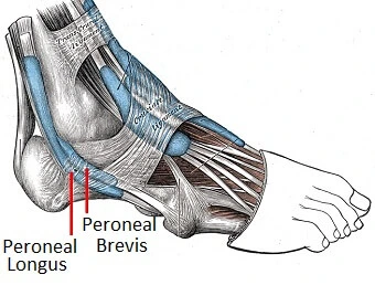
Let’s begin by peeking at the lateral ankle tendons located on the outer side of the ankle and foot, the peroneal tendons.
- Peroneal Tendons: There are two peroneal tendons, one from the peroneal longus muscle and the additional one from the peroneal brevis. The peroneal tendons go down together behind the outer side of the ankle and then break before connecting to different parts of the foot
- Peroneus Longus: starting from the upper part of the fibula, passes under the foot, and connects by the medial foot arch
- Peroneus Brevis: starting from the lower part of the fibula and connecting to the outer side of the midfoot. The peroneal tendons and their separate muscles support to pull the foot down into plantarflexion and outwards into eversion. Functionally, they are very important for supplying stability when running, especially on rough ground.
Medial Foot Tendons
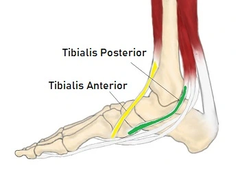
Several foot and ankle tendons go around the inner side of the ankle producing various foot moves.
- Tibialis Anterior Tendon: The tibialis anterior muscle arises from the outer side of the tibia and passes down the front of the shin. The muscle shifts into a tendon about two-thirds of the way down the shin and travels across the front of the ankle joint to the inner side of the foot under the medial foot arch. The tibialis anterior is a healthy ankle tendon that pulls the foot up into dorsiflexion. Functionally, it is really important when walking as it raises the foot to prevent it from catching on the floor as the leg swings forward and controls foot placement once the heel strikes the ground. It also functions with other medial ankle tendons to turn the foot inwards into inversion.
- Tibialis Posterior Tendon: The Tibialis posterior is the most serious muscle on the back of the leg. The tendon attaches to the tarsal bones beneath the foot and behind the medial malleolus, the inner ankle bone. The tibialis posterior tendon is the main invertor of the foot and also helps the calf muscles to plantarflex the foot. It plays an important role in supporting the medial arch and functionally maintains the position of the foot during walking and running.
Posterior Ankle Tendons
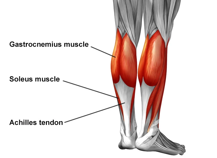
Several muscles and tendons pass down the back of the ankle, but the main one is the Achilles Tendon, aka Calcaneal Tendon.
- Achilles Tendon: The two main calf muscles, gastrocnemius, and soleus, run down the back of the calf and join jointly to form a strong, thick tendon, the Achilles tendon, that connects to the back of the heel. The calf muscles, through the Achilles tendon, are the main plantar flexors of the ankle that pull the foot down. Functionally, the Achilles ankle tendon supplies the large propulsive force required for walking, running, and jumping. The Achilles tendon is the most powerful and thickest in the body and can fight strains of up to 10 tons.
Extensor Tendons
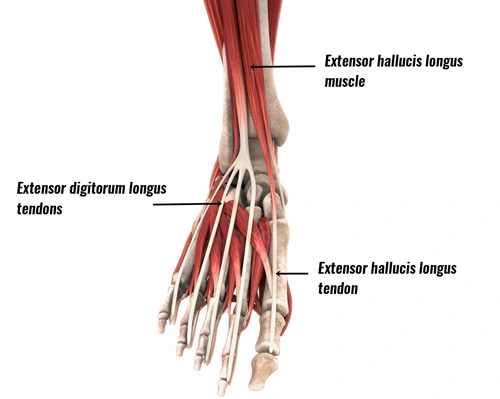
There are two sets of extensor tendons located in the foot from three muscles, extensor digitorum tendons and hallucis longus.
1. Extensor Digitorum Longus: arises just below the knee on the outer shin bones and forms a tendon before passing across the front of the ankle after which it splits into four-foot tendons, one to each of the outer four toes
2. Extensor Digitorum Brevis: It arises from the top part of the heel bone, goes across the top of the foot, and has small tendon connections at the base of the big toe and the tips of the second, third, and fourth toes.
3. Extensor Hallucis Longus: Arise from part way down the outer shin, passes medially across the front of the shin forming a tendon as it travels in front of the from the big toe tip to the ankle.
All three of these extensor tendons of the footwork to pull the toes up into extension. Functionally, they raise their toes clear of the floor during running and walking.
Flexor Tendons
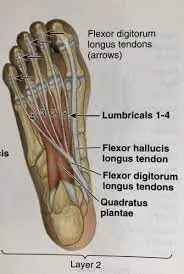
There are two groups of flexor foot tendons, each made up of two muscles and tendons, one set that bends the big toe, and the other group that bends the remaining four toes.
1. Flexor Digitorum Brevis: originates from the bottom of the heel bone and passes under the foot to the middle of the sole where it divides into four-foot tendons that attach to the base of the exterior four toes. The tendons connect to the mid-toe rather than extending to the tips.
2. Flexor Digitorum Longus: originates partway down the back of the calf, under gastrocnemius and soleus, constituting a tendon just above the medial malleolus. It passes behind the inner ankle bone and travels underneath the sole where it splits into four tendons that attach to the ends of the outer four toes.
3. Flexor Hallucis Longus: arises from the back of the lower fibula and crosses medially, where it develops into a tendon and hooks under the inner ankle bones as it travels along the back of the ankle. continues below the foot, connecting to the distal phalanx, which is the base of the big toe’s end bone.
4. Flexor Hallucis Brevis: A small muscle that starts deep within the sole, divides into two, and then forms brief tendons that join the proximal phalanx, the base of the first big toe bone, The flexor digitorum muscles and tendons extend the outer four toes. They functionally pull the toes down into the earth to help the foot grasp the ground for balance and propel off through the toes when walking, running, or jumping. The longus muscle helps in plantarflexing the foot and supporting the arches of the feet, while the flexor hallucis tendons and muscles bend the big toe downward. These great toe flexor tendons play an essential function in walking by producing the last push from the foot.
The main function of the Foot tendon
- Dorsal flexion (moving the foot and toes upward): The main tendon for this action is the Anterior Tibialis.
- Plantar flexion (moving the foot and toes downward): The main tendon for this is the Achilles Tendon.
- Inversion (bending the foot inward): The main tendon for this action is the Posterior Tibialis Tendon.
- Eversion (twisting the foot outward): The main tendons for this action are the Peroneal tendons (longus and brevis).
Additional tendons help in regulating toe motion. A solid and large tendon regulates flexion and extension in the big toe. Any foot tendon that is torn can cause extreme discomfort. An injured foot tendon might be the result of trauma. Although sprains of the ligaments are more common, both can occur in severe injuries. Walking may become more difficult if a tendon is completely torn. For example, nothing will prevent the foot from moving forward if the Achilles tendon in the back of the foot tears, which might make it impossible to stand at all.
Foot & Ankle Tendon Injuries
The foot and ankle tendons are usually injured by
- Repetitive Overloading: Excessive force frequently placed through the tendon e.g. sports leads to inflammation and degeneration
- Friction: Repetitious pressure rubbing on the tendon e.g. Moreover, wearing tight shoes causes degeneration and inflammation.
- Overstretching: Muscle and tendon extended beyond their elastic limit resulting in pulling
Foot tendon pain may be expected to:
- Tendonitis: When the foot or ankle tendons become inflamed inducing pain and swelling.
- Tendonosis: Wear and tear or deterioration of the foot tendon
- Tendon Tears: Where the foot or ankle tendon partially or completely breaks
What is causing my foot tendon pain?
Foot and ankle discomfort is usually due to tendon damage. The site of the pain will depend on which tendon is injured
- Outer Foot/Ankle Pain: Peroneal Tendon Injury
- Inner Foot/Ankle Pain: Posterior Tibial Tendon Injury
- Heel/Calf Pain: Achilles Tendon Injury
- Top of Foot Pain: Extensor Tendon Injury
- Front Of Ankle Pain: Tibialis Anterior Injury
You can find out loads more about each of these reasons for foot tendon pain, including symptoms, diagnosis, and therapy ideas by using the links above. Alternatively, visit the Foot Tendonitis page. The foot and ankle tendons play a really important role in day-to-day life, so any issues deserve immediate therapy.
Treating tendon injuries
Therapy for foot and ankle tendon damage will depend on which tendon is injured and the severity of the damage and foot tendon pain, but generally consists of
- Rest: from any exacerbating movements so as not to overload the tendon
- Ice: Regularly using ice packs can help relieve pain and inflammation with foot tendon damage.
- Compression Bandage: benefits to help the ankle and foot as well as decrease swelling
- Elevation: assist to decrease swelling
- Immobilization: A splint or cast may be worn to prevent movement so the tendon can recover. In extreme cases, crutches may be required to keep the weight of the injured foot.
- Medication: analgesics and non-steroidal anti-inflammatories can be a big benefit with foot tendon pain
- Physical Therapy: Friction massage and ultrasound can assist with tendon damage
- Exercises: Strengthening and stretching movements help the foot tendons to recover correctly and decrease the risk of future injury.
- Orthotics: Shoe inserts can be useful for offloading tendons and providing the foot is in the right position so as not to place excess strain on the foot tendons.
- Injections: Some ankle tendon damages respond well to steroid injections which relieve pain and inflammation.
- Surgery: In some examples, injury to the foot or ankle tendons may need surgery. This is usually because either the tendon has been ruptured completely and thus requires to be back together, or when symptoms fail to settle after six months of ongoing therapy.
FAQ
What is the most typical tendon injury in the leg?
Achilles tendonitis
The Achilles tendon is the biggest and most typical tendon to develop inflammation in the foot and ankle. Achilles tendonitis induces pain either at the site where the Achilles attaches to the calcaneus (insertional), or about two inches above this point (non-insertional).
Which tendons are seen in the foot?
Tendons are bands of connective tissue that connect the foot and ankle muscles to the bones. The two tendons in the foot are known as the peroneal tendons. One goes through the bottom of the foot and attaches to the inside of the arch, while the other runs down the outside of the foot and attaches to the fifth toe.
Can tendons recover naturally?
Most tendon damages get better on their own, but a complete recovery is difficult to achieve regardless of age. Young athletes typically experience tendon healing, but it takes a while, and function and elasticity never fully return to how they were before the injury.
What are the symptoms of tendon injury?
Pain, tenderness, redness, warmth, and edema near the injured tendon. Pain may get aggravated when you are active. Symptoms may involve just the spot where the injured tendon is located, or they may be spread out from the joint area. Crepitus, or a crunchy sound or sensation when the tendon is used.
What are the phases of tendon recovery?
Tendon regeneration, like fibrillogenesis, can be reduced to three interconnecting stages: (1) inflammation, (2) proliferation, and (3) remodeling. The inflammatory stage lasts about 48 hours and consists of erythrocytes, leukocytes, endothelial chemoattractant, and platelet infiltration.
References
- Foot Pain Explored. (n.d.). Foot & Ankle tendons: Anatomy, function & injuries. Foot-Pain-Explored.com. https://www.foot-pain-explored.com/ankle-tendons.html
- Wall, T., & Wall, T. (2023, February 21). Tendons of the foot. JOI Jacksonville Orthopaedic Institute. https://www.joionline.net/library/show/tendons-of-the-foot/
- Mbbs, L. K. (2022, June 23). Anatomy of the foot. https://patient.info/news-and-features/anatomy-of-the-foot
- Tendon injuries of the foot and ankle. (n.d.). Cal Sports & Orthopaedic Institute | Orthopaedic Specialists in Berkeley & Orinda. https://calsportsortho.com/specialties/foot-and-ankle/tendon-injuries-of-the-foot-and-ankle/