
Kienbock’s Disease
What is Kienbock’s Disease?
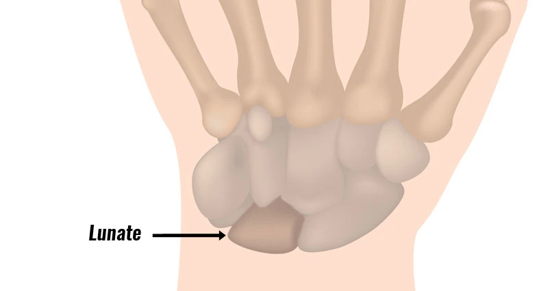
Kienbock’s disease is a condition where the blood supply to one of the small bones called the lunate is disturbed leading to Chronic wrist pain and other related symptoms. The lunate is a carpal bone of the wrist.
Bone is a living tissue that requires a steady supply of blood to survive. If a bone’s blood supply is cut off, the bone or regions of the bone may die. This is referred to as osteonecrosis.
A loss of blood supply to the lunate affects bone to lose its structural asset, and it will collapse, generating a painful, stiff wrist. Over time, these modifications can conduct to arthritis of the surrounding bones in the wrist
Epidemiology
Kienbck condition is the second most familiar variety of avascular necrosis of the carpal bones, preceded only by avascular necrosis of the scaphoid. The commonly involved population is men aged 20-40 years.
How do you get Kienbcks disease?
Although the actual reason for Kienbcks disease is unknown, several elements might contribute to the breakdown of your lunate bone, involving:
Slighter blood flow to your lunate. Specialists believe this is the most probable factor. Your bone can die if it doesn’t accept nourishing blood. The death of a bone is known as osteonecrosis.
Trauma to your lunate. It might have been injured in a car accident, for instance.
Uneven forearm bones. If your ulna and radius bones in your forearm aren’t the exact length, that can generate issues with your wrist.
Uneven lunate shape. You might’ve been birth with a lunate that’s the incorrect shape.
Stages of Kienbcks disease
Your healthcare provider might advise you on the phase of your Kienbcks condition. Stage 1 is the least extreme and stage 4 is the most extreme. MRI scans and CT scans assist define the phase by ruling out fractures and seeing blood flow.
Stage 1: In stage 1, you might sense pain equal to a wrist sprain. Although the reason might be unclear, it’s probably because the blood supply to your lunate has been delayed down or eliminated.
Stage 2: In stage 2, your lunate begins to stiffen because of the lack of blood flow. The method of hardening is known as sclerosis, and it suggests your bone is dying.
Stage 3: In stage 3, your hardened lunate will begin to break. This might generate further bones in your wrist to move around. You’ll sense more pain, work to grip things with as great power and your range of motion will be restricted.
Stage 4: In stage 4, the externals of the bones near your lunate will even weaken. Your wrist might evolve arthritically.
It might be several months or several years between phase 1 and phase 4.
Symptoms of Kienbcks disease
Not everyone with Kienbcks condition experiences symptoms. Normally reported symptoms contain:
Wrist pain that’s equal to a sprained wrist. A sprained wrist can be a dull ache that arrives and goes or a sharp pain that’s more even.
Pain straight over your lunate bone (regarding the center of your wrist).
Swelling of your wrist.
Stiffness of your wrist.
Weakness of your wrist.
Creaking, crackling, or scratching sounds when you move your wrist known as crepitation.
Incapability to move your wrist like you used to (a reduced range of motion).
Minor strength when you go to grip something.
Cause of Kienbock’s disease
The reason for Kienbck’s condition is not known. There may be one or more factors that disrupt blood flow to the bone.
Many people with Kienbck’s condition think they have a sprained wrist at first. They may have felt some state of trauma to the wrist, like a fall. This variety of trauma can damage the blood vessels that supply the lunate.
Some items may place you more at risk for the condition. For example:
Some people may have rarer blood vessels that supply the lunate, and most people have two arteries that supply blood to the lunate, but in some people, there is just one source.
Pressures acting on the lunate may be various relying on the relative sizes of the two bones in the forearm (the radius and the ulna). If the radius and ulna are various lengths, additional stress can be placed on the lunate during some wrist movements and when force is applied to the wrist during activity, like when doing a push-up. Over time, this additional pressure may conduct in a compromise of blood flow to the lunate and generate Kienbck’s condition.
What are the risk factors for Kienbock’s Disease?
Risk factors are:
Dissimilarities in the size and form of your forearm bones, the radius, and the ulna. This can place more stress on your lunate.
Just one blood vessel supplying blood to your bone, rather than the normal two. This can involve blood supply to your bone.
Other conditions, like sickle cell anemia, lupus, cerebral palsy, and conditions that involve blood supply, are associated with Kienbocks condition.
Kienbock’s condition happens most normally in males between 20 and 40 years old. You’re also at a risen risk if you regularly do serious manual labor.
Differential Diagnosis
Ulnar impaction syndrome: More familiar with ulnar positive variance and results from repetitious microtrauma to the lunate from a fairly long ulna. Parallel to the Kienbck condition, ulnar impaction syndrome effects in reduced T1-weighted signal coupled with raised T2 signal if hyperemia exists, or reduced T2 signal if the condition has already advanced to lunate sclerosis. The signal modifications in Kienbck disease, yet, are more diffuse or more extreme on the radial side of the lunate. Further, ulnar impaction syndrome involves the ulnar head and triquetrum, which are entirely in the Kienbck person.
Lunate intraosseous ganglion: Intraosseous ganglia are real cysts that have a low T1 signal and high T2 signal on MRI. Sharp, soft margins on both MRI and radiography permit recognition of Kienbck’s condition.
Bone contusion: May be hard to determine early-stage Kienbck condition on MRI. A record of current trauma and concomitant injuries to the wrist/hand, in this issue, would aid in creating the diagnosis.
Arthritis: Bone marrow signal modifications in inflammatory or degenerative arthritides may imitate those noticed in Kienbck’s condition. Specific features contain clinical presentation, demographics, and absence of ulna minus in arthritis.
Osteoid osteoma: Osteoid osteoma of the carpal bones is irregular, and just a few patient data exist in the literature. Clinical manifestation and the result of a lucent nidus within a sclerotic rim on CT determine it from Kienbck’s condition.
Enostosis/bone island: Has low signal intensity on all series, yet, displays maintained bone morphology and the region of sclerosis is stellate and interdigitated with the normal trabeculae.
Diagnosed
You’ll meet with your healthcare provider and note your symptoms. Be as precise as you can regarding the variety and site of your pain, and how long you’ve been feeling it. Your healthcare provider or doctor might order examinations, involving:
- X-ray
- MRI.
- CT scan.
- Bone scan.
Treatment of Kienbock’s disease
Medication
There are both nonsurgical and surgical therapies for Kienbcks’s condition. Measures of nonsurgical treatments contain:
Maintaining your wrist from moving by placing it in a cast for up to several months. If your wrist is relaxed, the blood flow might continue.
Occupational therapy. Your occupational therapist can guide you on how to use your wrist in a manner that decreases pain and delay the condition.
Taking drugs like cortisone injections and over-the-counter (OTC) anti-inflammatory drugs, involving ibuprofen (Advil, Motrin) and naproxen (Aleve).
Inspect your healthcare provider about the best drugs for you. Comment that ibuprofen and naproxen are not appropriate for a person with some health diseases, involving:
- Asthma
- Stomach ulcers
- High blood pressure
- Kidney problems
- Heart problems
Surgery for Kienbock’s Disease
There are five varieties of surgeries suggested for Kienbck’s condition. The right one for you relies on the phase of your condition and your healthcare provider’s suggestions. Possible surgeries contain:
Joint leveling. Joint leveling is required when the two bones that cause up your forearm (your radius and ulna) are various lengths. The various lengths placed additional force on the lunate bone, squeezing it. Your surgeon will extract a little bit of bone to shorten the radius or ulna or use bone grafts to extend it.
Revascularization. This method restores or raises the quantity of blood flowing to your lunate. Your surgeon will bring a little area of bone from another portion of your body and place it into your lunate. Pins will maintain your bones together while they recover, and a metal machine outside of your wrist will maintain everything in location. Revascularization can just be done during phases 1 or 2.
Fusion. You may have a partial fusion or a full fusion. This permanent method is where your surgeon connects (welds together, sticks together) some or all of the bones in your wrist. If you have a full fusion, you won’t be capable to move your wrist, but you’ll still be qualified of pivoting your forearm.
Implant arthroplasty. For this method, your surgeon substitutes your lunate with a prosthetic.
Proximal row corpectomy (PRC). If your lunate is damaged, it and some different carpal bones in your wrist might be withdrawn.
Physiotherapy treatment
Physical therapy is not a familiar treatment for this condition. There are too few analyses regarding the physical management of this disease. Analysis results
Mixed a bone marrow transfusion (medical), low-intensity pulsed ultrasound (PT), and an external fixation (surgery). For most of the people in this analysis, wrist pain enhanced to a pain-free level, wrist flexion enhanced, and average grip strength raised. This technique can be used as a less invasive surgical choice for Kienbcks’s condition likened to surgical methods.
An analysis compared various therapies after surgery. One group was managed according to a therapy protocol consisting of electrotherapy, kinesiotherapy, massage therapy, thermotherapy, therapeutic activities, and domiciliary guiding. The further group was managed by non-specialized Units in Hand Therapy and obtained just thermotherapy and activities without direct advice from the therapist. After treatment, 90% of group one were comfortable with the outcomes of the therapy, and 66% of the second group. Also, group 1 scored nicely on pain, prehension force, muscular strength, forearm articular range of movement for pronation and supination motions, wrist articular range of movement in abduction and adduction motions, and manual function performance.
Prognosis
Kienbock condition is always progressive, and joint destruction happens within 3-5 years of beginning.
The prognosis depends on:
Functional Staging: The more significant the size of viable bone, the better the prognosis.
Negative ulnar variance: The more significant the negative conflict, the more extreme the condition and the more probable it is to progress.
Age at diagnosis: Persons analyzed at a more senior age are more probable to have the advanced-stage condition and are more probable to progress.
It is important to mention that the severity of symptoms does not ever connect with the morphologic stage.
Complications
Kienbck condition can conduct scapholunate dissociation, secondary radiocarpal and mid-carpal degenerative arthrosis, and malalignment of the triquetrum.
FAQ
How long does it take to recover from Kienbck disease surgery?
Recovery. After surgery for Kienbck’s condition, you will require to wear a splint for three to four months. Your surgeon or doctor will like to observe you to see how your wrist bones are recovering.
Is wrist surgery serious?
As with any surgery, there is a risk of difficulties. These risks are little but involve the risk of residual weakness/ less function and the risk of bleeding.
What is the success rate of wrist surgery?
Graph displaying survival studies. A, Revision total wrist arthroplasty Kaplan-Meier survival study. The 5- and 10-year recurrence revision-free survival rates were 71% and 60%, respectively.
How many hours does wrist surgery take?
The entire method normally carries 30-90 minutes, relying on the severity of the fracture, and is conducted under either general anesthesia or regional anesthesia with sedation. In most patients, ORIF wrist surgery is an outpatient surgery, so you can go household the exact day.
Can you fully recover from wrist surgery?
For most people, wrist surgery healing times vary from a few weeks to several months. The bone itself may recover within a month or two, but complete healing from the surgery or the injury can take 4 to 6 months. Confer with your doctor to obtain a more precise healing plan for yourself.


6 Comments