
Obturator externus muscle
What is Obturator externus muscle?
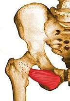
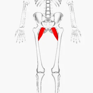
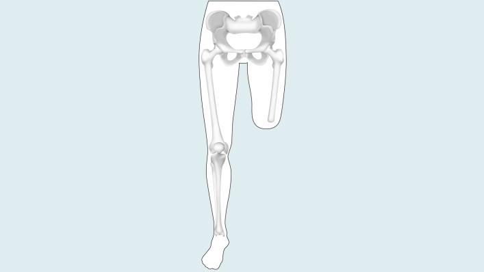
The obturator externus muscle is a flat, triangular muscle located in the hip region. It arises from the outer surface of the obturator membrane and the surrounding bones, including the pubic bone, ischium, and ilium. The muscle fibers then converge to form a tendon that passes through the lesser sciatic foramen and inserts into the greater trochanter of the femur bone.
The obturator externus plays out at least a couple of activities. When the hip is extended, it rotates the femur externally, but when the hip is flexed, it abducts the thigh. It helps stabilize the hip joint, along with other short muscles around it.
Origin of Obturator externus muscle
From midnight to the 10 o’clock position (right hip viewed from the front), the Obturator externus (OE) muscle originates from the rami of the pubis and ischium, the external bony margin of the obturator foramen, and a few fibers from the obturator membrane.
Insertion
It shaped a musculotendinous intersection at the level of the femoral neck. The fibers are inserted into the trochanteric fossa as a cylindrical tendon, with some fibers extending towards the piriformis fossa, passing laterally along the inferior margin of the acetabulum and acting as a sling at the inferior part of the neck.
Relations
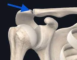
The obturator externus is situated in the pelvis on the anterior part of the innominate bones. It is deep within the pectineus and superior parts of the adductors of the thigh and covers the obturator foramen. Its ligament lies profound into the quadratus femoris muscle and isolates it from the neck of the femur. The obturator vessels (foremost and back parts of the obturator course and vein) are found somewhere down in the obturator externus muscle, on the outside surface of the obturator film. Additionally, nerves pass close to this muscle. Before both branches descend to innervate the muscles of the thigh, the anterior branch of the obturator nerve crosses the muscle’s anterior surface, and the posterior branch penetrates the muscle.
The obturator externus bursa is a bursa that can sometimes be found between the obturator externus tendon and the hip joint capsule. To lessen the amount of friction that exists between the tendon and the joint capsule, this bursa communicates with the hip joint.
Innervation
The lumbar plexus-derived posterior branch of the obturator nerve (L3 and L4) innervates the obturator externus.
Blood supply
The medial circumflex femoral artery and the anterior branch of the obturator artery supply the obturator externus. The muscle may receive blood supply from both or just one of these blood vessels, which forms a variable pattern.
Functions of Obturator externus muscle
Depending on where the thigh is, the obturator externus performs two primary functions. The contraction of the obturator externus results in lateral, or external, thigh rotation when the hip is extended (the body is in the anatomical position). This activity is particularly helpful in climbing. Additionally, it is believed to counteract the medial rotation caused by the anterior adductors of the thigh during walking.
The obturator externus muscle abducts the thigh when the hip joint is flexed, or when the thigh is closer to the body. It accomplishes this by moving the inferior part of the femur away from the body and pulling the superior part of the femur medially.
It additionally adds to the stability of the hip joint with other short muscles encompassing it (pectineus, piriformis, obturator internus, quadratus femoris, and the Gemelli inferior and superior). Although supporting the hip joint is for the most part defined as an optional capability, it has been proposed that it very well might be a higher priority than what’s viewed as the essential capability.
Clinical relevance
Through a posterior approach, the role of the short external rotators in hip stability after a total hip replacement (THR). They noted that the preservation of the external obturator and piriformis lowers the risk of dislocation following THR, suggesting alternative THR strategies. OE stabilizes the hip joint because of its fibers that reinforce the posterior capsule.
Between the cross-over acetabular tendon and the OE muscle, there was access to the obturator externus (OE) bursa, which contained bursal fluid. According to studies, hips with intra-articular pathology have OE bursa more frequently than normal hips.
Professional basketball players suffer from a musculotendinous tear as a result of repetitive eccentric contraction of the Obturator externus. Impingement syndrome after total hip replacement: If a transverse acetabular ligament is released or if the acetabular cup position protrudes beyond the caudal rim after arthroplasty, this close relationship between the musculotendinous portion of the OE muscle and the inferior margin of the acetabulum can result in impingement syndrome. Targeted rehabilitation will ensure a rapid return to competition without complications. The risk of impingement was reduced by releasing the OE insertion attachment from the posterior capsule.
obturator externus muscle pain
Pain in the obturator externus muscle can be caused by a variety of factors, including overuse, muscle strain or injury, and referred pain from other areas of the body. Symptoms may include a dull ache or sharp pain in the hip or groin area, difficulty walking or standing, and decreased range of motion in the hip joint.
Treatment for obturator externus muscle pain typically involves a combination of rest, ice or heat therapy, anti-inflammatory medications, and stretching or strengthening exercises. Physical therapy may also be recommended to help improve muscle function and alleviate pain.
Obturator externus muscle stretching
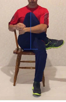
Lateral rotators stretching
Sit up straight in a chair and perform this exercise. To begin, cross your right thigh with your left ankle. Pull the left knee toward the right shoulder while holding it in both hands. It must be a painless, gentle stretch during exercise. Relax after holding this for 10 to 20 seconds. On another leg, repeat. Perform this exercise three to four times each week.
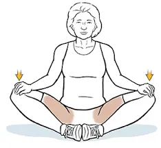
Hip adductor stretching
On a soft mat, you can kneel with your left foot in front of you and your right foot 90 degrees in front of you. Hold for 10 to 20 seconds, slide the left knee to the side, keep the chest up, and relax. On another leg, repeat. Perform this exercise three to four times each week.
Obturator Externus muscle strengthening exercise
Frog bridge
To strengthen your hip external rotators, the Frog Bridge exercise is a modified version of the Glute Bridge and Hip Thruster. As you raise the pelvis off the ground, you will feel the buttock muscles work. To set up place your feet along with your knees spread separated at shoulder distance. The knees are twisted to 90 degrees. As you lift your pelvis off the floor, engage your gluteus maximus muscle by bracing your core.

FAQ
Where is the pain in the obturator externus?
The hip area is the most common location for obturator externus bursa, which typically affects athletes and those over forty. The bursae may become inflamed as a result of a serious injury or as a result of joint motion, such as running.
How would you reinforce the obturator externus?
By abducting your knees while sitting on a seat or seat with no arms and wrapping the opposition band around your lower thigh, you can strengthen the obturator externus. Wrap weighted cuffs around your ankles and abduct your knee while lifting one foot at a time a few inches off the ground to increase the intensity.
What signs does obturator externus bursitis have?
Since patients with obturator internus bursitis have dubious side effects, for example, vague myofascial irritability, fever, and buttock enlarging, clinical analysis is made by direct palpation over the physical locus of the obturator internus bursa.
How much time does it take for an injury to the external obturator muscle to heal?
Most of the cases of deep hip external rotator muscle strains that have been reported have responded well to rest and physical therapy. In most cases, athletes can resume full sports participation within two to six weeks.
What is the obturator externus nerve’s root?
The thigh adductor muscles are primarily innervated by the obturator nerve, which originates in the anterior division of the L2-L4 nerve roots and travels through the psoas major before emerging from the medial border.

4 Comments