
Obturator Nerve: Anatomy, Function, Importance
Obturator Nerve Anatomy
The obturator nerve begins at the medial border of the psoas major muscle. It travels through the obturator foramen (an opening in the pelvic bone) before entering the thigh, where it branches into two parts, an anterior branch and a posterior branch.
The nerve is part of a group of nerves called the anterior lumbar plexus.
The nerve provides sensory perception to the skin on the medial side of the thigh. It also provides motor function to the hip and knee joints and the abductor muscles and gracilis.
Description
- The nerve arises from the lumbar plexus on the posterior abdominal wall and descends within the psoas muscle, emerging from the medial margin of the muscle to enter the pelvis.
- The nerve path continues by following along the lateral wall of the pelvis, passing through the obturator canal, to enter the medial compartment of the thigh.
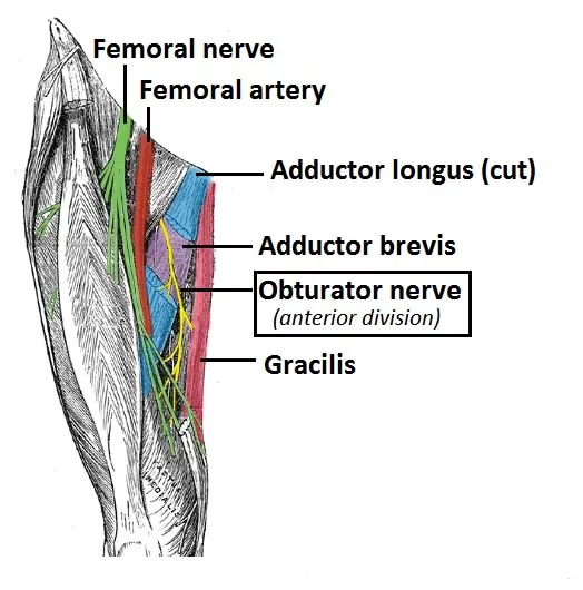
- From here the nerve divides into the anterior and posterior branches which are separated by the adductor brevis muscle.
- The posterior branch travels underneath the adductor muscle along the anterior surface of the adductor magnus muscle, innervating the obturator externus, adductor brevis, as well as part of the adductor magnus muscle that is attached to the linea aspera.
- On the anterior surface of the adductor brevis muscle, the anterior branch travels underneath the pectineus and adductor longus muscles to innervate the adductor longus, gracilis, and adductor brevis muscles.
- This branch also often contributes to the pectineus muscle. The cutaneous branches innervate the skin on the medial thigh.
Nerve roots:
L2-L4
Motor functions:
Innervates the muscles of the medial compartment of the thigh (obturator externus, adductor longus, adductor brevis, adductor magnus, and gracilis).
Sensory functions:
Cutaneous branches of the obturator nerve innervate the skin of the medial thigh.
Course
The obturator nerve (latin: nervus obturatorius) is a mixed nerve that originates from the lumbar plexus and innervates the muscles and skin in the medial region of the thigh.
The obturator nerve arises from the ventral rami of the second, third, and fourth lumbar nerves (L2 – L4). The obturator nerve emerges from the medial side of the psoas major, then it crosses the linea terminalis and passes by the lateral wall of the lesser pelvis.
The obturator nerve enters the obturator canal and divides into two terminal branches: the anterior and the posterior branch.
The anterior branch of the obturator nerve is larger than the posterior, it runs between the adductor brevis and adductor longus muscles, then penetrates the fascia lata at the middle third of the medial surface of the thigh and continues as the cutaneous branch.
The anterior branch of the obturator nerve innervates the adductor brevis, adductor longus, pectineus, and gracilis, and also the skin at the medial part of the thigh as far as the knee joint.
The posterior branch of the obturator nerve runs posterior to the adductor brevis. The posterior branch of the obturator nerve innervates the adductor magnus and external obturator muscles, and the hip joint.
Branches:
- Posterior Branch
- Anterior Branch
- Cutaneous Branch
Functions:
Motor Functions:
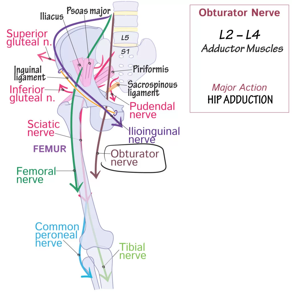
The obturator nerve innervates all the muscles in the medial compartment of the thigh – except the hamstring part of the adductor magnus (innervated by the tibial nerve).
They are collectively known as the hip adductors:
- Adductor longus – adducts thigh
- Adductor brevis – adducts thigh
- Adductor magnus:
- Adductor part – adducts and flexes thigh
- Hamstring part – extends thigh
- Gracilis – adducts thigh
- Obturator externus – laterally rotates thigh
Sensory Function:
- The cutaneous branch of the obturator nerve supplies the skin of the middle part of the medial thigh.
Embryology
Neuroepithelial cells give rise to the peripheral nervous system. From the pia mater to the periphery, these neuroepithelial cells move and undergo differentiation into glioblasts, neurons, and ependymal cells. These cells aid in the development of an eventual obturator nerve and other structures like the lumbar plexus.
Anatomical Variants
The bifurcation of the obturator nerve can occur anywhere in the medial thigh, obturator canal, or pelvic cavity. Normally, the obturator nerve splits into the anterior and posterior divisions when it exits the obturator canal and enters the thigh. On the other hand, the obturator nerve can bifurcate intrapelvic (23.22%), in the thigh (25%), or within the obturator canal (51.78%). Furthermore, the anterior branch has the potential to generate two, three, or four branches, the most common number being four. By contrast, the posterior branch has the ability to divide into one to four segments, with the most common variety having two divisions.
The obturator nerve’s articular branches showed nine distinct branching patterns. The obturator nerve supplies cutaneous innervation to the skin surface of the medial thigh, although studies have indicated that in over 50% of cases, this nerve gives no cutaneous innervation at all.
Surgical Considerations
When performing procedures that call for pelvic dissection and access, the obturator nerve may sustain damage. Regardless of the surgical technique used, the obturator nerve and ureter are particularly vulnerable when it comes to pelvic organ prolapse treatment. Laparoscopic pelvic procedures, including laparoscopic lymphadenectomy, can also cause damage to the obturator nerve. Furthermore, transvaginal mid-urethral sling deployments may cause injury to the obturator nerve.
When treating bladder wall masses, transurethral resection of bladder tumors, or TURBT, is frequently performed. In this procedure, the obturator nerve is especially useful. Due to the obturator nerve’s close proximity to the prostatic urethra, bladder neck, and inferolateral wall, excision of a tumor may stimulate the adductor muscles abruptly, resulting in a “adductor jerk.” This abrupt thigh movement could result in extravesical tumor seeding and bladder rupture. Furthermore, the obturator artery may be harmed by the adductor jerk. A safe and efficient TURBT can be made possible by eliminating the adductor jerk by the use of a local anesthetic during an obturator nerve block.
Clinical Relevance:
- Injury to the nerve is rare as it lies deep within the pelvis and medial thigh. It can be damaged through direct injury to the nerve or to surrounding muscle tissue.
- Mild damage to the obturator nerve can be treated with physical therapy. More severe cases may require surgery.
Injury may be caused by:
- Nerve being stretched during surgery
- Entrapment within the obturator canal
- Compression during pregnancy
- Car or household accident
- Abdominal surgery
- Athletes may present with pain that may be brought on by exercise, often sports involving a lot of running and twisting.
- They may have been predisposed to this injury by previous pelvic trauma or surgery.
- In mild damage, physiotherapy treatment is employed. More severe cases may require surgery with follow-up rehab physiotherapy. Axonal growth is at about 1mm per day.
Symptoms of Obturator Neuropathy:
- Sensory alteration in the medial thigh
- Pain & paresthesias may extend from hip to knee along the medial aspect of the thigh
- Extension or lateral leg movement can increase pain
- May have trouble walking or experience leg weakness due to problems adducting the ipsilateral hip
- Signs of Obturator Neuropathy:
- Weak hip adductors on the affected side
- Wasting of the medial thigh
- Abnormal abduction of the hip during ambulation resulting in a circumducting, wide-based gait
- Area of sensory loss or alteration in the mid and lower third of the medial thigh which sometimes may extend below the knee
- Ipsilateral loss of the hip adductor tendon reflex (test against asymptomatic leg as is not always present in healthy population)
FAQs
Where does the accessory obturator nerve come from?
Branches of the lumbar plexus: It is tiny and originates from the third and fourth lumbar nerves’ ventral divisions. It has been demonstrated recently that dorsal divisions are the source of this nerve.
Where is the obturator canal located?
The obturator canal, which joins the pelvis to the medial compartment of the thigh, is a tiny aperture in the superior face of the obturator foramen. The obturator membrane covers the obturator foramen otherwise.
What are the four muscles supplied by the obturator nerve?
The pectineus, adductor (longus, brevis, and magnus), gracilis, and external obturator muscles are supplied by the obturator nerve (L2-L4).
What is the location and function of the obturator nerve?
The obturator nerve is located in the groin. It makes your inner thigh’s muscles and feelings possible. Sports injuries and post-operative problems from medical procedures can cause nerve damage (obturator neuropathy).
What is the obturator nerve root?
The anterior division of the L2–L4 nerve roots gives birth to the obturator nerve, which passes through the psoas major and emerges from the medial border to innervate the thigh adductor muscles primarily.
References
Koh, M., & Markovich, B. (2023, July 24). Anatomy, Abdomen and Pelvis, Obturator Nerve. StatPearls – NCBI Bookshelf. https://www.ncbi.nlm.nih.gov/books/NBK551640/


13 Comments