
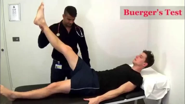
Straight Leg Raise Test – SLR test
The Straight Leg Raise (SLR) Test is a physical examination maneuver commonly used in medical assessments to evaluate the presence of sciatica or lumbar nerve root compression.
- This Straight Leg Raise Test is also known as the Lasegue’s test.
- This Straight Leg Raise Test is commonly used to identify the impairment in disc pathology /irritation of the nerve root.
- This test is also of specific importance in detecting disc herniation & neural compression.
- This test is classified as to neurodynamic evaluation test which detects excessive nerve root tension/compression.
- This Straight Leg Raise test was 1st described by the League, before the 100 years ago so this test is referred to as the Laseagues test.
- This Straight Leg Raise test places tensile stresses at the nerve of sciatic & gives traction at the lumbosacral nerve roots primarily from the L4 to S2.
- During the Straight Leg Raise test, these nerve roots are pushed anteriorly & inferiorly, pulling the dura mater is pulled caudally, anteriorly & laterally.
- When the occur Tension in the sciatic nerve, it pulls the sciatic foramen, then the sacrum & then the nerves are that cross over the pedicles& finally the intervertebral foramen.
- The patient feels pain /tenderness in the vicinity of the greater sciatic notch.
Purpose of the Straight Leg Raise Test:
- This test is used to check the impairment in disc pathology /irritation of the nerve root.
- This test is also used to check the disc herniation & neural compression.
The technique of performance of the Straight Leg Raise Test:
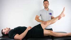
- The Straight Leg Raise Test is done with the patient completely relaxed.
- This test is one of the most common neurological tests of the lower limb.
- This test is passive & each leg is tested individually with the normal leg being tested first.
- With the patient in the supine position, the hip joint in medially rotated means internally rotated & adducted & knee joint in the extended position.
- Then the examiner flexes the hip until the patient complains of pain /tightness in the back/back of the leg.
- If the pain is primarily back pain, it is more likely a disc herniation from pressure on the anterior theca of the spinal cord,/ pathology causing the pressure is more central.
- Back pain only patients who have a disc prolapse have small, more central prolapses.
- If the pain is primarily in the leg, it is more likely that the pathology causing the pressure on neurological tissue is more lateral.
- Disc herniation or pathology causing pressure between the two extremes are more likely to cause pain in both areas.
- The examiner then slowly & carefully drops the leg back slightly until the patient feels no pain/tightness.
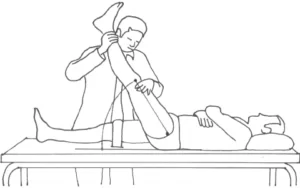
- The patient is then asked to flex the neck so the chin is on the chest, or the examiner may dorsiflex the patient’s foot, or both actions may be done simultaneously.
- Most commonly foot dorsiflexion is done first.
- Both of these maneuvers are considered to be provocative/sensitizing tests are for neurological tissue.
- Modification of the Straight Leg Raise Test than can be used to stress different peripheral nerves to a greater degree, these are referred to as Straight Leg Raise Tests with particular nerve roots.
- The neck flexion movement has also have been called Hyndman’s sign, Brudzinski sign, Linder sign,& Soto – Hall test.
- If the examiner desire, neck flexion may be done by itself as a passive movement.
- Tension in the cervicothoracic junction is normal & should not be considered production of symptoms.
- If lumbar, leg or arm symptoms are produced, the neurological tissue is involved,
- The ankle dorsiflexion movement has also been called the Bragard’stest.
- Pain that indicates stretching of the dura mater of the spinal cord or a lesion within the spinal cord[disc herniation, tumor, meningitis].
- Pain that does not increase with neck flexion may indicate a lesion in the hamstring area [ tight hamstring muscle]/in the lumbosacral or sacroiliac joints.
- Sicard’s test involves straight leg raising & then an extension of the big toe instead of foot dorsiflexion.
- Turyn’s test involves an only extension of the big toe.
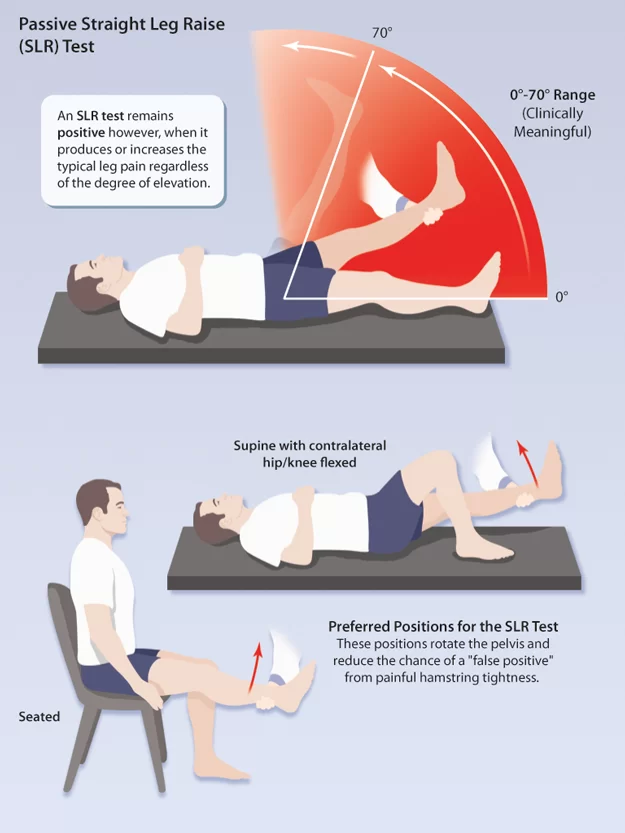
- With unilateral straight leg raising, the nerve roots, primarily the L5’S1&S2 nerve roots[ sciatic nerve ].are normally completely stretched at 70′,having an excursion of approximately 2 to 6 cm[ 0.8 to 2.4 inches ].
- Pain after 70′ is probably joint pain from the lumbar area/sacroiliac joints.
- However, if the examiner suspects hamstring tightness, the hamstrings must also be cleared by examination.
- The examiner should compare the two legs for any differences.
- Although the sciatic nerve roots are commonly stretched at 70′ hip flexion, the ROM for Straight Leg Raising &stress placed on the neurological tissue vary greatly from person to person.
- For example, very hypermobile patients may not show a positive straight leg raising test until 110′ to 120′ of hip flexion, even in the presence of nerve root pathology.
- It is more important to compare the left & right sides for symptoms before deciding whether a lesion is caused by stretching of the neurological tissue or arises from the joints or other soft tissue.
- During the unilateral straight leg raising test, tension develops sequentially.
- It first develops in the greater sciatic forearm, then over ala of the sacrum, next in the area where the nerve roots crossed=s over the pedicle,& finally in the intervertebral foramen.
- The test causes traction on the sciatic nerve, lumbosacral nerve roots,& dura mater.
- Adhesions within these areas may result from herniation of the interpretable disc or extradural or meningeal irritation.
- Pain comes from the dura mater, nerve roots, adventitial sheath of the epidural veins, or synovial facet joints.
- The test is positive if the pain extends from the back down into the leg in the sciatic nerve distribution.
- A central protrusion of an intervertebral disc leads to pain primarily in the back with the possibility of bowel & bladder symptoms; a protrusion in the intermediate area causes pain in the posterior aspect of the lower limb& low back & a lateral protrusion cause pain primarily in the posterior leg with pain below the knee.
- Having said that examiner must realize that the intervertebral discs are only one cause of back pain.
- For patients who have difficulty lying supine, a modification straight leg raising test has been suggested.
- Patient in the side-lying position with the test leg uppermost & the hip& knee at 90′.
- The lumbosacral spine is in neutral but may be positioned for the patient.
- The examiner then passively extended the patient’s knee, noting pain, resistance & reproduction of the patient’s symptoms for a positive test.
- The knee position on the affected side compared with the on the good side.
- The examiner should then both legs simultaneously means bilateral straight leg raising.
- The test must be done with care because the examiner is lifting the weight of weight both lower limbs & thereby placing large stress on the examiner’s lumbar spine.
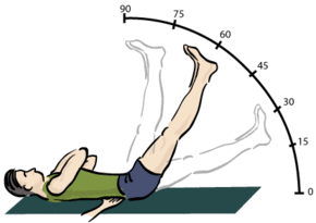
- With the patient relaxed in the supine position & knees extended, the examiner lifts both the leg by flexing the patient’s hip until the patient complains complain of pain/tightness.
- Because both legs are lifted the pelvic is not stable, so on hip joint flexion movement, the pelvic is free to rotate, thereby decreasing the stress on the neurological tissue.

- If the test cause pain before 70′ of hip flexion, the lesion is probably in the sacroiliac joints.
- If the test cause pain after 70′ the lesion is probably in the lumbar spine area.
- With the unilateral straight leg raising test,80’to 90′ of hip flexion is normal.
- If one leg is lifted & the patient complains of pain on the opposite side, it is an indication of a space-occupying lesion.
- This finding of pain when the examiner is testing the opposite leg may be called the good leg raising test of Fajersztajn.
- It typically indicates an intervertebral disc protrusion, usually medial to the nerve root.
Sensitizing Maneuvers of the Straight Leg Raise Test:
- After the elicitation of the symptoms, the examiner[ therapist] is slowly & carefully lowers the leg till the patient is no longer feels pain/tightness.
- Then, either instruct the patient is for to bring their chin to the chest/ the examiner [therapist ] may dorsiflex the patient’s foot,& both actions are done simultaneously;
- However, the foot dorsiflexion is most commonly performed first.
- Both maneuvers are considered provocative/sensitizing tests for the neurological tissue.
- When the increase of Pain with neck flexion/foot dorsiflexion; this both condition indicates a lesion within the spinal cord like as disc herniation, tumor, meningitis & stretching of the dura mater of the spinal cord.
- When the Pain does not increase with the movement of neck flexion, it is indicated to a lesion in the hamstring area means tight hamstrings muscle / in the lumbosacral/sacroiliac joint.
Modifications of the Straight Leg Raise Test :
| SLR(BASIC) Straight Leg Raise Test [basic] | SLR2 Straight Leg Raise Test2 | SLR3 Straight Leg Raise Test3 | SLR4 traight Leg Raise Test 4 | Cross leg [ well leg ] SLR 5 Straight Leg Raise Test 5 | |
| Hip joint | Flexion & adduction | Flexion | Flexion | Flexion &medial Rotation [ internal rotation ] | Flexion |
| Knee joint | Extension | Extension | Extension | Extension | Extension |
| Ankle joint | Dorsiflexion | Dorsiflexion | Dorsiflexion | Plantar flexion | Dorsiflexion |
| Foot | ——- | Eversion | Inversion | Inversion | ———– |
| Toes | ———- | Extension | ——— | ———– | —— |
| Nerve bias | Sciatic Nerve &Tibial Nerve | Tibial Nerve | Sural Nerve | Common Peroneal Nerve | Nerve Root (Disc Prolapse) |
FAQs
What is the straight leg raising test SLRT?
A neurodynamic examination known as the Lasegue sign or straight leg raising test (SLRT) measures nerve root irritation in the lumbosacral region. It is a crucial component of the neurological examination for individuals who report with either radicular or low back pain. Lazarevic sign is another, less popular name for this sign.
What is a positive SLR test?
The test is regarded successful when the patient feels pain along the lower limb in the same distribution of the lower radicular nerve roots (often L5 or S1) after the examiner gently elevates the patient’s leg by flexing the hip with the knee extended.
What is the SLR range for sciatica?
The most often used physical exam for diagnosing sciatica and lumbar disc hernia is the straight leg raise (SLR) test. When the SLR elicits pain below the knee and along the course of the sciatic nerve between 30 and 70 degrees of hip flexion, it is regarded as positive.
Is SLR passive or active?
The straight leg raise (SLR) test is used to evaluate the neural tissues’ mechanosensitivity. The SLR was first classified as a passive test, but in the clinical setting, it is frequently carried out actively.
What is a negative straight leg test?
Straight Leg Raise Test Negative
A negative Straight Leg Raise (SLR) test result means there were no noticeable neurological symptoms, pain, or discomfort during the test. In the SLR test, the patient is lying on their back and the healthcare professional or clinician raises their straightened leg. When the patient does not feel any radiating pain, tingling, or discomfort down the elevated leg, the test is considered to be negative.
One Comment