
Suboccipitals Muscle
Suboccipitals Muscle Anatomy
The suboccipital muscles are a group of four muscles situated at the base of the skull. The suboccipital muscles are important because they have a key role in providing motor control of the head and neck. The four muscles are the rectus capitis posterior major, obliquus capitis superior, obliquus capitis inferior, and the transversospinalis. Each muscle originates from a different part of the occiput.
The suboccipital region is a muscle compartment, located inferior to the external occipital protuberance and the inferior nuchal line.
These are anatomical landmarks on the occipital bone of the skull. It is of a pyramidal shape and includes the posterior aspects of the atlas and axis (C1 and C2 vertebrae respectively).
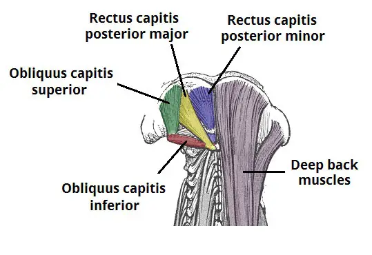
This region comprises four pairs of small muscles:
- Rectus capitis posterior major
- Rectus capitis posterior minor
- Obliquus capitis inferior
- Obliquus capitis superior
These muscles mainly function as postural muscles but can also contribute to movements of the head.
Suboccipital triangle
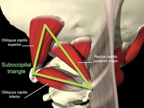
Three of the four muscles contribute to the formation of the boundaries of the suboccipital triangle rectus capitis posterior major, obliquus capitis inferior and obliquus capitis superior.
This triangle is a region of the posterior neck containing the vertebral artery and the dorsal ramus of the first cervical nerve (suboccipital nerve).
The superomedial and superolateral boundaries are formed by the rectus capitis posterior major and obliquus capitis superior respectively.
The obliquus capitis inferior forms the inferolateral boundary. The roof of the triangle is formed by semispinalis capitis muscle, which is one of the three parts of a deep intrinsic back muscle.
The floor consists of the posterior atlanto-occipital membrane and the posterior arch of the atlas. Located deep to the semispinalis capitis is a layer of fibrous adipose tissue.
The suboccipital muscles are a group of four muscles situated underneath the occipital bone. All the muscles in this group are innervated by the suboccipital nerve.
They are located within the suboccipital compartment of the neck; deep to the sternocleidomastoid, trapezius, splenius and semispinalis muscles. They collectively act to extend and rotate the head.
In this article, we shall look at the anatomy of the suboccipital muscles – their attachments, actions and innervation.
Suboccipitals Muscle
Rectus Capitis Posterior Major:
- The rectus capitis posterior major is the larger of the rectus capitis muscles. It is located laterally to the rectus capitis posterior minor.
- Attachments: Originates from the spinous process of the C2 vertebrae (axis), and inserts into the lateral part of the inferior nuchal line of the occipital bone.
- Actions: Extension and rotation of the head.
- Innervation: Suboccipital nerve (posterior ramus of C1).
Rectus Capitis Posterior Minor:
- The rectus capitis posterior minor is the most medial of the suboccipital muscles.
- There is a connective tissue bridge between this muscle and the dura mater (outer membrane of the meninges) which may play a role in cervicogenic headaches.
- Attachments: Runs from the posterior tubercle (a rudimentary spinous process) of the C1 vertebra to the medial part of the inferior nuchal line of the occipital bone.
- Actions: Extension of the head.
- Innervation: Suboccipital nerve (posterior ramus of C1).
Obliquus Capitis Inferior:
- The obliquus capitis inferior is the most inferiorly positioned of the suboccipital muscles. Additionally, it is the only capitis muscle that has no attachment to the cranium.
- Attachments: Originates from the spinous process of the C2 vertebra, and attaches into the transverse process of C1.
- Actions: Extension and rotation of the head.
- Innervation: Suboccipital nerve (posterior ramus of C1)
Obliquus Capitis Superior:
- The obliquus capitis superior is located laterally in the suboccipital compartment.
- Attachments: Originates from the transverse process of C1 and attaches to the occipital bone (between the superior and inferior nuchal lines).
- Actions: Extension of the head.
- Innervation: Suboccipital nerve (posterior ramus of C1
Clinical Importance
Suboccipital syndrome:
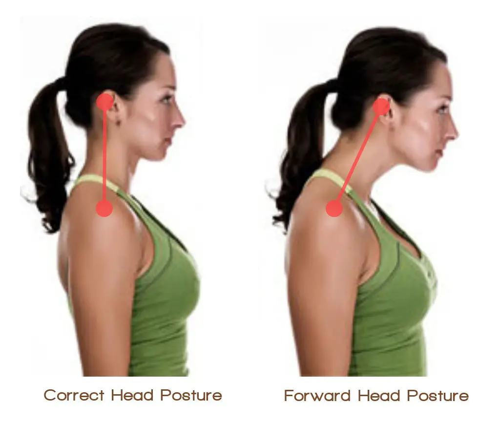
Suboccipital syndrome is a disorder that can occur due to the shortening of the muscles in the suboccipital triangle or spasms of these muscles.
This may be due to poor posture or due to the effects of underlying spinal degeneration. Forward Head posture is also the cause of the syndrome.
The suboccipital syndrome causes pain in the posterior neck and skull, which can radiate to the side of the head and in the back of the eye.
Treatment of this syndrome is patient-specific, but can involve:
- Postural correction
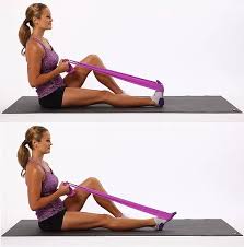
- Suboccipital muscle stretching
- Massage
- Non-steroidal anti-inflammatory medications (NSAIDs) to relieve the pain
- Strengthening exercises
- Avoiding activities that cause the pain
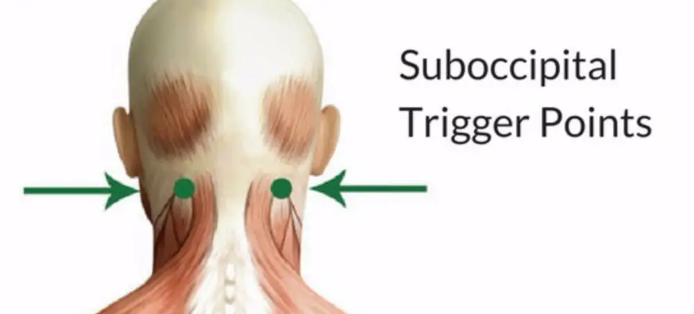
Tension headaches (Cervicogenic Headache)
- Tension headaches are one of the most common headache disorders, and can be caused by trigger points in the suboccipital muscles.
- Trigger points are hyperirritable regions in the fascia surrounding the muscles that are painful on compression.
- These trigger points in the suboccipital muscles can cause referred pain over the temporal and occipital bones, which can be interpreted as a bilateral headache.
FAQ
What are suboccipital muscles?
A set of four muscles in the back of the neck, below the occipital bone, are called the suboccipital muscles. The obliquus capitis superior, obliquus capitis inferior, rectus capitis posterior major, and rectus capitis posterior minor are these four muscles.
What causes tight suboccipital muscles?
In addition to common causes like whiplash and slouching, other factors that can induce tension and stiffness in the area include eye strain and teeth grinding. Your suboccipital muscles are probably strained if you have suboccipital pain, stiffness, or a dull aching near the top of your neck.
How do I loosen my suboccipital muscles?
You can try gentle neck stretches, massage, and relaxation techniques to loosen your suboccipital muscles. If you’re experiencing persistent discomfort, consider consulting with a healthcare professional for personalized advice.
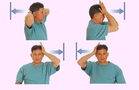
How do I strengthen my suboccipital muscles?
This very basic exercise is one approach to develop these very crucial muscles. In essence, you make a double chin by retracting your chin straight back and in the opposite direction of the direction your nose points. For one to two seconds at the height of the contraction, hold that position, and then release.
What overworked the suboccipital muscles?
A number of things, including eye strain, wearing new spectacles, bad posture, poor ergonomics at a computer workstation, and trauma, can cause the suboccipital muscles to become stiff and painful. In addition, overwork may result from them correcting for instability or injury to the ligaments. The base of the skull may hurt as a result of this.
What technique is suboccipital release?
Using the suboccipital release technique, healthcare professionals can address musculoskeletal issues like headaches and neck pain with relative ease. Because the occipital-atlanto region is being manipulated, this approach is also flexible and can aid in the regulation of the body’s autonomic system.
What is the pain pattern of the suboccipital muscles?
Referred pain generated by trigger points in the suboccipital muscles (TrPs) typically manifests as a bilateral headache and extends over the occipital and temporal bones to the side of the head.
Can suboccipital muscles cause dizziness?
Suboccipital muscles are those of the head and neck that support and govern the head. Dizziness can be brought on by modifications to the suboccipital muscles’ structure and function.

4 Comments