
Supinator muscle
What is the Supinator muscle?
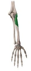
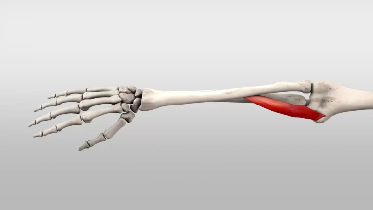
The supinator muscle is a muscle located in the forearm that plays an important role in the movement of the radius bone of the forearm. It arises from the lateral epicondyle of the humerus (upper arm bone) and the supinator crest of the ulna (one of the two bones in the forearm) and attaches to the lateral surface of the radius (the other bone in the forearm).
The main function of the supinator muscle is to rotate the forearm from a pronated (palm-down) position to a supinated (palm-up) position. This movement is important for tasks such as turning a doorknob or using a screwdriver. The supinator muscle also assists in flexing the elbow joint and stabilizing the head of the radius bone during movement.
The supinator muscle is innervated by the radial nerve, which also provides sensory innervation to the skin on the back of the hand and forearm. Injuries to the radial nerve can result in weakness or paralysis of the supinator muscle, leading to difficulties with supination and other forearm movements.
The supinator muscle connects the ulna to the proximal part of the radius by curling around it. The muscle crosses the forearm’s sagittal axis in this manner. The forearm supination movement is made possible by the supinator muscle’s ability to rotate the radius laterally thanks to this fascinating anatomy. The pronator quadratus muscle, which rotates the radius medially and causes the opposite movement of pronation, is its well-known antagonist.
Origin of Supinator muscle
The ulna and humerus are the broad sources of the supinator muscle. The supinator crest of the ulna, the lateral epicondyle of the humerus, the radial collateral ligament, and the annular radial ligament all contribute to the common origin of the two layers of fibers. The upper portion of the radius is surrounded by the superficial fiber layer, which has a tendinous origin. Above the radial tuberosity, the deeper layer of fibers encircles the neck of the radius.
Insertion
On the anterolateral and posterior surfaces, the proximal third of the radius is distal to the radial tuberosity.
Relations
The supinator is tracked down somewhere down in the lower arm, shallow just to the pieces of the range and ulna over which the muscle lies. It forms the cubital fossa’s floor with the brachialis muscle. Extensor carpi radialis brevis and longus cover the muscle spiral surface, while the second rate piece of the anconeus muscle overlies it from the ulnar side.
Just proximal to the supinator muscle, the deep branch of the radial nerve releases the posterior interosseous nerve. The nerve then enters the forearm’s extensor compartment by passing between the muscle’s superficial and deep heads. At times a conflicting fascial band called the sideways rope disregards the profound top of the supinator. By extending between the ulnar and radial tuberosities, the cord connects the ulna and the radius.
Innervation
The supinator is innervated by the back interosseous nerve (C7, C8), a part of the radial nerve.
Blood supply
Two distinct sources supply the supinator muscle’s superficial and deep layers:
The radial artery supplies the superficial layer with blood via its radial recurrent branch, while the ulnar artery supplies the deep layer via its posterior interosseous and posterior interosseous recurrent arteries.
Function of Supinator muscle
At the proximal radioulnar joint, the supinator muscle rotates the radius laterally. The hand is brought into the supine position as a result of this action, which aligns the radius with the ulna and positions the palm up, similar to holding a soup bowl.
While creating a sluggish and unopposed supination development, the supinator muscle gets the job done all alone and is the main player. The straightforward rotation of the hand when grabbing popcorn from a bowl is an illustration of such a movement. Notwithstanding, for a speedy, solid, powerful supination development, or when the development occurs against the opposition, the supinator muscle is helped by the biceps brachii muscle. The most powerful supination occurs when the elbow joint is flexed to 90 degrees, as the biceps brachii cannot supinate when the forearm is fully extended. This behavior can be observed, for instance, when turning a screwdriver or removing the wine cork from a bottle.
Clinical Relevance
Just proximal to the supinator muscle, the radial nerve divides into deep and sensory superficial branches. The deep branch may become entangled and compressed as a result, which has the potential to cause selective paralysis of the muscles served by this nerve. This nerve syndrome, also known as supinator entrapment syndrome, is known to have several potential causes, including stress brought on by repetitive supination and pronation as well as compression caused by various soft-tissue masses surrounding the nerve.
Assessment
With the forearm in a mid-position, extend the patient’s arm and elbow. Palpate along the posterior portion of the proximal third of the radius and actively resist supination.
Supinator muscle stretching
Supination and pronation of the forearms: Maintaining your elbow at your side, bend the injured arm’s elbow 90 degrees. Hold for five seconds while raising your palm. Then hold for five seconds while slowly lowering your palm. During the exercise, make sure to keep your elbow at your side and bent 90 degrees.
Supinator strengthening exercise
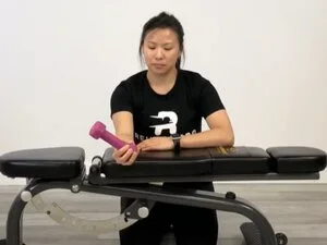
Eccentric wrist supination
The supinators of the wrist can be strengthened with this exercise. Start by holding one end of a dumbbell with your palm facing upward. A bench, table, or thigh can be used to support the forearm. At a tempo of 5-7 seconds, slowly rotate the wrist inward (pronation). To return the weight to its starting position, you can do so with your free hand.
Half dumbbell supination
The supinators of the wrist can be strengthened with this exercise. Start by holding one end of a dumbbell with your palm facing upward. A bench, table, or thigh can be used to support the forearm. Using pronation, rotate the wrist inward until the weight is parallel to the ground. Supinate the wrist to raise the load back up to the beginning position.
Dumbell supination
The wrist and forearm supinators can be strengthened with this exercise. Begin by supporting your forearm against a table, bench, or thigh. Start with your palms facing the floor and hold a small dumbbell from one end. Take a 5-second tempo and slowly rotate the wrist back down into the palms-up position. During the upward (concentric) phase of the motion, you can support yourself with the other hand.
Kettlebell bottoms up
This is an exercise for strengthening the wrist and forearm. Begin by supporting your forearm against a table, bench, or thigh. Start with your palms facing the ground while holding a kettlebell by the handle. Take a 5-second tempo and slowly rotate the wrist back down into the palms-up position. During the upward (concentric) phase of the motion, you can support yourself with the other hand.
Banded supination
The supinator of the wrist can be strengthened with this exercise. Place a band in your palm beginning from the thumb side and move it towards the pinky side and circle it once around the rear of your hand. Use your thumb to hold onto the band. With the hand that is not the target, hold the other end of the band. Rotate the band so that your palm is now facing upward after starting with your palm facing downward. To complete a repetition, and reverse the rotation.
Isometric wrist supination
This exercise exploits the uneven weight dispersion of a portable weight to give opposition to this enemy of turn wrist workout. This exercise specifically targets wrist supinators. Bend your elbow to 90 degrees of flexion and place a yoga block or rolled-up towel between your body and elbow to ensure proper form. Holding a kettlebell’s handle for 5-7 seconds in a thumbs-up position is similar to holding a cup’s handle.
FAQ
What controls the hand’s supination?
Forces and Muscles The forearm muscles known as the pronator teres, pronator quadratus, and flexor carpi radialis are responsible for pronation. The biceps brachii, which is attached to the ulna and provides a powerful supination moment, and the supinator, which is located in the forearm, work together to produce supination.
Which muscle in the arm is most essential for supination?
The biceps is the main supinator of the forearm, assisting the brachialis and brachioradialis in bending the elbow and allowing us to rotate the palm up and down. The long head tendon and the distal tendon that inserts into the radius are particularly susceptible to injury in the biceps.
How is supination evaluated?
The biceps should move proximally during supination and distally during pronation, as shown in Kai here, the examiner will observe. Comparing the visual with the uninvolved side is suggested, and if the muscle’s contours are hard to recognize, the muscle belly may be palpated for motion.
What makes supination so effective?
The simultaneous action of the triceps keeps the biceps flexing action under control when it is performing its supinator function. Supination is more powerful than pronation because it uses a lot of strength from the biceps.
What advantages does supination offer?
Because it enables lifters to pull more weight down to engage the lat muscles, the supinated grip can handle more weight because it recruits the bicep and tricep muscles. The lat muscles that are necessary to build a stronger back can be strengthened to their fullest potential with this grip.


6 Comments