
Radial Nerve
The radial nerve is a major peripheral nerve of the upper limb. The radial nerve is a nerve in the arm that provides sensation to the skin on the back of the hand and the thumb side of the forearm. It is the largest nerve in the upper arm, and is a mixed nerve. The radial nerve is a continuation of the posterior cord of the brachial plexus, which is formed when the axons from the spinal cord segments C8, T1, and a small amount from C7 are joined together.
The radial nerve is a nerve in the forearm that provides sensation and some motor function to the skin and muscles in the thumb, index finger, and part of the middle finger. The radial nerve is a mixed nerve, containing both sensory and motor fibers.
Anatomy of Radial Nerve
The nerve is one of the terminal branches of the posterior cord. In the axilla, it lies behind the axillary and upperbrachial arteries and passes anterior to the tendons of teres minor, latissimus dorsi, and subscapularis.
It enters the posterior compartment of the arm passing through a triangular space, formed by the lateral humerus, long head of triceps and teres minor.
Through this space, the nerve enters the spiral groove of the humerus and descends obliquely between lateral and medial heads of triceps where it reaches the lateral border of the humerus in the distal third of the arm.
The nerve enters the anterior compartment by piercing through the lateral intermuscular septum where it continues between brachialis and brachioradialis.
The superior branch of the radial nerve continues on from the radial nerve anterior to the lateral epicondyle. It travels along the anterolateral side of the forearm. It is only function is sensory.
In the distal third of the forearm, the nerve rises posteriorly from below the tendon of brachioradialis and pierces the deep fascia to become superior. It further divides into the digital nerves.
The deep branch of the radial nerve or posterior interosseous nerve, is entirely motor.
It begins anterior to the lateral epicondyle of the humerus and enters the posterior compartment of the forearm through the two heads of supinator where it curves around the lateral and posterior surfaces of the radius.
It descends between the deep and superficial extensor muscles and lies on the interosseous membrane and ends in a flattened expansion.
Descriptions:
- The nerve runs down the underside of your arm and controls the movement of the triceps muscle, which is located at the back of the upper arm.
- The nerve is responsible for extending the wrist and fingers. It also controls sensation in part of the hand. nerve injury can cause pain, weakness, and loss of function in the wrist, hand, and fingers. A common term for this is radial nerve palsy.
- The radial nerve is close to the bone in the upper arm, so it is vulnerable to injury, especially if the arm breaks.
- Injury to the radial nerve can lead to radial nerve palsy.
Root value:
- It originates from the brachial plexus, carrying fibers from the ventral roots of spinal nerves C5, C6, C7, C8 & T1.
- Radial nerve passes from the Posterior cord of the brachial plexus:
- Branches:
- Superficial branch of the radial nerve.
- Deep branch of the radial nerve (posterior interosseous nerve).
Structure
The radial nerve originates as a terminal branch of the posterior cord of the brachial plexus. It goes through the arm, first in the posterior compartment of the arm, and later in the anterior compartment of the arm, and continues in the posterior compartment of the forearm.
Arm:
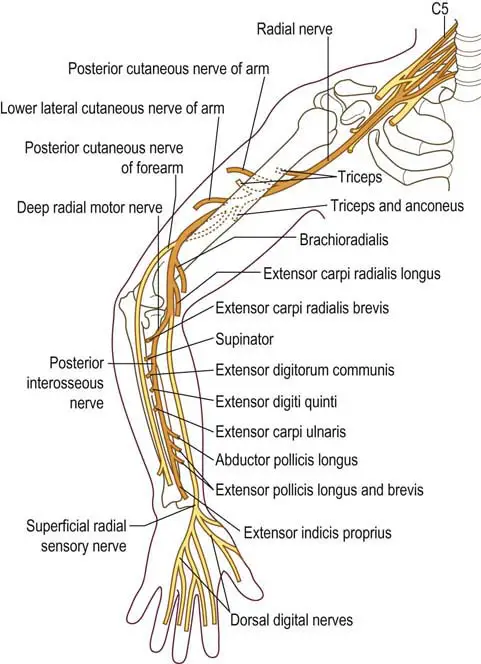
The radial nerve derives from the posterior cord of the brachial plexus and exits the axilla posteriorly to the brachial artery. It passes with the deep brachial artery and gives two motor branches and one sensory branch before traversing the triangular interval. These motor branches innervate the medial and long heads of the triceps. This sensory branch is called the posterior cutaneous nerve of the arm which supplies cutaneous sensory innervation to a portion of the distal posterior arm.
After passing through the triangular interval, the radial nerve descends the radial groove before laterally wrapping around the humerus. At this point, the radial nerve gives a motor branch to the lateral head of the triceps brachii followed by two sensory branches: the inferior lateral cutaneous nerve of the arm which perforates through the lateral head of the triceps, and the posterior cutaneous nerve of the forearm.
Forearm
The posterior cutaneous nerve of the antebrachium also perforates through the lateral head of the triceps but continues to innervate a posterior strip of the forearm. After giving these two sensory branches, the radial artery passes through the lateral intermuscular septum to infiltrate the anterior compartment of the forearm between the brachialis and brachioradialis muscles.
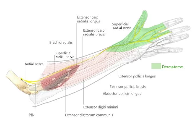
The radial nerve then passes over the lateral epicondyle into the cubital fossa and forearm. Here, the radial nerve separates into deep and superficial branches. The deep branch is a motor branch that passes between the heads of the supinator muscle and becomes the posterior interosseous nerve to innervate the muscles of the posterior compartment of the forearm.
The superficial branch follows the radial artery inferiorly to the anterolateral portion of the radius, deep to the brachioradialis muscle. The superficial branch then courses dorsally over the distal radius over the anatomical snuffbox to innervate the posterior lateral three and a half digits (the thumb, index, middle, and lateral half of the ring fingers) and the associated hand area.
Function of Radial Nerve
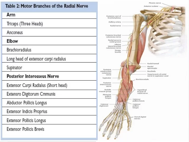
Motor function of Radial Nerve
- The Radial Nerve branches off to the Deep Branch after it passes through the cubital fossa and then continues as the Posterior Interosseous Nerve after it passes between the supinator muscle heads.
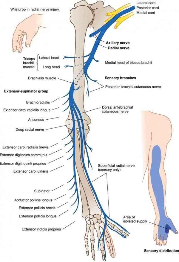
Radial Nerve: - Triceps brachii (medial and lateral heads) — provides an extension of the forearm.
- Extensor carpi radialis longus — provides for an extension of the wrist.
- Brachioradialis — provides flexion of the elbow as well as pronation and supination, depending on the position of the forearm.
- Anconeus — provides for elbow extension.
Deep Branch of the Radial Nerve: - Extensor carpi radialis brevis — extends and abducts the wrist.
- Supinator — supinates the forearm.
- Posterior interosseous nerve:
- Abductor pollicis longus — abduct the thumb at the wrist.
- Extensor carpi ulnaris — extends and adducts the wrist.
- Extensor digiti minimi — extends the wrist and small finger.
- Extensor digitorum — extends the medial four digits of the hand.
- Extensor indicis — extends the index finger and to some extent wrist extension.
- Extensor pollicis brevis — extends and abducts the thumb at the carpometacarpal and metacarpophalangeal joints.
- Extensor pollicis longus — extends the terminal phalanx of the thumb.
Sensory:
Anterior aspect:
- Inferior lateral cutaneous nerve of the arm – provides sensation to the anterior lateral aspect of the mid-arm.
Posterior aspect:
- Posterior cutaneous nerve of the arm – sensation to the posterior distal arm.
- Posterior cutaneous nerve of the forearm – sensation to a strip posterior aspect of the forearm
- Superficial branch – sensation to the posterior aspect of the thumb, index, middle, and lateral half of the ring fingers as well as the associated dorsal hand area.
Dorsum of the wrist
- Lateral dorsal surface of the hand
- Dorsum of the thumb
- Dorsum of the index and middle fingers
- Dorsum of the lateral aspect of the ring finger
Embryology
The neural crest, or the strips of ectodermal cells at the edges of the neural tube in the developing embryo, is the source of the radial nerve. It is bilaterally paired. Peripheral nerves, including the radial nerve, are the result of neural crest cell migration to the upper limb.
Anatomical Variations
Normally, the radial nerve’s deep branch travels between the heads of the supinators, where it transforms into the posterior interosseous nerve, supplying innervation to the posterior forearm muscles. Impingement risk may rise in certain individuals, though, as the deep branch of the radial nerve passes via the Supinator Arch, or arcade of Frohse.
Clinical Relevance:
Injury to the Radial Nerve:
Injury to the radial nerve can be broadly categorized into four groups – depending on where the damage has occurred.
In the Axilla:
- The radial nerve can be damaged in the axilla region by a dislocation at the shoulder joint, or a fracture of the proximal humerus. Occasionally, it is injured via excessive pressure on the nerve within the axilla (e.g. a badly fitting crutch).
- Motor functions – the triceps brachii and muscles in the posterior compartment are affected. The patient is unable to extend the forearm, wrist, and fingers. Unopposed flexion of the wrist occurs, known as wrist drop.
- Sensory functions – all four cutaneous branches of the radial nerve are affected. There will be a loss of sensation over the lateral and posterior arm, posterior forearm, and dorsal surface of the lateral three-and-a-half digits.
In the Radial Groove:
In the Forearm
There are two terminal branches of the radial nerve located within the forearm.
- Superficial Branch:
- Mechanism: Stabbing or laceration of the forearm
- Motor functions: None
- Sensory functions: Sensory loss affecting the lateral 3 ½ digits, and associated palm area
- Deep Branch:
- Mechanism: Fracture of radial head, or posterior dislocation of radius
- Motor functions: The majority of the muscles in posterior forearm are affected. Wrist-drop does not occur, as the extensor carpi radialis longus is unaffected, and maintains some extension at the wrist
- Sensory functions: None
- Wartenberg’s syndrome: (not to be confused with Wartenberg’s sign), due to nerve entrapment beneath the tendinous insertion of brachioradialis, tight jewellery, and watch bands.
Radial Tunnel Syndrome
The radiocapitellar joint, or lateral elbow, marks the start of the 5-centimeter-long radial tunnel, which ends at the supinator’s proximal border. This region is not far from where the forearm branches of the radial nerve originate. This level of the radial nerve is affected by a rare compressive neuropathy called radial tunnel syndrome. The disorder is linked to specific jobs or sporting activities and is usually caused by repetitive forearm movements, especially supination and pronation.
Weariness or a dull, throbbing pain at the proximal part of the forearm during use are among the symptoms. Pain in the dorsal aspect of the wrist or hand is less common. Due to the cutaneous branches of the radial nerve being most frequently damaged, extensor and supinator weakness are typically not detected.
Radial tunnel syndrome and posterior interosseus nerve compression syndrome are related conditions. The posterior interosseus nerve, which innervates the wrist and hand extensors as well as the supinator, is affected by this neuropathy. As a result, motor impairments are the most typical presentation.
Treatment options for radial tunnel syndrome include medication and surgery. The palpable proximal portion of the radial head serves as the primary surgical marker for the correction of radial tunnel syndrome. 7.4 cm distal to this landmark, the superficial head of the supinator muscle is where the posterior interosseus nerve exits.
Posterior Interosseus Nerve Compression Syndrome
The motor deficits to the muscles innervated by the posterior interosseus nerve distinguish posterior interosseus nerve compression syndrome from radial tunnel syndrome. There is compression of the nerve at the Frohse arcade. This neuropathy is frequently caused by inflammation, tumors, and trauma.
Because it is located in the radial groove of the humerus, the radial nerve is susceptible to damage following a fracture of the humeral shaft. This degree of radial nerve injury can result in wrist drop, a disorder characterized by the inability to extend the wrist. The radial nerve can also be harmed by a lateral supracondylar fracture, although the presentation is distinct from that of a midhumeral injury.
Above this fracture, nerve branches that supply the brachioradialis and extensor carpi radialis longus arise, leaving these muscles mainly untouched. When requested to extend the hand, the patient’s wrist weakly extends radially as opposed to showing a wrist drop.
FAQs
What is radial nerve?
The radial nerve is one of the major nerves in the human body and plays a crucial role in providing motor and sensory innervation to the upper limb. It is one of the main nerves of the brachial plexus, a network of nerves that originates from the cervical and thoracic spinal cord and supplies the shoulder, arm, forearm, and hand.
Anatomy:
The radial nerve originates from the posterior cord of the brachial plexus, which is formed by nerve fibers from the spinal nerve roots C5 to T1. It courses down the arm and forearm, passing through the axilla (armpit) and along the back of the humerus bone (upper arm bone). It then wraps around the humerus in the radial groove before branching out into various branches in the forearm.
What is the origin of the radial nerve?
The radial nerve originates from the brachial plexus, which is a network of nerves formed by the anterior rami (major branches) of the spinal nerves C5 to T1. The brachial plexus is responsible for innervating the muscles and providing sensory information to the upper limb.
The brachial plexus is divided into three main cords – the lateral cord, the medial cord, and the posterior cord – based on their position relative to the axillary artery. The posterior cord is where the radial nerve originates.
The nerve fibers that contribute to the formation of the radial nerve originate from the spinal nerve roots C5, C6, C7, C8, and T1. These nerve fibers come together to form the radial nerve as it courses down the arm. The nerve then travels through the axilla (armpit) and along the back of the humerus (upper arm bone) before branching out into various smaller nerves in the forearm.
What happens when the radial nerve is damaged?
Radial nerve injuries can be caused by trauma (such as humerus bone fractures), compression, or entrapment, among other things. Radial nerve palsy is a condition that may result from an injury, depending on where it occurs and how severe it is. The muscles that the radial nerve innervates may become weak or paralyzed as a result of this illness, and the affected areas may experience sensory impairments.
This nerve regulates the extension of the elbow, wrist, and fingers as well as movement and sensation in the arm and hand. When the radial nerve is damaged, a condition known as radial nerve palsy can develop. As a result, the muscles this nerve controls may become weak, numb, or uncontrollable.
Which nerve causes wrist drop?
The radial nerve, which runs down the arm and regulates the movement of the wrist extensor’s muscle at the back of the forearm, is damaged and the result is wrist drop.


30 Comments