
Strength Duration Curve (SDC)
What is the Strength Duration Curve?
- The strength-duration curve is a graph between electrical stimuli of different intensities and records the time needed by each stimulus to start the response.
- S-D curve should be plotted after 20th day of injury/lesion.
- It indicates the strength of impulses of various durations required to produce muscle contraction by joining the points that graphically represent the threshold value along the ordinate for various duration.
- After 21st/22nd day, regeneration of nerve will start, generally it take about 270 days to regenerate.
- The purpose of S-D curve plotting is to know whether the stimulated muscle is innervated, denervated or partially denervated.
- There are also other method for this purpose like EMG and NCV.
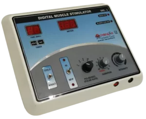
APPARATUS:
- The apparatus used for plotting S-D Curve supplies rectangular impulses of different duration.
- Impulse with duration of 0.01, 0.03, 0.1, 0.3, 10, 30, 100, 300 ms are required.
- The stimulator may be of either the constant current or constant voltage type.
- The constant current stimulator was thought to produce the more accurate result but constant voltage stimulator is rather more comfortable for patient.
OPTIMUM TIMING OF SDC:
- SDC test can be done 10 – 14 days after the lesion has occurred.
- The degeneration of nerve from the proximal to distal is called Wallerian degeneration.
- When the motor end plate is no longer functioning, it is done weekly under the same condition until there is recovery and decision has been reached on the eventual final state of the muscle.
- SDC is used to identify denervation, partial innervation, and compression.
Methods of Strength Duration Curve
- The patient must be warm, fully supported and in sufficient light.
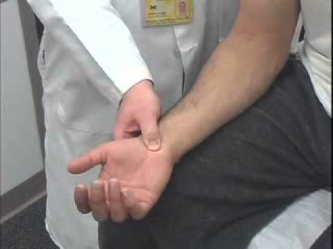
- The indifferent electrode may be applied over some convenient area usually on the midline of the body or the origin of the muscle group.
- Active electrode placed over fleshy part of muscle. (Sometime two small electrode may be used, one over each end of muscle belly).
- Current is applied using the longest stimulation first and increased until a minimal contraction is obtained.
- Intensity of current (or voltage) is noted and impulse is shortened. This procedure is repeated with each stimulation in turn, the intensity of current being increased as required.
- A minimal contraction is used, as this make it easy to detect any change in strength, and electrode should be placed in same point over the muscle throughout the test.
- The S-D Curve is plotted from the result of the test, and although it will be further to the left with constant voltage than with the constant current stimulator.
- The shape of the curve is the essential feature.
Characteristics of Strength Duration of Curve :
- Innervated muscles:
- When all the nerve fibers supplying the muscles are intact, the strength duration curve has a shape characteristic of normally innervated muscles as shown in the figure.
- The same strength of stimulus is required to produce a response with all the impulses of longer duration, while those of shorter duration require an increase in strength of the stimulus each time the duration is reduced.
- Denervated muscles:
- When all the nerve fibers supplying a muscle have degenerated, the strength duration produced is characteristic of complete denervation as shown in the figure.
- For all impulses with duration of 100 ms or less the strength of the stimulus must be increased each time the duration is reduced and no response is obtained to impulses of very short duration. The curve rises steeply and is shifted to the right than that of normally innervated muscle.
- Partially denervated muscles:
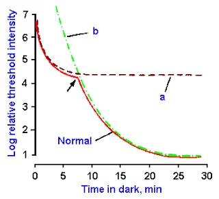
- For the stimulation of denervated fibers impulses of longer duration are required while for the stimulation of innervated fibers impulses of shorter duration are required. The kink produce shows partial denervation which disappears after 10 -20 days or a month.
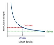
RHEOBASE:
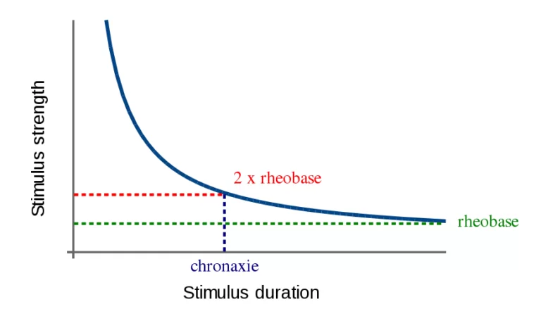
- When stimulus is given using the maximum pulse width available on the stimulator, the intensity of the current required to produce a twitch is called rheobase of the muscle.
- Mainly 100 to 300 ms duration are used to record rheobase.
- The pulse is always rectangular measured in miliamperes or volts.
- Rheobase is measured using the cathode on the motor point of the nerve or by using bipolar technique.
- Normal values of rheobase are 2 to 18 mA or 5 to 35 volts.
- Normal value of rheobase of different muscle:
- Deltoid-14 volts,5mA.
- Triceps-18 volts,5mA.
- Abductor digiti minimi-30volts,8mA.
- Frontalis-14volts,4mA.
FACTORS RESPONSIBLE FOR RHEOBASE:
- Resistance of skin and subcutaneous tissue.
- Edema and inflamation.
- Ischemia and underlying pain.
- Temperature variation.
- Position of electrode.
- Amount of subcutaneous tissue.
- Degeneration.
- Deneravtion.
- Partial denervation generally produce no changes in rheobase.
- Re-innervation can show a sharp rise in rheobase which herald clinical recovery.
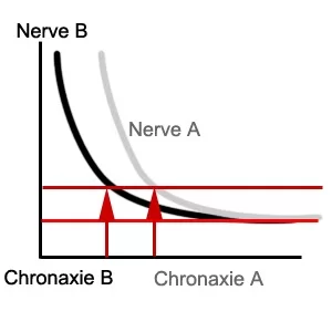
CHRONAXIE:
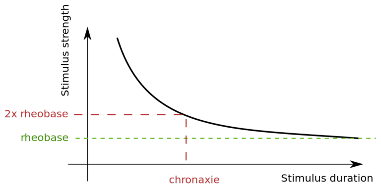
- At the double intensity of rheobase, the minimal pulse width required to produce the twitch is called chronaxie of muscle.
- Chronaxie is an index of excitability and is time in millisecond, necessary to induce minimal visible contraction with a stimulus of twice the strength of the rheobase.
- Normal values of chronaxie are less than 1ms (0.05 to 0.5 ms).
Variation in chronaxie depends on whether a constant current machine or a constant voltage machine is used. - At birth chronaxie is 10 times higher than normal and 18 to 20th month, the chronaxie falls to normal values.
FACTORS RESPONSIBLE FOR CHRONAXIE:
- Texture of skin.
- Ischemia.
- Edema.
- Fatigue.
- Position of stimulating electrode.
- Denervation.
- Partial denervation.
- Re-innervation.
- Nerve root lesion.
- Peripheral neuropathy.
- Myopathy (No significant change).
EQUIPMENT REQUIRED FOR S-D CURVE:
- Low-frequency generator with varying pulses from 0.02 to 1000ms.
- Moist saline pad.
- Electrodes.
- Leads.
- Bandage.
- Plastic protactors.
DETERMINATION OF ACCOMMODATION:
- Accommodation is the property of nerve or muscle membrane to react less strongly to a slowly increasing current intensity by accommodating the electrical impulse.
- Measure of the constant of accommodation is lambda.
- Accommodability are calculated from ratio of rectangular wave rheobase and the value of the progressive current rheobase.
- Normal- 3 to 6.
- Denervated- Below 3.
- No accommodation- 1 or below.
ACCOMMODATION QUOTIENT(AQ):
- When repeated stimulation are given to a muscle after some time it accommodates. If the muscle is stimulated by triangular impulses and rectangular pulses at different time frames, the triangular impulse requires more intensity to produce contraction of the same strength as that of rectangular ones. The intensity of current used by triangular impulse divided by rectangular is called A.Q.
- A.Q. values show you the state of innervation, denervation, and partial innervation.
- Value of A.Q:
- 1: Dysfunction of the muscle persists for more than 6 months then there is difficult to regenerate the nerve by electrotherapy, and no further plotting is required.
- 2-4: The nerve has degenerated but it will respond to electrotherapy.
- 4-6: The neuromuscular system is unimpaired and it will respond to electrotherapy and muscle get back to almost within 3 weeks.
ADVANTAGES OF THE S-D CURVE:
- It is simple, reliable, and cheaper.
- Indicate proportion of denervation.
- Less time consuming.
DISADVANTAGES OF THE S-D CURVE:
- In large muscles, only a proportion of fibers may respond hence picture is not clearly shown.
- It is a qualitative rather than quantitative method of testing innervation.
- It won’t point out the site of lesion.
FAQs
What is the strength duration curve of a neuron?
The relationship between the power of an electrical stimulation delivered to a muscle’s motor point and the time needed to cause a minimum contraction is depicted graphically by the strength-duration curve.
What is rheobase and chronaxie?
Chronaxie is the pulse width at twice the rheobase threshold current, and rheobase is the threshold stimulus current for an active response with a lengthy pulse.
What is the strength curve of a muscle?
With force as the y-axis and joint angle as the x-axis, strength curves are graphical representations of the amount of force needed at increasing joint degrees for a specific exercise. This describes the amount of force needed at each point throughout the range of motion (ROM) of a certain movement.
What is chronaxie time?
When the amplitude is two times the rheobase, the time it takes for a fibre to activate is known as the chronaxie. The chronaxie provides a measure of the relative excitability of two neurons with similar rheobases; shorter chronaxies apply to more excitable neurons.
Author: Nitesh Patel - Physiotherapist
Physiotherapist in Samarpan Physiotherapy Clinic Ahmedabad Bapunagar Amaraiwadi Vastral Mobile Physiotherapy Clinic Dr. Nitesh Patel ( Physiotherapist ) : Mo No : 09898607803


Very informative
It’s really good
It’s really good .I want more other notes pls how can I get it
GOOD INFORMATIIN
thank you
i want more details about sd curve
its very eaSY TO STUDY
Very good
Really awesome bro
Can s d curve be carried out at home of the patient? What will be charges?
Yes ! Physiotherapist are able to do S D Curve at Home and in Clinic. For That Diagnostic Electrical Stimulator Machine ( ES ) is Required and this machine is small and able to carried out at home.
In kink ,above kink which type of nerve fibres is there and it starts from 300 to 0.01 or 0.01 to 300??