
Torticollis: Physiotherapy Treatment
What is a Torticollis?
- Torticolis is a condition (also known as ‘wryneck’) in which the baby’s head is tilted. The head often rotates towards one shoulder and tilts away to the opposite side. The term ‘congenital’ is also sometimes used when describing torticollis. This means that it is present at or shortly after birth. Babies treated early with physiotherapy programmes usually respond well to treatment.
- Torticollis is a symptom related to turning or bending of the neck. Many different causes are possible. In newborns, torticollis usually results from injury during labor and delivery or the infant’s position in the womb. Less often, it is caused by birth defects. In older children, torticollis may result from injuries to the neck muscles, common infections, or other causes.
- Painful spasms of the neck muscles may occur.
- Other symptoms may be present, depending on the cause. For example, there may be a tender lymph node (gland) if the cause is infection.
Types of Torticollis:
- Congenital torticolis.
- Acquired torticolis.
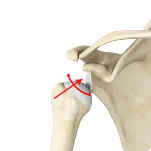
Anatomy Related To Torticollis:
- The normal physiologic range of rotation of the atlas on the axis is 25-53 degrees to either side. The transverse ligament is the primary stabilizer of the atlanto-axial joint and prevents excessive anterior motion of the atlas on the axis. It extends behind the dens, between the medial portions of the lateral masses of C1.
- The paired alar ligaments act as secondary stabilizers to prevent anterior shift. The alar ligaments extend from the lateral aspect of the dens tip to the medial aspect of the occipital condyles, with a lower portion attaching to the medial aspect of the lateral masses of C1.
- The sternocleidomastoid muscle has a sternal and clavicular head. The sternal head is directed from the manubrium sterni superiorly, laterally and posteriorly and the clavicular from the medial third of the clavicle vertically upward. It runs to the mastoid process.
- It enables an ipsilateral lateral flexion and a contralateral rotation. The muscle extends the upper part of the cervical spine and flexes the lower part.
Causes of Torticollis:
- Muscular in more than 80% of the cases. Types muscular torticollis:
- – Fibromatosis colli: torticollis with palpable mass in the SCM;
- – Tightness of the SCM without an apparent mass;
- Postural torticollis with neither mass or tightness.
- Birth trauma: facet dislocation, tears in the sternocleidomastoid muscle
- Congenital anomalies of the craniovertebral junction: occipitoatlantal fusion or Klippel-Feil syndrome.
- Sternocleidomastoid tumour.
- Ocular abnormalities.
- Intrauterine mechanical factors
Other Causes of Torticollis:
- Sitting or sleeping in an unusual position without adequate neck support.
- Poor posture when looking at a computer screen.
- Carrying heavy unbalanced loads (for example, a briefcase or shopping bag).
- Allowing certain muscles of the neck to be exposed to cold (sleeping in a draught).
Epidemiology
Ten to twenty percent of cases of toricollis are posttraumatic; the remaining cases are idiopathic. Posttraumatic cervical dystonia often manifests days after the injury, and in its delayed version, it may take up to 12 months.
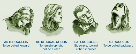
Typically, torticollis involves a variety of movements. Torticollis is the most prevalent kind, involving some degree of rotation. Rotational torticollis is followed by laterocollis, retrocollis, and anterocollis, which is the least common kind.
There is a 2:1 preference for females over males. Idiopathic cervical dystonia usually manifests in people between the ages of 30 and 50. The prevalence of congenital muscular torticollis in infants is less than 0.4%.
Which Symptoms are seen in Torticollis?
- The twisting of your neck (torticollis) occurs when your muscles supporting the neck on one side are painful.
- The pain is usually on one side of your neck and stiffness of the muscles in that area twists the neck to one side.
- You may find it very difficult when you try to straighten your neck, due to pain. Occasionally, the pain is in the middle of your neck.
- The pain may spread to the back of your head or to your shoulder. The muscles of your affected side may be tender. Pressure on certain areas may trigger a ‘spasm’ of these muscles. Movement of your neck is restricted, particularly on one side.
Differential Diagnosis
- Essential tremor
- Myasthenia gravis
- Multiple sclerosis
- Neuroleptic agent toxicity
- Parkinson disease
- Peritonsillar abscess
- Rehabilitation and cerebral palsy
- Retropharyngeal abscess
- Spinal hematoma
- Tardive dyskinesia
- Wilson disease
How to Diagnosis Done in Torticollis?
- A thorough neurologic examination should be performed, and anteroposterior and lateral radiographs of the cervical spine should be obtained.
- A CT scan or MRI of the head and neck is necessary for any patient with persistent neck pain or with neurologic signs and symptoms.
Treatment of Torticollis:
Medicines:
- Pain killer are often helpful. such as,
- Paracetamol at strength is often sufficient.
- Anti-inflammatory painkillers.
- A stronger pain killer such codeine.
- A muscle relaxant such as diazepam.
- Other treatments such as:
- Rest.
- A good posture.
- A firm supporting pillow.
- Heat pack.
PHYSIOTHERAPY TREATMENTS AND EXERCISES:
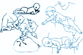
- Positioning.
- Gentle range of motion exercises for neck.
- Stretching of sternocleidomastoid muscle.
- Stretching the muscle in a prone position both actively and passively.
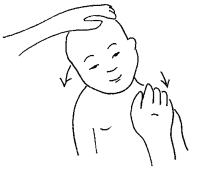
- Stretching the muscle in a lateral position supported by a pillow (have infant lie on the side with the neck supported by pillow). Affected side should be against the pillow to deviate the neck towards the non-affected side.
- Active rotation exercises in supine, sitting, or prone position (use toys, lights, and sounds to attract the infant’s attention to turn the neck and look toward the non-affected side).
- Passive cervical rotation.
- Strengthening exercises.
- Activities to encourage active head movement.
- Visual tracking.
- Lateral head tilt.
- Therapy ball exercises.
- Side sitting exercises.
- Hands and knees.
- Kneeling to standing.
- Assisted rolling.
- Proped sidelying.
Torticollis treatment at home for babies:
- The best method of torticollis treatment is to encourage your baby to turn his or her head in both directions. This will help to loosen tense neck muscles and tighten the loose ones. Here are some exercises to try:
- When your baby wants to eat, offer the bottle or your breast in a way that encourages your baby to turn away from the favored side. (Use your child’s desire to eat to encourage him or her along!)
- When putting your baby down to sleep, position him or her to face the wall. Since babies prefer to look out onto the room, your baby will actively turn away from the wall and this will stretch the tightened muscles of the neck.
- During play, draw your baby’s attention with toys and sounds to make him or her turn in both directions.
Doctor Team
For the treatment of these patients, specialized consultation in physiotherapy, orthopedic surgery, plastic surgery, neurology, neurosurgery, or otorhinolaryngology may be required because of the wide variety of etiologies of torticollis.
The cause determines the results. Every treatment has the possibility of recurrences. Torticollis that persists causes isolation and poor appearance. Therefore, a mental health nurse consultation may be beneficial for some of these patients.
The best results for patients with torticollis can be obtained through an interprofessional team approach involving doctors, mid-level practitioners, nurses, pharmacists, physical therapists, and even chiropractors who collaborate with one another.
How Can I Prevent Torticollis?
Torticollis cannot be prevented, as far as is known. However, prompt medical attention can help save your baby’s illness from getting worse. It might also save your infant from requiring surgery down the road.
Within a few months of your baby’s birth, start regular torticollis stretches with them. Additionally, by getting started right away, you can avoid any potential long-term issues.
Prognosis
A rather frequent disorder in infants and young children is torticollis. Treatment for the condition usually involves adjusting your baby’s head and stretching their neck. You should see some improvement in your baby after a few months. If treatment is initiated early, there shouldn’t be any long-term problems.
Possible Complications
Your infant may get a facial deformity if they have torticollis because of the restricted movement of their muscles. Plagiocephaly, often known as flat head syndrome, is another possible consequence. The skull of your infant is malleable and soft. The pressure from frequently resting the same part of their head on a surface can cause that area to become flat.
Conclusion
It could be distressing to see your newborn with their head twisted or tilted to one side. However, infants with torticollis usually don’t feel pain. Stretches and positioning are typically effective treatments for the problem. Get in touch with your baby’s healthcare physician if these at-home workouts and other therapies don’t seem to be helping. They’ll be able to assess whether your child requires more care.


8 Comments