Great Cardiac Vein
Introduction
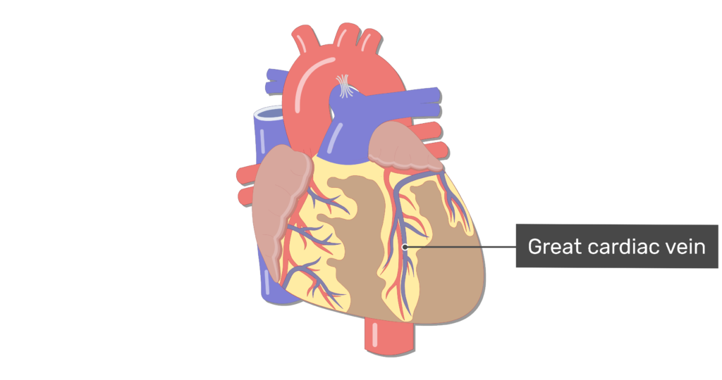
The great cardiac vein is a significant anatomical feature of the heart’s venous system, playing a crucial role in the circulation and function of the myocardium (heart muscle). Located in the anterior interventricular groove of the heart, alongside the left anterior descending artery (LAD), it serves as a major conduit for deoxygenated blood drainage from the front of the heart.
The coronary sinus does indeed have a massive tributary in the great cardiac vein (GCV), which would be the longest vein in the heart. It travels down the anterior chamber of the left ventricle and travels to the base of the heart via the anterior interventricular groove having to carry deoxygenated blood. The great cardiac vein has two other names which are the anterior interventricular vein as well as left coronary vein.
Anatomy of the Great Cardiac Vein
These are some of the best cardiac vein consistent coronary venous frameworks among specimens.
- Location and Course:
- Position: The anterior interventricular groove of the heart will be where the left anterior descending artery (LAD) and the great cardiac vein are stored.
- Origin: Usually starts near the heart’s apex, where it gathers blood from the myocardium that has lost oxygen.
- Pathway: ascends toward through the use of the coronary sulcus anterior interventricular groove.
- Termination: the GCV traditionally decides to join the coronary sinus, which is actually currently situated in the posterior coronary sulcus of the heart.
- Relationships with Adjacent Structures:
- Left Anterior Descending Artery (LAD): The great cardiac vein closely parallels the left anterior descending artery, forming a significant anatomical and functional relationship in the coronary circulation. There are significant variations in the left anterior descending artery’s location with respect to the great cardiac vein. The great cardiac vein is currently located above the artery in 60 to 70 percent of the overall cases.
- Coronary Sulcus: Runs adjacent to this groove, providing a pathway for venous drainage towards the coronary sinus.
Which veins and arteries focus on providing the huge cardiac vein?
Tributaries and Branches: The tributaries of the great cardiac vein include several smaller veins that drain blood from specific regions of the heart’s anterior surface. These tributaries typically contribute to the overall venous drainage of the anterior myocardium and eventually feed into the great cardiac vein itself. Here are the main tributaries:
Anterior Interventricular Veins (Anterior Cardiac Veins):
- These veins drain the anterior segment of the interventricular septum and the surrounding regions of the left and right ventricles. They implement the anterior interventricular artery, also recognized as the left anterior descending artery, or LAD.
- They merge to contribute significantly to the blood flow directed towards the great cardiac vein.
Left Marginal Veins:
- The veins, also referred to as the left diagonal veins, run alongside the marginal branches of the left anterior descending artery
- They make a significant contribution of plasma to the anterior venous drainage system and vacant the lateral aspect of the left ventricle.
Left Atrial Veins:
- The blood from the left atrium is drained by these veins, specifically from the anterior and lateral walls.
- They play a role in the venous drainage of the left atrium, contributing to the overall blood flow toward the great cardiac vein.
Veins of the Left Ventricle:
- Various smaller veins from the left ventricle, including veins from the anterior and lateral walls, contribute to the drainage into the great cardiac vein.
- These veins collect blood from the myocardium of the left ventricle and transport it toward the coronary sinus via the great cardiac vein.
These tributaries collectively ensure that the great cardiac vein receives deoxygenated blood from the anterior portions of both ventricles and parts of the left atrium, ultimately facilitating its function in the venous drainage system of the heart. Understanding these tributaries is essential in clinical contexts such as cardiac surgery and interventions where precise knowledge of cardiac anatomy helps in managing and treating cardiovascular conditions effectively.
Understanding the relationships and contributions of these veins helps in comprehending the complex anatomy of cardiac circulation, which is essential in clinical practice, cardiac surgery, and the treatment of cardiovascular diseases.
Variations of great cardiac vein
About one-third of the time, the great cardiac vein’s route from the coronary sulcus to the inferolateral wall of the left atrium is shaped like an s. The coronary sinus obtains direct drainage from this spot.
- Anatomical Variability: The great cardiac vein can vary in size, course, and termination pattern. A significant difference between the two in the great cardiac vein’s orientation with respect to the circumflex artery was ended up finding in a cadaver heart that was otherwise nutritious. While the vessels originated and terminated as expected, they intertwined in their midcourse, each making a complete loop around the other. This variation did not appear to be linked to any underlying abnormalities. Until recently, there has been limited exploration of the different paths of the great cardiac vein, underscoring the uniqueness of our interconnected situation. Variations in the great cardiac vein were indeed generally related to the existence, placement, and shallow or deep relationship between personality anterior interventricular and circumflex artery crossings.
- Anomalies: Occasionally, variations may include multiple branches or connections with other cardiac veins, which are important considerations in surgical and interventional procedures.
What are the functions of the great cardiac vein?
The cardiac veins, including the great cardiac vein, play crucial roles in the circulatory system of the heart. Here’s a detailed exploration of their functions. The great cardiac vein’s main function is to assist in the removal of venous blood from the myocardium’s outer layer.
Functions of Cardiac Veins:
- Venous Drainage of the Myocardium: Primarily responsible for collecting deoxygenated blood from the anterior part of the heart’s myocardium.
- Great Cardiac Vein: The left ventricle and some areas of the left atrium are the main sources of deoxygenated blood that are collected by the great cardiac vein from the anterior region of the heart.
- Other Cardiac Veins: Include the middle cardiac vein, small cardiac veins, and anterior cardiac veins, which collectively drain blood from different regions of the heart muscle.
- Transport of Deoxygenated Blood:
- These veins transport deoxygenated blood containing metabolic waste products (such as carbon dioxide) away from the myocardium.
- The collected blood flows through the cardiac veins towards larger venous structures like the coronary sinus.
- Coronary Circulation: Blood samples were drawn into the coronary sinus by channels, which then returned the blood to the right atrium.
- The great cardiac vein and other cardiac veins play a role in coronary circulation by carrying oxygen-depleted blood back to the right atrium of the heart.
- Through pulmonary circulation, blood is circulated throughout the body to reduce carbon dioxide emissions and also provide oxygen.
- Integration with the Coronary Sinus:
- The coronary sinus is a significant venous channel located in the coronary sulcus (atrioventricular groove) on the posterior aspect of the heart.
- It receives blood from the cardiac veins, including the great cardiac vein, and acts as a reservoir before emptying into the right atrium.
In summary, the cardiac veins, including the great cardiac vein, fulfill essential roles in the cardiac circulation system by efficiently draining deoxygenated blood from the heart muscle and facilitating its return to the systemic circulation for oxygenation and metabolic waste removal. It is essential to comprehend their anatomy and function in order to treat and manage a wide range of cardiac conditions.
What is the end of the great cardiac vein?
The great cardiac vein typically ends by draining into the coronary sinus. The atrioventricular groove, which partitions the left ventricle and left atrium in the posterior region of the heart, comprises a significant vein known as the coronary sinus. serves as the main venous channel collecting blood from the myocardium (heart muscle).
From the coronary sinus, blood flows into the right atrium of the heart, completing the venous return pathway from the cardiac muscle back to the systemic circulation. One membranous leaflet found in 60% of hearts, the valve of Vieussens, marks the end of the Great cardiac vein.
Conclusion
In conclusion, the great cardiac vein is a vital component of the heart’s venous system, intricately involved in the efficient drainage of deoxygenated blood from the anterior myocardium. Situated alongside the left anterior descending artery in the anterior interventricular groove, it collects blood from the left ventricle and portions of the left atrium. This venous blood is crucially transported through the great cardiac vein towards the coronary sinus, located in the coronary sulcus on the heart’s posterior surface.
- Functionally, the great cardiac vein supports coronary circulation by ensuring the removal of metabolic wastes such as carbon dioxide from the myocardium. Its integration with the coronary sinus facilitates the eventual return of deoxygenated blood to the right atrium, completing the circulatory cycle necessary for sustaining cardiac function.
- Clinically, the anatomy and function of the great cardiac vein are significant in various medical contexts, including cardiac surgery where it may be utilized in procedures like coronary artery bypass grafting (CABG). Additionally, its role in cardiac electrophysiology underscores its relevance in diagnosing and treating arrhythmias.
Understanding the anatomy, physiology, and clinical implications of the great cardiac vein is essential for medical professionals involved in cardiology, cardiac surgery, and cardiovascular research, emphasizing its critical role in maintaining optimal heart health and function.
Disclaimer: The information provided here about the Great Cardiac Vein is intended for general educational purposes only. It is not intended to take the place of expert medical advice, diagnosis, or care. Never forget to ask your doctor or another licensed healthcare professional any questions you may have pertaining to a medical condition. It’s important not to ignore professional medical guidance or postpone seeking it due to the information you have read here. We cannot guarantee the content’s completeness, adequacy, or currency, despite our best efforts to assure its accuracy and dependability. Therefore, we accept no responsibility for any consequences, direct or indirect, that may arise from the use of this information.
References
The Great Cardiac Vein is an important anatomical structure in the human heart, primarily known for its role in coronary circulation. Here are some references and sources where you can find detailed information about the Great Cardiac Vein:
- Gray’s Anatomy: This classic anatomical textbook provides detailed descriptions and illustrations of the Great Cardiac Vein and its anatomical relationships within the heart.
- Medical Physiology by Guyton and Hall: This book covers cardiovascular physiology and includes information on coronary circulation, which involves the Great Cardiac Vein.
- Netter’s Atlas of Human Anatomy: A highly visual resource that includes detailed illustrations of the cardiovascular system, including the Great Cardiac Vein.
- Journal of Anatomy and Physiology: Peer-reviewed journals often publish detailed anatomical studies and research articles that may include information on the Great Cardiac Vein.
- PubMed: A search on PubMed using keywords like “Great Cardiac Vein anatomy” or “coronary circulation” will yield research articles and reviews that discuss the anatomy, physiology, and clinical relevance of the Great Cardiac Vein.
- Cardiology textbooks: Textbooks specifically focused on cardiology often cover the anatomy of the heart in detail, including the Great Cardiac Vein.
- Medical school anatomy and physiology courses: Lecture notes, textbooks recommended by instructors, and online resources provided during medical school courses often contain comprehensive information on the Great Cardiac Vein.
These resources should provide you with a solid foundation of knowledge regarding the anatomy, function, and clinical significance of the Great Cardiac Vein.
FAQ
How do you identify a great cardiac vein?
Its position can be recognized next to the left anterior descending artery artery as well as its pathway from the tip to the bottom of the heart. The vein follows the left circumflex coronary artery parallel to the left AV groove before encircling the left side of the heart.
What is the great cardiac vein?
The major venous structure in the heart that runs alongside the left anterior descending artery (LAD) in the anterior interventricular groove is known as the great cardiac vein. plays a crucial role in draining deoxygenated blood from the anterior part of the myocardium.
How does the great cardiac vein contribute to coronary circulation?
By collecting deoxygenated blood from the myocardium, the great cardiac vein participates in coronary circulation by facilitating the removal of metabolic waste products (e.g., carbon dioxide) from the heart muscle. This blood is eventually oxygenated through the pulmonary circulation.
What are the great cardiac vein’s possible consequences?
The great cardiac vein is important in cardiac surgeries, such as coronary artery bypass grafting (CABG), where it may be used as a conduit to bypass blocked coronary arteries. Furthermore, it has an impact on cardiac electrophysiology research and operations, specifically on arrhythmia mapping and therapy.
What relevance does the great cardiac vein’s anatomy have for medical practice and education?
Knowledge of the anatomy of the great cardiac vein is crucial for medical professionals involved in cardiology, cardiovascular surgery, and interventional procedures. It helps in understanding the normal physiological processes of the heart and in diagnosing and treating various cardiovascular conditions.
What imaging techniques are used to visualize the great cardiac vein?
Imaging techniques such as echocardiography, cardiac MRI, and coronary angiography are used to visualize the great cardiac vein and assess its function.
What happens if the great cardiac vein is compromised?
Compromise of the great cardiac vein can lead to impaired coronary circulation and potentially serious cardiac complications.