Cephalic Vein
The cephalic vein is a superficial vein of the upper limb and one of the arm’s two primary veins. Because the vein goes up to the shoulder, its name is derived from the word ‘cephalic,’ which means “head.”
The superficial venous network provides blood for most blood tests and is the most convenient location to obtain venous blood. The anatomy and clinical significance of the cephalic vein will be discussed in this article.
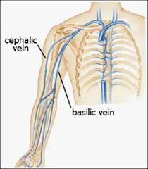
The cephalic vein drains the radial part of the hand, forearm, and arm and interacts with the basilic vein, which drains the ulnar part, along its path. The axillary vein receives the cephalic vein’s drainage.
Anatomy of Cephalic Vein
The cephalic vein is a vein that runs through the hand, forearm, and arm. There are valves between the superficial and deep vein networks that limit the possibility of leakage from the deep venous system.
Course
The cephalic vein drains the dorsal venous network of the hand, which crosses the anatomical snuffbox, runs superficial to the radial styloid process, and then ascends in the superficial fascia of the forearm. At the cubital fossa, the cephalic vein communicates with the basilic vein via the median cubital vein. At this angle, the vein is positioned in the lateral region of the elbow joint crease.
The cephalic vein now passes between the brachioradialis (forearm supinator and elbow flexor) and biceps brachii (forearm supinator and elbow flexor) muscles. The vein continues to climb in the superficial fascia anterolateral to the biceps brachii and superficial to the lateral cutaneous nerve of the forearm, a sensory branch of the musculocutaneous nerve (C5-7) that innervates the muscles of the arm’s anterior compartment.
The cephalic vein ascends farther in a groove formed by the pectoralis major and deltoid muscles. The cephalic vein is accompanied in this location by the deltoid branch of the thoracoacromial trunk.
Origin
The cephalic vein arises from the radial side of the superficial venous network of the dorsum of the hand in the anatomical snuffbox.
Tributaries
Accessory cephalic veins might develop from a venous plexus on the forearm’s dorsum or the medial aspect of the dorsal venous network of the hands. These accessory veins subsequently join the cephalic vein slightly below the elbow. It may also be supplied with blood via the variable median cubital vein.
Termination
At the clavipectoral triangle, the cephalic vein drains into the first part of the axillary vein.
Structure and location of cephalic vein
The cephalic vein, like the basilic vein, is one of the main superficial veins of the arm and is sometimes visible through the skin. It “communicates” (the clinical term for “connects”) with deep veins since it runs along the surface. The little connecting veins contain specific valves to avoid backflow.
Anatomical Variations
Congenital variations in the anatomy of the cephalic vein have been identified clinically, as with all veins in the body. These are largely divided into two categories:
Variations in the number and structure of the tiny branches that connect the cephalic vein to deeper-seated veins: These are the most prevalent variants.
Size distinctions: The cephalic vein, which is usually smaller, can occasionally be larger than the basilic vein.
Accessory cephalic veins: The cephalic vein may have two additional branches that emerge from the hands or from a piece of the forearm in some situations. These branches then connect to the main branch near the elbow.
Function of Cephalic Vein
One of the primary functions of the circulatory system is to transport oxygen to the rest of the body via blood cells. In the heart, oxygen is introduced to the blood. In contrast to arteries, which carry blood away, veins such as the cephalic vein return it.
This vein is one of the primary routes for deoxygenated blood from the hands and arms to the heart. This vein specifically transports blood from the radial region of the hand (around the thumb), the inner forearm, and the upper arm.
Clinical Importance
In the clinical and medical setting, the cephalic vein, like other superficial veins in the arm, has numerous functions and can be affected by a variety of health issues. Here’s a quick recap:
Blood sample collection: This vein, or more commonly, the median cubital vein that branches from it, is used to take blood samples. This is partly due to the ease of access it provides just beneath the skin.
Some procedures, such as the placement of a heart pacemaker or a venous catheter (to supply medication, drain blood, or provide other help to surgery), necessitate the use of a healthy, safe vein. When the body’s central veins are insufficient, the cephalic vein is used via a cephalic vein cutdown surgery.
Varicose veins occur when blood accumulates in the veins, causing them to swell and become uncomfortable. It develops in the cephalic vein due to insufficient activity of the valves in the short veins that connect the surface to deeper veins. These primarily affect the lower limbs, but cases have occurred in the arms as well.
A blood clot in a surface vein, such as the cephalic vein, can be caused by cancer, heredity, injury, being overweight, smoking, or other factors. If blood-thinning medications or lifestyle changes such as elevation do not help, surgical options such as sclerotherapy or endovenous ablation may be considered.
Drainage
It drains into the axillary vein below the clavicle after crossing the clavipectoral fascia and the axillary artery. When the axillary vein penetrates the lateral border of the first rib, it is dubbed the subclavian vein, and it joins the internal jugular vein to form the brachiocephalic vein.
The thoracic duct drains lymph from the lower limbs, pelvis, belly, left thorax, left upper limb, and left side of the head and neck into the angle formed by the left jugular vein and the subclavian vein.
The lymphatic drainage of the right upper limb, the right thorax, and the right side of the head and neck drains into the confluence of the right subclavian vein and the internal jugular vein, which unite to form the right brachiocephalic vein. The superior vena cava is formed by the union of the two brachiocephalic veins and drains into the right atrium of the heart.
FAQ
What vein connects the cephalic?
In the elbow region, the median cubital vein combines with distinct types of these two veins. Cephalic and basilic veins typically communicate with one another via a median cubital vein.
What are the three main veins for drawing blood?
The larger median cubital, basilic, and cephalic veins are most commonly used, but others may be required and will become more visible if the patient’s hand is tightly closed. Phlebotomists must never perform venipuncture on an artery.
What is the length of the cephalic vein?
The cephalic vein length within the deltopectoral triangle ranged from 3.5 cm to 8.2 cm, with a mean of 4.8+/-0.7 cm. According to the morphometric analysis, the mean cephalic vein diameter was 0.8+/-0.1 cm, with a range of 0.1 cm to 1.2 cm.
What is the significance of the cephalic vein?
These findings indicate that the cephalic vein is a stable structure that functions as a drainage vein of the hand and a reliable cannulation site in the forearm. The venous confluence could be used as a new landmark to anticipate the SBRN’s running path.
What is an animal’s cephalic vein?
The cephalic vein is the primary superficial vein of the forelimb, found on the cranial side of the antebrachium.
References
- Wikipedia contributors. (2023a, September 5). Cephalic vein. Wikipedia. https://en.wikipedia.org/wiki/Cephalic_vein
- Mbbs, S. S. (2023, October 10). Cephalic vein. Kenhub. https://www.kenhub.com/en/library/anatomy/cephalic-vein
- Gurarie, M. (2023, August 21). The anatomy of the cephalic vein. Verywell Health. https://www.verywellhealth.com/cephalic-vein-anatomy-5097153
- Accessory Cephalic vein | Complete Anatomy. (n.d.-b). www.elsevier.com. https://www.elsevier.com/resources/anatomy/cardiovascular-system/veins/accessory-cephalic-vein/17302

