
Diaphragm Muscle
Anatomy of Diaphragm Muscle
The Diaphragm muscle is the primary muscle utilized in respiration, which is the action of breathing. This dome-shaped muscle is found just below the lungs and heart. It also contracts continually as breathes in and out.
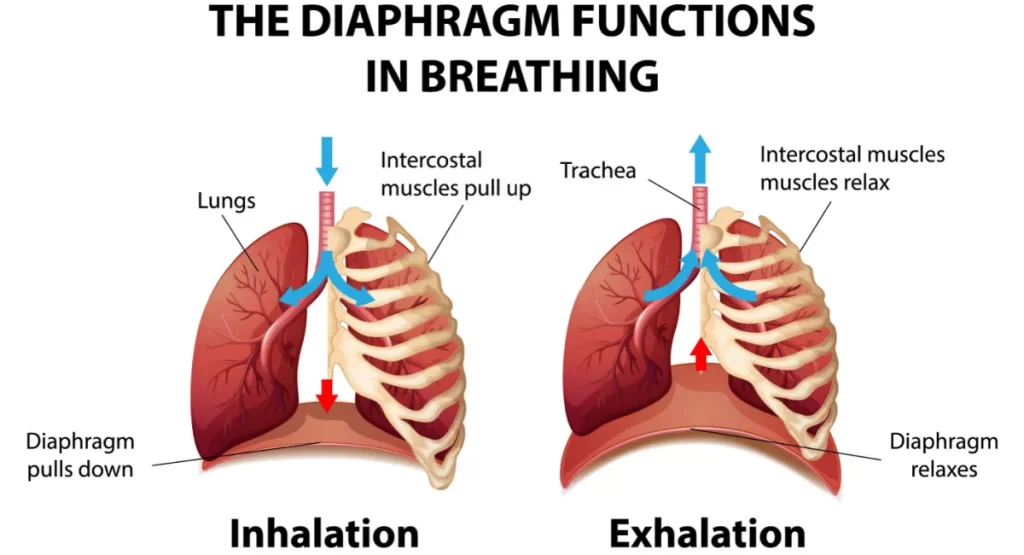
The diaphragm muscle is a thin skeletal muscle that seats at the base of the chest and separates the abdomen from the chest. It contracts and flattens when inhale. This creates a vacuum impact that pulls air into the lungs. When exhaling, the diaphragm muscle relaxes and the air is pushed out of the lungs.
It even has some non-respiratory functions. The diaphragm increases abdominal pressure to allow the body to get rid of vomit, urine, and feces. It also positions pressure on the esophagus to control acid reflux. The phrenic nerve, which runs from the neck to the diaphragm, prevents the movement of the diaphragm.
Origin and insertion of Diaphragm Muscle
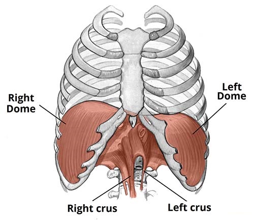
The diaphragm muscle is located at the inferior-most element of the ribcage, filling the inferior thoracic aperture. The diaphragm muscles originate from the lower part of the sternum bone area(breastbone), also the lower sixth ribs area, and also the lumbar (loin) vertebrae of the spine and are connected to a central membranous tendon area. It functions as the floor of the thoracic hole and also the roof of the abdominal cavity. The attachments of the diaphragm muscle can be divided into peripheral and central attachments. It has three peripheral attachments:
Lumbar vertebrae and arcuate ligaments; Costal cartilages of ribs 7-10 (connect directly to ribs 11-12); Xiphoid process of the sternum. The portion of the diaphragm that originates from the vertebrae is tendinous in structure, and is also known as the right and left crura:
Right crus – It Originates from L1-L3 and their intervertebral discs. Some fibers from the right crus cover the oesophageal opening, functioning as a physiological sphincter to contain the reflux of gastric contents into the esophagus.
Left crus – It Originates from L1-L2 and their also intervertebral discs.
The muscle fibers of the diaphragm connect to form a central tendon. This tendon ascends to connect with the inferior surface of the fibrous pericardium. On either side of the pericardium, the diaphragm climbs to form left and right domes. At rest, the right dome fibs a little higher than the left – this is thought to be due to the presence of the liver.
Three large openings in the diaphragm allow certain structures to pass between the chest and the abdomen.
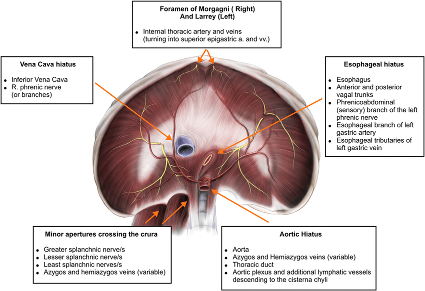
These openings include the:
Esophageal opening. The esophagus and vagus nerve, which maintain much of the digestive system, pass through this opening.
Aortic opening. The aorta, the body’s major artery that transports blood from the heart, passes via the aortic opening. The thoracic duct, a major vessel of the lymphatic system, also gives through this opening.
Caval opening. The inferior vena cava, a big vein that transports blood to the heart, passes through this opening.
Nerve supply
The halves of the diaphragm acquire motor innervation from the phrenic nerve. The left half of the diaphragm muscle (known as a hemidiaphragm) is supplied by the left phrenic nerve, and evil versa. Sensory innervation (pain and proprioception) at the central tendinous portion is supplied by the phrenic nerves, while the peripheral muscular portions are innervated by the 6th to 11th intercostal nerves. Each phrenic nerve is created in the neck within the cervical plexus and includes fibers from spinal roots C3-C5.
Blood supply
The respiratory diaphragm is a big complex muscle and thus its blood supply arrives from various arteries. The costal portion of the diaphragm muscle is supplied by the subcostal arteries and also the five most inferior pairs of intercostal arteries. The majority of the arterial supply to the diaphragm is delivered through the inferior phrenic arteries, which originate directly from the abdominal aorta.
The superior phrenic artery, pericardiacophrenic artery, and musculophrenic arteries provide the remaining blood supply. The final source of blood supply is superior phrenic arteries. They supply the superior surface of the diaphragm muscle. The draining veins observe the aforementioned arteries.
Function of Diaphragm Muscle Anatomy
The diaphragm is the prior muscle of respiration. The diaphragm muscle is the primary muscle that functions in inspiration motion. During inspiration, it contracts and flattens, improving the vertical diameter of the thoracic cavity. This creates lung expansion, and the air is drawn in. During expiration motion, the diaphragm passively relaxes and returns to its initial dome shape. This decreases the volume of the thoracic cavity.
When the diaphragm muscles function with the anterolateral abdominal muscles, and then diaphragm muscle contraction supports increasing intra-abdominal pressure. This is needed in activities such as expelling vomit, defecation, micturition (urination), and parturition (childbirth). Another function of the diaphragm muscle is to supply a passageway for certain structures from the thorax to the abdomen (inferior vena cava, esophagus, and aorta) as mentioned earlier.
Clinical relations
Hiatal hernia
A hiatal hernia happens when the upper portion of the stomach bulges through the esophageal opening of the diaphragm. Experts aren’t sure why it occurs, but it could be caused by:
Age-related changes in the diaphragm: injuries or birth defects; chronic pressure on enveloping muscles from coughing, straining, or heavy lifting; They’re more common in individuals who are over the age of 50 or obese.
Small hiatal hernias usually don’t induce any symptoms or require treatment. But a larger hiatal hernia may cause some symptoms, including heartburn; acid reflux; trouble swallowing; chest pain that occasionally radiates to the back.
Larger hiatal hernias occasionally need surgical repair, but other cases are usually manageable with over-the-counter antacid medicine. Proton pump inhibitors can also assist to reduce acid production and heal any damage to the esophagus.
Diaphragmatic hernia
A diaphragmatic hernia happens when at slightest one abdominal organ bulges into the chest cavity through an outlet in the diaphragm. It’s sometimes current at birth. When this happens, it is also called a congenital diaphragmatic hernia (CDH).
Injuries from an accident or surgery can also induce a diaphragmatic hernia. In this circumstance, it’s named an acquired diaphragmatic hernia (ADH). Symptoms can differ depending on the size of the hernia, the cause, and also the organs affected. They may include:
- difficulty breathing
- rapid breathing
- rapid heart rate
- blueish-colored skin
- bowel sounds in the chest
- Both acquired diaphragmatic hernia and congenital diaphragmatic hernia instruct immediate surgery to remove the abdominal organs from the chest cavity and repair the diaphragm.
Cramps and spasms
A diaphragmatic cramp or spasm can cause chest pain and also shortness of breath which can be misinterpreted as a heart attack. Some people also encounter sweating and anxiety during a diaphragm spasm. Others express feeling like they can’t accept a full breath during a spasm.
During a spasm, the diaphragm doesn’t awaken back up after exhalation. This inflates the lungs, yielding the diaphragm to tighten. This can also induce a cramping sensation in the chest area. Vigorous exercise can cause the diaphragm to spasm, which usually results in what people call a side stitch.
Diaphragm spasms usually go out on their own within a few hours or days.
Diaphragmatic flutter
Diaphragmatic flutter is an occasional condition that’s often mistaken for a spasm. During an episode, somebody might feel the fluttering as a pulsing sensation in the abdominal wall. It can also cause:
- shortness of breath
- chest tightness
- chest pain
- abdominal pain
- Phrenic nerve damage. The phrenic nerve can be harmed by a number of problems, including multiple sclerosis, autoimmune diseases, severe injuries, surgery, cancer of the lungs or adjacent lymph nodes, spinal cord issues, and some viral infections. This impairment can cause dysfunction or paralysis of the diaphragm muscle. But also phrenic nerve damage does not always cause symptoms. When it does, possible symptoms include:
- shortness of breath when lying flat position or exercising
- morning headaches
- trouble sleeping
- chest pain
Exercise of Diaphragm Muscle
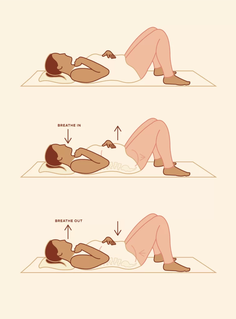
Strengthen your diaphragm with special exercises. Diaphragmatic breathing or abdominal breathing is the most useful way to do this. It affects inhaling deeply and slowly through the nose so that your lungs serve with air as your belly expands. Along with strengthening your diaphragm muscle, diaphragmatic breathing can also reduce stress and lower blood pressure.
Balloon Exercises
Balloons arrive in different strengths depending on their quality but the stiffer, higher-quality balloons with additional resistance are the ones to look out for. Usually, these will be the 10-pack of balloons sold at the exact price as the 25-pack…There is nothing wrong with the 25-pack balloons but just be familiar as progress you will want the stiffer balloons to inquire you further so you can continue to progress…
Exhale into the balloon and hold it inflating as if there was no hole in it. Maintain your exhalation for as prolonged as possible keeping the balloon inflated to its maximum potential with the hole in it. This allows the training of the abdominal muscles subconsciously while lengthening the diaphragm and mobilizing the ribcage.
Once you have finished an exhaling action, hold the balloon in your mouth and also pause for three to four seconds before lightly inhaling via the nose. Focus your attention on the air as it passes through the nostrils and delay the air down as it passes through.
At the top of the inhalation… pause for a second and then replicate the exhalation via the balloon. Override the subconscious mind at the base of the exhalation motion and make certain all air is out of your lungs before waiting for three seconds at the base.
Slow the air down via the nostrils as you inhale again. Repeat this exercise for a total of six full breaths before carrying a short break of two to three minutes and replicating twice for a total of three sets.
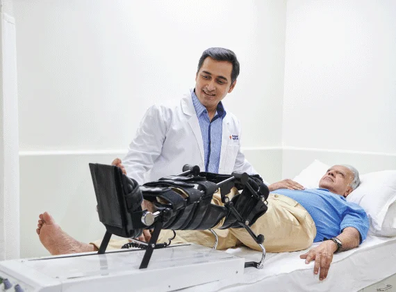
Devices
In this also try using a vibrating device to relax your diaphragm muscle. This device is placed over your diaphragm and vibrates, assisting to relax the muscle and reduce tension. This can be done while lying down or/and while standing. If you opt for this technique, be sure to confer with a qualified healthcare professional to ensure that you are using the device correctly.
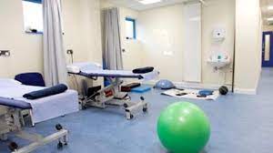
Massage
Massage can also be used to relax a tight diaphragm muscle. Massage helps to relieve tension and increase blood flow to the area, which can also assist relax the diaphragm muscle. It is important to find a qualified massage therapist who has an understanding of performing with diaphragm muscles. Additionally, you can utilize a foam roller to massage the diaphragm muscle.
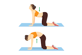
Stretching
Stretching can be an acceptable way to loosen your diaphragm. Diaphragmatic stretching is a technique that targets the muscles that maintain your diaphragm, such as your abdominal muscles and your intercostal muscles. To do this type of stretch, fib on your back on a flat shell with your both knees flexed. Take a few slow, deep breaths and also concentrate on relaxing your body. Place one hand on your upper chest and the additional on your belly, just below your rib cage.
FAQs
What is the action of the diaphragm muscle?
The diaphragm is the primary muscle of respiration. During inspiration motion, it tightens and flattens, increasing the vertical diameter of the thoracic cavity. This creates lung expansion, and the air is drawn in. During expiration motion, the diaphragm passively relaxes and returns to its initial dome shape.
What controls the diaphragm muscle?
The phrenic nerve regulates your diaphragm (the large dome-shaped muscles between your abdominal and chest holes). It’s essential to breathing. Your nerve transmits signals that cause your diaphragm to contract (become more stagnant and flatter).
What is the diaphragm made of?
The periphery of the diaphragm muscle is made of strong muscular fibers that have their origin in the surroundings of the inferior thoracic aperture. These muscle fibers then converge and insert into the central tendon area.
What happens if your diaphragm stops working?
Symptoms of significant, usually bilateral side diaphragm weakness or paralysis are shortness of breath when lying flat, walking, or with immersion in water up to the lower chest area. Bilateral diaphragm paralysis can create sleep-disordered breathing with reductions in blood oxygen levels.
Does exercise help the diaphragm?
It’s the muscle that’s accountable for 80 percent of your breathing. This muscle’s main role is to support breathing, which can assist your body adapt to increases in intensity during your workout. Like your other muscles, you can do workouts to train your diaphragm and boost your overall aerobic implementation.
7 Comments