Dorsal Midfoot Exostosis
Introduction
Dorsal midfoot exostosis, also known as dorsal midfoot bump or dorsal midfoot osteophyte, is a medical condition characterized by the formation of a bony outgrowth on the upper surface of the midfoot bones. This condition is often associated with chronic mechanical stress or inflammation of the midfoot region.
The dorsal midfoot exostosis can lead to discomfort, pain, and restricted mobility, affecting an individual’s ability to engage in activities of daily living.
Exostosis: What is it?
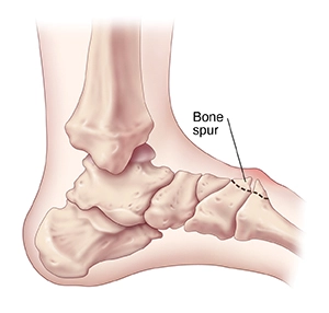
The development of new bone on a bone’s surface is called an exostosis, sometimes referred to as a bone spur. Depending on the form, size, and location of the lesion, exostoses can result in persistent discomfort ranging from hardly noticeable to incapacitating. Small bone growths like those in the ribs are where it is most frequently discovered, but bigger growths can also develop in areas including the ankles, knees, shoulders, elbows, and hips. They seldom ever appear on the cranium.
Exostoses, like calcaneal spurs, can occasionally resemble spurs. A bone infection called osteomyelitis may cause the neighboring bone to develop an exostosis. In addition to leaving bone spurs, Charcot foot, a neuropathic breakdown of the feet most commonly observed in diabetics, can also become symptomatic.
They can grow vertically and typically develop on the bones of joints. For instance, an additional bone that developed on the ankle may have grown up to the shin.
The word is thought to be similar to osteochondroma when used in the phrases “cartilaginous exostosis” or “osteocartilaginous exostosis”. Even without qualifiers, some sources believe the two phrases indicate the same thing, but this understanding is not shared by everyone.
What is dorsal midfoot exostosis?
There is a noticeable, frequently painful hump on top of the foot known as dorsal midfoot exostosis. The hump, which is frequently noticeable, is caused by bone development on top of the arch, which is medically referred to as the metatarsal-cuneiform joint.
Midfoot bone spurs can provide a unique set of challenges caused by discomfort, as well as visual issues and shoe sensitivity. Trauma, arthritis, or biomechanical foot types with high or low arches can all cause bone spurs in the midfoot.
One of our doctors will do a clinical evaluation of the “bony bump” during a workup before taking radiographs to look at the underlying joints that could be the source of these particular spurs. Numerous superficial tendons and nerves leave the leg and sit on top of the foot; having a bone spur can make these soft tissue structures rub up against the bone spur, which can result in numbness, tingling, or discomfort. Ganglion cysts are frequently created in the region of complaint as a result of friction caused by these tendons pressing on the bone spur.
Dorsal midfoot exostosis: Causes
Weak foot mechanics are usually seen in the overpronated foot. Due to its propensity for excessive movement, the first metatarsal can irritate the metatarsal-cuneiform joint and cause bone development.
Because of the first metatarsal’s greater angle of inclination towards the ground in the supinated foot, the base of the metatarsal may protrude above the top of the foot. Other factors that contribute to dorsal midfoot exostosis include inflammation, trauma, and long-term irritation to the region, such as that caused by tight footwear.
The overpronated foot frequently has weak foot mechanics. The first metatarsal has a tendency to move about excessively, which might irritate the metatarsal-cuneiform joint and lead to a bone growth. The first metatarsal is inclined further towards the ground in the supinated foot, which can make the base of the metatarsal stand out towards the top of the foot.
Other factors that contribute to dorsal midfoot exostosis include inflammation, trauma, and long-term irritation of the area, such as that caused by tight shoes.
What symptoms and signs indicate dorsal foot exostosis?
The following are typical dorsal foot exostosis symptoms and signs:
There is a noticeable, frequently painful hump on top of the foot known as dorsal midfoot exostosis. The hump, which is frequently noticeable, is caused by bone development on top of the arch, which is medically referred to as the metatarsal-cuneiform junction.
Dorsal midfoot exostosis is indicated by a prominent, solid hump on the arch of the foot. Bone growth is not painful in and of itself; rather, the challenges that the bony expansion causes are often uncomfortable.
Patients find it difficult to wear certain shoes because of the bony protrusion. Open-toed footwear generally poses no problems, but enclosed footwear can compress and rub against the bony protrusion, frequently resulting in discomfort, redness, and swelling.
In addition to the bump itself, compression and rubbing can aggravate neighbouring nerves. Because these nerves are irritated, aching, tingling, and pins-and-needles sensations are usually experienced in the top of the foot and the toes.
Even though the bone development is not specifically stimulated, pain may be experienced in more extreme circumstances. This generally shows up as a very painful soreness, usually after weight-bearing exercise.
The medial dorsal cutaneous nerve, which supplies parts of the foot and leg, may be compressed over the region, resulting in paresthesias (an unnatural prickling or burning sensation), tingling, and numbness. The medial dorsal cutaneous nerve almost invariably crosses the metatarsocuneiform joint.
What kinds of studies are done on dorsal foot exostosis?
- Sensory Testing: In the dorsal first web space in particular, sensory testing can be used to identify nerve involvement.
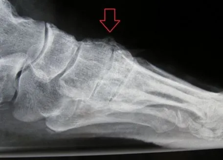
- X-ray: A lateral X-ray will show the dorsal prominence, and dorsal hypermobility may result in a plantar gap in the foot’s metatarsocuneiform joint.
How is dorsal foot exostosis classified?
What Subtypes of Dorsal Foot Exostosis Exist?
The many forms of dorsal foot exostosis include:
- Localized dorsal exostosis is type 1.
- Type 2: Dorsal exostosis and osteoarthritis of the joints.
- Type 3: Dorsal exostosis and an angular deformity.
- Exostosis that spans additional metatarsocuneiform joints is type 4.
- Type 5: Pseudoexostosis of the dorsal hump of the front pes cavus.
Conservative treatment
One or more of the therapies could be:
Modifications to the footwear and the use of padding can both alleviate direct bump discomfort and lower compressive pressures. The use of orthotics can help with underlying problems such as excessive pronation and high arches in the foot.
Suitable footwear is crucial, so wear that. Finding comfortable shoes that do not aggravate the bump can be challenging for many people. Do not fear; we will propose the right shoes to you based on our extensive combined knowledge of footwear.
Nonsteroidal anti-inflammatory medicines (NSAIDs), such as Ibuprofen, Naproxen, or aspirin, will reduce pain and swelling when used as directed by a doctor.
- Injection therapy: This might relieve problems associated with the nerves and the joints.
Dorsal midfoot exostosis surgery
In cases when conservative treatment fails, surgical intervention may be required.
- Surgery may be necessary if conservative treatment is ineffective in a certain situation. The midfoot osteotomy procedure, which involves surgically removing the hump, may be beneficial if there are no indications of underlying arthritis. The discomfort may stay the same or occasionally even worse if there is arthritis present and the lump is just removed. A surgical solution might be suggested in this situation.
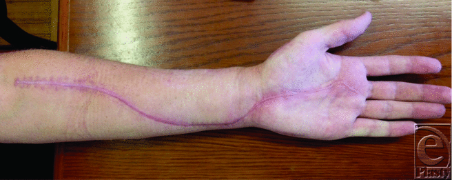
- Midfoot exostectomy: Surgery is frequently recommended if a person is in discomfort, is constrained or incapacitated, and experiences difficulty putting on shoes despite wearing appropriate shoes. The surgery will be performed while the patient is awake because a local anaesthetic is frequently utilised. Patients can eat normally and take their regularly prescribed medications on the day of the operation. The local anaesthetic is injected close to the exostosis and the ankle using injections. Most people find this to be more pleasant than a dental injection.
On occasion, a behind-the-knee injection may be advised. Although the treatment takes approximately an hour, patients can expect to stay in the day surgery unit for around four to five hours. This allows recovery time following the surgery while the doctor offers any required discharge instructions and materials. Patients must have a responsible adult at home for the first day and night following surgery for their security.
What advantages might surgery offer?
The benefits of surgical intervention are:
- To relieve pain and an apparent bone deformity.
- To improve the footwear’s comfort and fit. The method is regional.
What are the surgery’s complications?
The pre-operative information brochure, which details the basic risks of foot surgery, should have been given to everyone by now.
Additionally, this method entails the following specific dangers:
- Due to nerve damage, there is numbness on the top of the foot.
- Ongoing pain in the underlying joint.
- The hump is returning.
- Rigid joints.
- Arthritis-related joint discomfort.
- A vulnerability to scars.
- Constant swell
Summary
Modifying shoe accessories, creating custom orthotics to address the underlying biomechanical issue, cortisone injection therapy, using topical pain relievers, and physical therapy are some of the treatment options for midfoot exostosis. When all other treatment options have failed, the patient is informed about the surgical option of performing a “exostectomy” to remove the exostosis.
Come in for a consultation with one of our podiatrists at one of our locations if you’ve discovered a bulge on the top of your foot and have been experiencing pain. There is no need for you to continue to endure suffering, and there is always a method to make your exit more comfortable.
FAQs
How do you treat dorsal exostosis in the foot?
Exostosis treatments:
To reduce swelling, use ice. Use NSAIDs, such as ibuprofen, which are nonsteroidal anti-inflammatory medicines. For bone spurs in your feet, put shoe inserts in them. Reduce your weight to ease the strain on your joints.
What is midfoot exostosis?
In medicine, the term “exostosis” refers to the growing of new bone over previously existing bone. It and the term “spur” are interchangeable. The big toe, midfoot, and heel joints are where exostoses most frequently develop in the foot.
What is the surgery for dorsal midfoot exostosis?
Surgery as a Treatment: When non-surgical treatments are unsuccessful, surgery may be required. If there are no signs of underlying arthritis, the midfoot osteotomy treatment, which includes surgically eliminating the hump, may be useful.
Does exostosis need to be removed?
You might not require medical attention. Whether an exostosis is painful depends on which bone it is growing on. Exostoses of a certain sort require surgical removal. As soon as you detect a growth or lump close to one of your bones, see a doctor.
Is exostosis surgery painful?
The recovery following surgery should be relatively painless, and any discomfort can be treated with over-the-counter pain relievers. Surgery frequently results in swelling, but all adverse effects should go away in a week.
Can you walk after bone spur surgery?
After minor surgery, you’ll wear a bandage for one to two weeks. After open surgery, you could need a cast, walking boot, or ankle splint for up to three weeks. You could also get a cane or crutches. You may need to take at least a few days off from walking since the surgery region will be sore and swollen.