Introduction to Neuroanatomy
Overview
The nervous system is formed by the vast neural networks; signaling within these circuits allows thinking, language, feeling, learning, memory, and all function and sensation. It is well-established that via the plasticity of existing cells our nervous systems can adjust to situations or circumstances not previously encountered, but it also has been shown that cells (NSCs) are plastic and involved in producing new connections in modifying and responding to injury.
The Nervous System has three specific functions:
- Sensory Input – Sensory receptors located in the organs and skin reply to external & internal stimulants by generating or manufacturing nerve impulses to the central nervous system (CNS)
- Integration – The brain and spinal cord of the Central Nervous System (CNS) combine and sum up all the data collects from the body and send out nerve impulses.
- Motor Output – The nerve impulses from the Central Nervous System (CNS) go to the effectors or individuals (muscles and glands). Muscle contractions and gland secretions are responses or show reactions to stimuli collected by sensory receptors.
The Nervous System is split into two 2 main divisions.
- Peripheral Nervous System (PNS)
- Central Nervous System (CNS)
The CNS comprises two parts: The brain; Spinal Cord
Brain
The Brain is mainly divided into four parts
The brain stem, include the medulla, pons, and midbrain
Cerebellum
Diencephalon, with the thalamus and hypothalamus
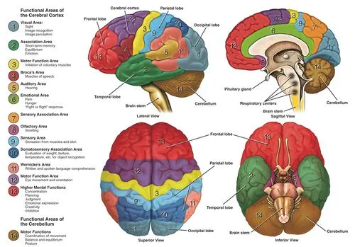
Cerebral hemispheres (comprised of the hippocampi, basal ganglia, cerebral cortex white matter, hippocampi, and amygdalae). The left and right hemispheres are connected to each other by the corpus callosum which facilitates communication between both sides of the brain. The Hemispheres are then further divided into four lobes (frontal lobe, parietal lobe, occipital lobe, and temporal lobe).
- The Spinal Cord (the caudal extension of the Central Nervous System).
Neurological Conditions:
Many neurological conditions affect the central nervous system (CNS). They range greatly in scope, impact, and nature of the effect. Some conditions lead to progressively impaired (affected)movement eg Parkinson’s disease. The demyelination in multiple sclerosis, Huntington chorea. can cause acute attacks, and over time, chronic degradation of the functions. Others may affect cognition such as the various type of dementias. Epilepsy can cause uncontrolled excitation. Headaches often impair the daily function or ADL activity of patients. Traumatic injuries can cause plegia or paresis and may cause a wide range of deficiencies depending on the location and expansion of the lesion or injury.
Neurons
Neurons are cells of the nervous system, present within the grey matter, and responsible or answerable for all neurological functions of the brain.
They are any of the impulse-conducting cells that constitute the brain, spinal column, and nerves in vertebrates, comprising a nucleated cell body with one or more dendrites and a single axon.
Cerebrum
The cerebrum is comprised of two cerebral hemispheres, the right hemisphere, and the left hemisphere which is connected by the corpus callosum which facilitates conveyance or communication between both sides of the brain, with each hemisphere in the main connection to the contralateral (opposite)side of the body i.e. the left hemisphere of the cerebrum collects information from the right side of the body resulting in motor control of the right side of the body and conversely or vice versa.
The hemispheres are then again divided into four lobes;
- Occipital lobe
- Parietal lobe
- Temporal lobe (medial part of which is a series of structures including the Hippocampus)
- Frontal lobe
Cerebral Cortex
The outer layer of the cerebral hemisphere is named the cerebral cortex. This is interconnected through pathways (tracks) that run sub-cortically. It is these connections as well as the connections from the cerebral cortex to the spinal cord, brainstem, and nuclei deep within the cerebral hemisphere that form the white matter of the cerebral hemisphere. The deep nuclei include structures such as the thalamus and basal ganglia
Basal Ganglia
The “basal ganglia” refers to or mentions a group of subcortical nuclei within the brain responsible principally for motor control, as well as other roles such as motor executive functions, learning, and emotional behaviors, and play an important role in addictive behaviors and habit formation. reward and reinforcement.
The Hypothalamus
The hypothalamus is an organ central to many autonomous functions of the human body, notably the regulation or management of homeostasis. It has an importantly large efferent output to the Autonomic nervous system and has a highly important role in the control of pituitary endocrine function.
The hypothalamus is situated on both sides of the 3rd ventricle, below the thalamus, and between the optic chiasm and the midbrain. It sustains a large input from limbic structures.
Meninges
The Central nervous system is surrounded by the skull and vertebral column. These structures are separated by a series of membranes known as the Meninges. The Pia Mater is separated from the delicate arachnoid membrane by the subarachnoid space, which is then separated from the Dura mater by the Sub-dural space.
Neuroglial
The brain is made up of more than only neurons. even though there are about 86-100 billion neurons in the brain, there are around a similar number of glial cells in the brain. neuroglia, or Glial cells neuroglia, are cells that surround the neurons of the central nervous system inserted between them, giving both structural and physiological support.
There are four main classes of neuroglial cells within the CNS.
- Oligodendrocytes
- Astrocytes.
- Microglial Cells
- Ependymal cells
Schwann Cell:
Found only in the Peripheral nervous system. accountable for the myelination of the peripheral nerves by wrapping or sheathing the cell around the axon. There are multiple layers of Schwann cell membrane wrapped or sheath around the nerve. One Schwann cell wraps or sheaths around one axon and gives myelin for one internode. They are very important for the regeneration of the damaged peripheral axons
Limbic System
The limbic system gives high-level processing of sensory information. The major outflow of the limbic system is to the prefrontal cortex and the hypothalamus as well as to the cortical region. It seems to have a role in attaching behavioral importance and response to a given stimulus.
Damage to this area has intellectual effects on emotional responses.
Long-term potentiation (LTP) is the increase in the strength of a synaptic transference with repetitive use, it can be seen to be affected in the hippocampus (primarily involved with memory) and is thought or convinced to be important for memory acquisition.
Brainstem
The Brainstem is located at the base of the brain & the top of the spinal cord. the brainstem is the structure that connects the cerebrum of the brain (mind) to the spinal cord and cerebellum. It conducts 3 sections in descending (downward) order: the midbrain, pons, and medulla oblongata. It is answerable for numerous vital functions of life, like blood pressure, breathing, consciousness, breathing, heart rate, and sleep.
CNS Blood Supply
- Brain: Arterial blood supply to the brain comes from four vessels;
- Right and Left Internal Carotid artery
- Right and Left Vertebral Arteries
The internal carotid arteries branch to form two major cerebral arteries, the anterior cerebral and middle cerebral arteries. The right vertebral artery and left vertebral arteries come together at the region of the pons on the ventral surface of the brainstem to form the midline basilar artery.
Circle of Willis: The basilar artery connects the blood supply from the internal carotids in an arterial ring at the base of the brain (in the vicinity of the hypothalamus and cerebral peduncles) called the circle of Willis. The posterior cerebral arteries get arise at this junction as do two small bridging arteries, the anterior and posterior communicating arteries. Combining the two major sources of cerebral vascular supply through the circle of Willis absolutely improves the chances of any area of the brain continuing to receive or collect blood if one of the major arteries becomes occluded or blocked.
Spinal Cord: The spinal cord is supplied by a single anterior spinal artery and paired posterior spinal arteries. Anterior spinal artery: get arises from the vertebral arteries and extends from the level of the lower brainstem to the tip of the conus medullaris. It supplies the ventral medial surface of the medulla and the anterior two – third of the spinal cord. The posterior spinal arteries supply the dorsal one-third of the cord. There are support branches from other arteries along the length of the cord.
If occlusion or blockage occurs, it is normally of the anterior spinal artery, producing loss of power and spinothalamic sensory deficiency, but dorsal column sensory capabilities are maintained.
Venous Drainage
The cerebrum, cerebellum, and brainstem are drained by many veins, which empty (unload) into the dural venous sinuses. The spinal cord is supplied by the anterior and posterior spinal veins, which drain into the internal and external vertebral plexuses.
If occlusion or blockage of either of these venous systems then raised intracranial pressure can occur.
The Cerebellum
The cerebellum is vital in the human brain as it plays important role in motor movement regulation and balance control. The cerebellum is neuron-rich, containing 80% of the brain’s neurons arranged in a dense cellular layer, and it is surface area when unfolded is nearly 75% of the surface area of the cerebrum.
Spinal Cord
The spinal cord is part of the central nervous system (CNS) and comprises a tightly packed column of nerve tissue that extends downwards from the brainstem via the central column of the vertebral spine. It is a comparably very small bundle of tissue (weighing 35g and just about 1cm in diameter) but is critical in facilitating our daily activities.
The spinal cord conducts nerve signals from the brain to other parts of the body (importantly the muscles we use to move) and collects sensory input from the body, partly processes it, and then conveys that information to the brain.
Peripheral Nervous System
The peripheral nervous system comprises the nerves and ganglia that are outside of the central nervous system (CNS). The peripheral nervous system (PNS) is made up of two parts the somatic nervous system and the autonomic system. Each part of this system plays a vital function in how information is communicated throughout the whole body.
Autonomic Nervous System (ANS)
The autonomic system is part of the peripheral nervous system (PNS) that’s answerable for regulating involuntary body functions. Functions of the ANS comprise the regulation of “circulation, secretion, respiration, body temperature, metabolism, and reproduction.
The ANS is divided into two Divisions:
Sympathetic: Preganglionic neurons present in the lateral horn of the spinal cord from the upper thoracic to the mid-lumbar cord (T1-L3). Postganglionic cell bodies are present in vertebral and prevertebral ganglia. Uses Noradrenalin as the postganglionic transmitter.
Parasympathetic: Preganglionic neurons have cell bodies in the brainstem and sacrum bone .. Postganglionic cell bodies are present close to or within the walls of the organ they supply. Uses acetylcholine (ACh) as the postganglionic transmitter.
Somatic Nervous System
The somatic system is part of the peripheral nervous system(PNS) answerable for conducting sensory and motor information to and from the central nervous system (CNS). The somatic nervous system obtains its name from the Greek word soma, which means “body
Cranial and spinal nerves contribute to the somatic nervous system. Cranial nerves give voluntary motor control and sensation to the head and face. Spinal nerves supply the trunk and limbs. The posterior rami run backward to supply the vertebral muscles, vertebral column, and skin of the back whilst the anterior rami give supply to the limbs and anterior trunk. The majority of anterior rami get combined to form nerve plexuses from which many major peripheral nerves stem. The anomaly to this is the anterior rami of the thoracic region which runs relatively independently from one another without forming any plexuses, as the intercostal and subcostal nerves of the trunk.
Nervous plexuses are as follows:
- C1-C4 forms the cervical plexus
- C5-T1 combine into the brachial plexus
- T12-L4 form the lumbar plexus
- L4 – S4 combine into the sacral plexus
Sensory Systems
The sensory nervous system is a part of the nervous system accountable for processing sensory information where information is transferred to the spinal cord and brain from peripheral sensory receptors. The sensory receptors are specialized neurons or nerve endings.
There are five main sensory systems in mammals.
- vision
- taste
- touch/pressure
- hearing and balance
- smell/olfaction
Pain System
Pain is defined as an unpleasant sensory or emotional experience, related to potential or actual tissue damage. Nociception defines the processing of information about damaging stimulants by the nervous system up to the level or up to the region of the cortex. Potentially damaging mechanical, thermal, and chemical stimulants are exposed by nerve endings called nociceptors, which are present in the skin, on internal surfaces such as the joint surfaces, periosteum, and in some internal organs.
There are two types of nociceptors: Delta fibers: are activated by high threshold mechanoreceptors. thinly myelinated; Unmyelinated C-fibres: activated by polymodal nociceptors(PMN) and respond or react to intense mechanical stimulation, irritant chemicals, and high temperatures.
There are three main pathways that transmit or convey the nociceptive signals to the brain:
- Spinothalamic tract
- Spino reticular tract.
- Spino mesencephalic
Motor Systems
Motor systems are the areas of the nervous system responsible for controlling or managing movement. Neural control of the somatic motor system includes complex feedback mechanisms between the brain, spinal cord, peripheral nerves, and musculoskeletal structures. Each component or constituent is functionally and structurally capable of adaptation and modulation to maintain or sustain as much efficiency as possible
Grey and White Matter.
The CNS is made up of grey matter and white matter.
Grey matter: named for its pinkish-gray color, is home to neural cell bodies, dendrites, and, axon terminals, as well as all nerve synapses. This brain tissue is plentiful in the cerebrum, cerebellum, and brain stem. It also makes a butterfly-shaped portion in the central part of the spinal cord.
White matter: comprised of bundles of axons. These axons are coated or covered with myelin sheath, a mixture of proteins and lipids, that helps conduct or carry nerve signals and protect the axons. White matter conducts, processes, and sends nerve signals up and down the spinal cord.
FAQ
What is the importance of studying neuroanatomy?
Neuroanatomy is the key to localization or center. understanding of neuroanatomy includes not only the morphology of the structure or construction but also its function.
What is the basis of neuroanatomy?
Neuroanatomy is the study of the structure and arrangement of the nervous system. In contrast to animals with radial symmetry, whose nervous systems comprise a distributed network of cells, animals with bilateral symmetry have separated, defined nervous systems.
What is the most superior neuroanatomical structure?
The most superior or upper brainstem structure, the diencephalon, is supplied anteriorly by the vascular branches of the anterior cerebral artery.
What are the different types of Fibres in neuroanatomy?
The nerve fibers are split into three categories: commissural, projection, and association fibers.
What are the 3 main shapes of neurons?
1) multipolar; 2) unipolar and 3)bipolar. Bipolar neurons have only two processes that expand in opposite directions from the cell body. One process or part is called a dendrite, and another process or part is called an axon.
What are the 3 main nervous systems called?
The PNS (peripheral nervous system) is cleaved into the somatic nervous system and the autonomic nervous system. (ANS)
Somatic Nervous System. The somatic nervous system is made up of motor neurons and sensory neurons that assist the body perform voluntary action.
Autonomic Nervous System.
Sympathetic Nervous System.
Parasympathetic Nervous System.
Which area of the brain is responsible for memory?
A curved or bent seahorse-shaped organ on the undersurface of each temporal lobe, the hippocampus is part of a larger structure called the hippocampal formation. It supports navigation, learning, memory, and perception of space. It collects information from the cerebral cortex and may play a major role in Alzheimer’s disease.
Which part of the brain has the highest function?
The Cerebrum
The Cerebrum: Also known as the cerebral cortex, the cerebrum is the largest or biggest part of the human brain, and it is connected or related to higher brain functions such as function, action, and thought.
Which brain segment is the largest?
Cerebrum: is the largest part of the brain and is comprised of the right and left hemispheres. It performs higher functions like interpreting or comparing touch, vision, and hearing, as well as reasoning, speech, emotions, learning, and fine control of movement. Cerebellum: is situated under the cerebrum.
Which part of the brain contains a high amount of fiber?
The two hemispheres are interconnected mainly by a thick bundle of nerve fibers called the corpus callosum, or “hard body,” because of its tough or hard stability or consistency. A smaller bundle, the anterior commissure, connects or links just the two temporal lobes.
Where is gray matter found in the brain?
Unlike the structure or construction of the spinal cord, the grey matter in the brain is situated in the outermost layer. The grey matter surrounding the cerebrum is also called the cortex of the brain. There are two major cortexes in the brain, the cerebellar cortex and, the cerebral cortex.
What is the brain’s function?
Memories and emotions.
Thoughts and decisions.
Automatic behavior such as breathing, sleep, heart rate, and temperature control.
Movements (motor function), balance, and coordination.
Perception of various sensations including pain.
Speech and language functions.
Regulation of organ function.
What is 75% of the brain made of?
About 75% of the brain consists of water
This means that dehydration (loss of water), even as small as 2%, can have a negative effect on brain functions. Dehydration and a loss of electrolytes and sodium can cause acute changes in memory and attention.
What is blood-brain?
A network of blood vessels and tissue that is made up of closely spaced cells and helps keep harmful or dangerous substances from reaching or extending the brain. The blood-brain barrier (BBB) lets some substances or some materials, such as water, oxygen, carbon dioxide, and general anesthetics, pass into the brain.
What are the 3 types of the brain?
The brain can be split into three basic units: the forebrain the midbrain, and the hindbrain. The hindbrain comprises the brain stem, the upper part of the spinal cord, and a creased ball of tissue called the cerebellum. The hindbrain regulates the body’s vital functions such as heart rate. and respiration
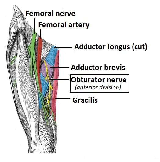
What is the largest nerve in the body?
The sciatic nerve
The sciatic nerve is the longest, largest, and thickest nerve in the body. the sciatic nerve roots start in the lower back and run down toward the back of each leg. Sciatica is the pain and discomfort in the back of the leg if the sciatic nerve gets compressed or pinched