
Adductor Magnus muscle
What is the Adductor magnus muscle?
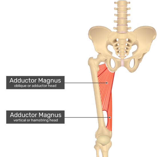
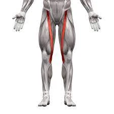
The adductor magnus muscle is a large muscle of the lower limb with three sides. Its base is on the linea aspera of the femur and its apex is on the hip bone. It can be found in the thigh’s medial and posterior fascial compartments. The fact that it receives a supply from two nerves reflects that this muscle is divided into two parts. No matter what the situation, the adductor magnus muscle has divided muscles in the middle compartment of the thigh.
As compared to the adductor longus, adductor brevis, pectineus, and gracilis muscles, the medial thigh’s adductor magnus is the largest and strongest muscle. Even though their actions are somewhat more complex than that, these five muscles are collectively referred to as the adductors of the thigh because of their function.
These muscles also involve thigh flexion, extension, external and internal rotation, pelvis stabilization while walking, and thigh adduction.
Origin
The pubis’ inferior ramus and the ischium’s ramus serve as the proximal attachment points for the portion of the muscle that is referred to as the adductor portion.
The ischial tuberosity serves as the hamstring portion of the muscle’s proximal attachment.
The distal attachment points of the muscle are different for each part.
Insertion
Adductor part: the middle of the linea aspera, the medial supracondylar line, and the posterior surface of the proximal femur
Hamstring part: the supracondylar line and the adductor tubercle of the femoral medial condyle.
Relations
The adductor magnus occupies most of the medial part of the thigh. Anteriorly to the adductor magnus muscle are the adductor longus, pectineus, and adductor brevis. The femoral artery and vein, the deep femoral artery and vein, and the posterior branches of the obturator artery, nerve, and vein are all neurovascular structures located around the anterior surface of the adductor magnus.
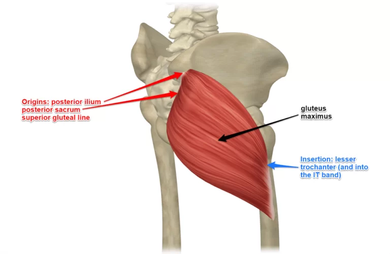
The adductor magnus muscle controls the sciatic nerve, semimembranosus, and gluteus maximus on the posterior aspect. The gracilis and sartorius muscles are associated with the adductor magnus muscle’s medial border, while the quadratus femoris muscle and the obturator externus are associated with the superior border. The femoral circumflex artery travels through a gap created by the adductor muscle’s parallelism with the quadratus femoris muscle. These muscles may occasionally fuse without a gap.
The boundaries of the adductor canal, also known as Hunter’s canal, are formed by the adductor magnus muscle. The saphenous nerve, the descending genicular and muscular branches of the femoral artery and their corresponding veins, and the nerve to the vastus medialis are all transmitted through the adductor canal, a narrow passageway in the middle third of the thigh. The adductor canal enters the popliteal fossa after passing through the adductor hiatus. The adductor canal is constrained by:
Anterolaterally: Vastus medialis muscle
Posterimedially: Adductor magnus and longus muscles
Anteromedially (also known as a roof): Subsartorial fascia and the sartorius muscle
There are numerous clinical applications for the adductor canal, one of which is providing access to anesthesia for leg and foot procedures. A saphenous nerve block at the adductor canal’s level can accomplish this.
Innervation
The adductor magnus muscle’s position in the medial and posterior compartments reflects the nerve supply. The adductor part, like other muscles in the medial compartment, is innervated by the obturator nerve’s posterior division (L2, L4). The tibial portion of the sciatic nerve (L4) also innervates the hamstring part, which is sometimes considered a part of the hamstring group of muscles. The adductor canal has a great deal of clinical importance, one of which is that it permits admittance to sedation for leg and foot methods.
Blood supply
Through its oseo-aponeurotic openings, the puncturing portions of the significant femoral course provide the adductor magnus muscle with its super vein blood supply. Additionally, the medial femoral circumflex artery supplies the superior portion of the muscle, while the femoral, popliteal, and genicular arteries supply the inferior portion.
Function
Together with the adductor longus, brevis, and pectineus muscles, the adductor magnus muscle is a powerful adductor of the thigh. All of these muscles are important stabilizers of the pelvis on the lower limb during walking, in addition to thigh adduction.
The adductor and hamstring parts of the adductor magnus muscle share some similarities but also perform distinct functions. The adductor part contributes to the flexion of the thigh in addition to adducting it, particularly its superior horizontal portion. The part of the hamstring that adducts the thigh also works with the hamstring group of muscles to help the thigh extend.
The adductor magnus doesn’t partake in that frame of mind of the snatched thigh while remaining since gravity alone is enough for that movement. The adductor magnus is generally dynamic during adduction of the flexed thigh while remaining, for instance, while kicking with the medial side of the foot in soccer, or during supine positions. Due to their angled connection, the adductor muscle fibers that connect to the linea aspera can also function as parallel rotators. This muscle is also thought to serve as a medial thigh rotator, along with the adductor longus. However, the thigh’s position and the mechanical axis of the femur play a role in these actions.
Clinical significance
Adductor Tendinopathy
Athletes frequently suffer from groin pain and injury as a result of adductor tendinopathy. They are most common in football, ice hockey, and sportspeople. Prevalent in male competitors. Due to the involvement of numerous muscles, it is difficult to determine the cause of groin pain; hip adductors, and gluteals, and because of proximity to the pelvis, hip joint, and sacrum.
Joint pain
Knee pain can be caused by tight adductors, especially in runners. The adductor muscles stabilize the hip and pull the thighs together as well as rotate the upper leg inward.
Although the adductor magnus has a greater proportion of type I muscle fibers, its muscle fiber composition appears to be fairly balanced.
When under chronic stress, type 1 postures typically shorten.
These muscles can be torn at the pelvis or in their bulk on the inside of the thigh, depending on where they originate.
Adductor Canal Compression Syndrome
A rare cause of acute arterial occlusion in younger men is adductor canal syndrome. It is the consequence of blood vessel pressure by an unusual musculotendinous band emerging from the adductor magnus muscle and lying neighboring and better than the adductor ligament.
The adductor hiatus, or the end of the adductor canal, is located close to the base of the adductor magnus muscle. The femoral artery, femoral vein, nerve to the vastus medialis, and saphenous nerve all use the canal as a means of communication between the anterior and posterior legs.
Assessment
Palpation
The Adductor Longus tendon is the most proximal of the hip joint’s adductors; the Gracilis tendon is medially adjacent to the Adductor Longus. The Adductor Magnus muscle is behind the Gracilis muscle. Adductor Magnus is touched on the medial part of the thigh while opposing the hip adduction against pressure and feeling for the commitment of the muscle structure.
Power
Position: Side-lying.
Test: Adduction of the lower extremity from the table without hip rotation, flexion, extension, or tilting. Strength is reviewed by pressure applied over the medial part of the thigh toward abducting i.e. descending towards the thigh.
Length
A hip adduction deformity known as a contracture of the adductors is caused by muscles that are too short.
Standing, the pelvis is lateral-tilted and high on the contracture’s side; making it important to plantarflex the foot on a similar side so the toes contact the ground. As another option, if the foot is level on the floor, the contrary limb is either flexed at the hip joint or abducted to make up for the obvious shortness of the adducted side.
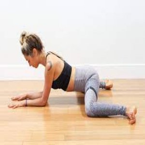
Adductor magnus muscle stretching
Frogger stretch
Frogger extends with your knees and lower arms on the ground with your knees and feet more extensive.
Attempt to keep the internal piece of your feet on the floor.
Sit the buttock back to the heels, sensing the stretch on the inner thighs.
After a brief pause of about 10 to 15 seconds, return to the stretch.
Recount 8-10 times.
Standing Adductor Stretch
Standing approximately three feet apart, get started.
Move the load aside and twist your knee.
To feel a stretch on the inside of the thigh, keep the other knee straight.
Hold the stretch for 15-30 seconds.
Calm down and recount on the opposite side.
Adductor magnus muscle strengthening
Side-Lying Hip Adduction
While isolating only one leg at a time, this is the best ground exercise for the hip adductors.
To lift your leg off the ground, all you need to do is concentrate on contracting the hip adductor muscles.
To perform this exercise, you must lie on your left side with your arms extended in front of you and your elbows and forearm supported on the floor. Place your heels against the thigh of your bottom leg as you raise your right leg above your lower leg.
Keep your leg extended and raise it as high as you can.
Return slowly to your starting position.
Perform 10 to 25 repetitions on the left side before switching to the right leg. By attaching an ankle weight to the leg or a resistance band to an anchor, you can make your exercise more challenging.

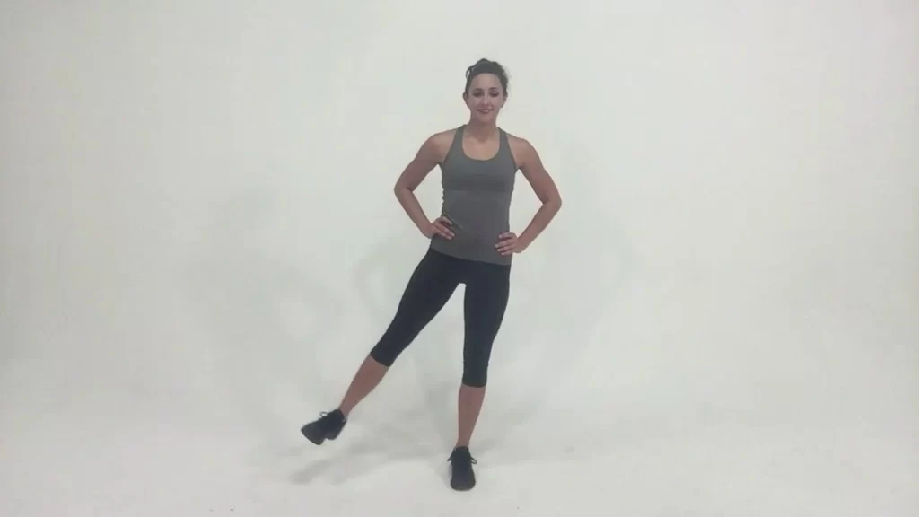
Standing side Leg Raise
Due to the fact that you will be maintaining an isometric hold on your other leg in order to keep it in the air, this is the best bodyweight exercise that can simultaneously target the hip adductors of one leg and the hip abductors of the other leg.
For this activity, you need to remain with your shoulders hip-width separated. Now, away from the body, on the left leg.
Then, at that point, Lift the right leg to the extent that is agreeable, and hold it there for 4-6 seconds.
After bringing your left leg and right leg up to touch, return to the starting position. Complete two sets of 10 to 18 repetitions.
Note: Attach ankle weights to both legs for a more challenging version of this exercise.
Cable Hip Adductor
You might see ladies at the gym center doing this activity while the men disregard it.
The time has come to move the disgrace that links hip adductions are not fundamental, everybody may be doing this activity to reinforce the adductors to lessen the gamble of injury.
Before beginning this exercise, warm up with some dynamic stretches. Then, try starting with less weight and maximum repetitions until you feel confident enough to increase the weight.
Find a connection that you can use to lash onto the lower leg nearest to the pulley.
Set the pulley so that it is at calf level. Keep your back to the pulley. Using your hand against the machine in a secure location where your fingers won’t be pinched, brace yourself. Your active leg might be moving toward the pulley from the floor.
Test your leg away from the pulley towards the center of your body.
Completed the desired number of repetitions by slowly allowing the leg to return to its starting position. Note: Using the same method, you can also exercise by attaching a resistance band to a fixed anchor point.
FAQ
What causes adductor magnus ache?
A common cause of groin pain and injury among athletes is adductor strain. Risk factors include age, weak adductors, muscle fatigue, reduced range of motion, inadequate adductor muscle complex stretching, and previous hip or groin injuries.
How is the adductor magnus treated?
The majority of adductor muscle strains can be treated conservatively. Beginning treatment incorporates action to change, which may briefly incorporate braces. Ice and anti-inflammatory medicine are proper for intense muscle strains. As side effects improve, delicate stretching is suitable to reinforce working out.
How can adductor magnus pain be alleviated?
To treat it you can press the muscle anyplace in that internal thigh space from your ischium down toward your knee, or press it between the fingers of each hand behind the gracilis line by pulling up the huge tummy of the muscle with the fingers of one hand and squeezing into it with the fingers of the contrary hand.
What yoga models for adductor magnus?
Adductor magnus could be utilized in back-bowing yoga presents like Scaffold Posture and Wheel Posture to assist with lifting the pelvis. With your hips raised in both of these postures, focus on squeezing the inward thighs toward the floor to actuate your adductor magnus.

One Comment