
Lumbrical muscles of the hand
What are the Lumbrical muscles of the hand?
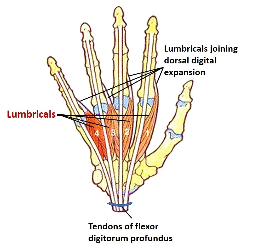
The four short intrinsic muscles of the hand that are arranged between the metacarpal bones and deep into the palmar fascia are referred to as the lumbrical muscles due to their worm-like appearance (lumbrical meaning in Latin is earthworm). The lumbrical muscles are one of the short muscles of the hand, along with the dorsal and palmar interossei.
At the metacarpophalangeal (MCP) and interphalangeal (IP) joints, the hand’s lumbrical muscles flex and extend the fingers. Many hand functions, like gripping movements, depend on these actions. On the toes, their counterparts, the lumbricals, perform a similar function.
Structure of the Lumbrical muscles
Each hand has four small, worm-like muscles called lumbricals. The fact that these muscles do not attach to bone makes them unique. Instead, they connect distally to the extensor expansions and proximally to the tendons of the flexor digitorum profundus.
The first number of head
Origin
It begins from the radial side of the most outspread ligament of the flexor digitorum profundus (compared to the pointer finger).
Insertion
It inserts on the extensor expansion close to the metacarpophalangeal joint after passing posteriorly along the index finger’s radial side.
The second number of head
Origin
It comes from the flexor digitorum profundus’s second-most radial tendon, which is on the radial side of the middle finger.
Insertion
It inserts on the extensor expansion near the metacarpophalangeal joint and travels posteriorly along the middle finger’s radial side.
The third number of head
Origin
One head starts on the radial side of the flexor digitorum profundus ligament compared to the ring finger, while the other begins on the ulnar side of the ligament for the center finger.
Insertion
To insert its extensor expansion, the muscle travels posteriorly along the radial side of the ring finger.
The fourth number of head
Origin
One head originates on the little finger’s radial side of the flexor digitorum profundus tendon, and the other on the ring finger’s ulnar side of the tendon.
Insertion
To insert its extensor expansion, the muscle moves posteriorly along the little finger’s radial side.
Innervation
The median nerve (C8-T1) supplies the first and second lumbricals. The ulnar nerve (C8-T1) supplies the third and fourth.
A quick and easy way to remember the lumbrical innervation is provided here.
1 2 me, 3 4 u (One two me, three four you)
1st and 2nd lumbricals – median nerve
3rd and 4th lumbricals – ulnar nerve
Blood supply
The dorsal carpal arch, an anastomotic network on the hand’s dorsal surface, provides the majority of the lumbricals’ arterial supply. The first and second lumbricals are supplied by their branches, the first and second dorsal metacarpal arteries, and the dorsal digital arteries.
The third and fourth lumbricals are supplied by the third and fourth dorsal digital arteries, as well as the second and third common palmar digital arteries, which are branches of the superficial palmar arch.
Function of Lumbrical muscles
The proximal (PIP) and distal (DIP) interphalangeal joints are in extension, while the metacarpophalangeal joints are in flexion. The way that the ligaments cross the MCP on the palmar side however insert distally at the dorsal side of the finger is the reason for the opposite activities. For instance, when holding a pen, these combined movements are a component of the hand’s complex movement and contribute to its overall dexterity.
Additionally, the hand’s lumbrical muscles are found to have a large fiber length and a large number of muscle spindles, suggesting that they are involved in proprioception.
Clinical relevance
Crush injuries to the hand can sometimes result in damage to the lumbrical muscle. Adhesions between the lumbrical and interosseous muscles may form after these injuries. Adhesions distal to the inter-palmar plate ligament restrict the proximal movement of the interosseus and lumbrical muscles because the lumbrical muscle passes volar to the ligament while the interosseus muscle passes dorsal. The patient experiences intermetatarsal pain when making a fist due to this deformity. Typically, it can be treated by relaxing the affected muscles. It has also been demonstrated that carpal tunnel syndrome is related to the lumbrical muscles.
Lumbrical-plus finger
A fascinating phenomenon occurs when the tendons of the flexor digitorum profundus detach distally from the lumbricals’ origin points: while attempting to close the clenched hand, the fingers unusually reach out all things considered.
The lumbricals now serve as the new flexor digitorum profundus insertion surface due to the distal tendons’ detachment. This indicates that rather than moving the flexor muscle, the individual moves the lumbricals. Also, because the lumbricals and the flexor digitorum profundus fight in the proximal interphalangeal (PIP) and distal interphalangeal (Plunge) joints, the expected clenched hand conclusion strangely causes the fingers to grow. The lumbrical-plus finger, clinically known as this anomaly, can develop following amputations or injuries.
Lumbrical muscle stretching
Bend your fingers forward to form an L shape with the hand and fingers while maintaining straight knuckles. Repeat using the opposite hand after holding for a few seconds. Five times, complete the stretch.
Lumbrical muscle strengthening exercise
Isometric for lumbricals
In a chair, sit up straight.
Try to grab the edge of the table with your knuckles bent and the middle and end joints of your fingers straight.
Squeeze the edge of the table with your thumb and fingers without bending your fingers’ middle and end joints.
FAQ
What are the numbers of the lumbric?
The lumbrical is one of the intrinsic muscles of the human hand that allows for these movements. From the lateral to the medial side, there are four lumbricals, referred to as the 1st, 2nd, 3rd, and 4th lumbricals.
What do the lumbricals look like?
The lumbricals of the upper appendage are four little muscles looking like the state of worms and subsequently, they are named so. From the lateral to the medial side, they are numbered.
What is the lumbrical’s position?
The hand’s lumbrical muscles are intrinsic, meaning they come from and insert within the hand. The lumbrical muscles insert into the lateral band of the extensor tendon mechanism after emerging from the flexor digitorum profundus (FDP).
What are lumbricals’ strengths?
After an ulnar nerve injury, the lumbricals have a mean strength of 0.8 kg or about 12% of the intrinsic muscle strength of a normal hand.
What causes hand lumbrical damage?
Pain in the palm, up to or near the base of a finger, and possibly into the area near the A1 or A2 pulley are typical symptoms of a lumbrical injury. As a result, it may appear to be a pulley injury even though it is not.

One Comment