Lumbrical plus finger
What is a lumbrical plus finger?
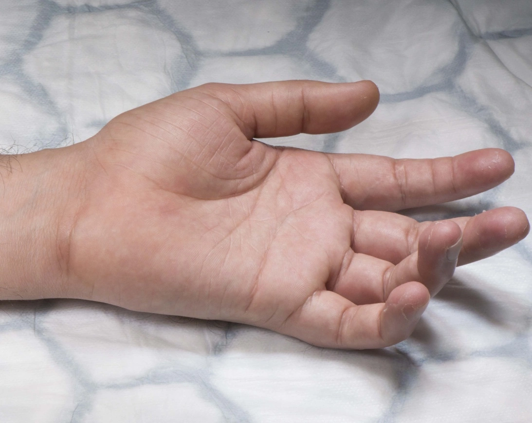
A lumbrical plus finger is a paradoxical extension of the interphalangeal joints during the flexion of the fingers. In the case of a lumbrical plus finger secondary to a distal interphalangeal amputation, the proximal interphalangeal will extend upon attempted finger flexion.
The lumbrical plus finger phenomenon occurs most commonly in the middle finger. the origin of the lumbricals is to be most common in the index.
Causes of a lumbrical plus finger:
- Human Bite.
- Dog and Cat Bites.
- Nail Bed Injury.
- High-Pressure Injection Injuries.
- Frostbite.
- Repetitive injury to the ulnar side of the palm.
- Severance of flexor digitorum profundus.
- Avulsion of flexor digitorum profundus.
- Over-long flexor tendon graft.
- FDP laceration.
Signs and symptoms of a lumbrical plus finger:
- Paradoxical finger extension
- Increased metacarpophalangeal flexion
- Notices that when attempting to grip an object or form a fist, the first digit sticks out or gets caught on clothes
Epidemiology:
Anatomic location:
- A lumbrical plus finger is most common in the middle finger.
- Flexor digitorum profundus three, four, and five is a common muscle belly.
- Index finger has independent flexor digitorum profundus belly.
Etiology:
- pathophysiology
- pathoanatomy
Pathophysiology of a lumbrical plus finger:
- Mechanism:
- Flexor digitorum profundus disruption is distal to the origin of the lumbricals.
- Can be due to flexor digitorium profundus transection, flexor digitorium profundus avulsion.
- Distal phalangeal amputation.
- Amputation through the middle phalanx shaft.
- Too long tendon graft.
Pathoanatomy of a lumbrical plus finger:
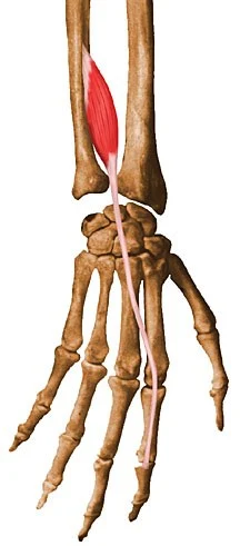
- Lumbricals originate from flexor digitorum profundus.
- With flexor digital profundus laceration, flexor digitorum profundus contraction leads to pull on lumbricals.
- Lumbricals pull on lateral bands leading to proximal interphalangeal and distal interphalangeal extension of the involved digit.
- With the middle finger, when the flexor digitorum profundus is cut distally, the flexor digitorum profundus shifts clearly.
Anatomy of a lumbrical plus finger:
Lumbricals:
- First and second lumbricals
- Unipennate.
- Median nerve.
- Originate from the radial side of flexor digitorum profundus two and flexor digitorum profundus three respectively.
- Third and fourth lumbricals
- Bipennate.
- Ulnar nerve.
- The third lumbrical originates from flexor digitorum profundus three and four.
- Forth lumbrical originates from flexor digitorum profundus four and five.
All insert on the radial side of the extensor expansion.
Physical examination:
- Paradoxical interphalangeal extension with grip.
- Fingers extend while holding a beer can.
Diagnosis of the lumbrical plus syndrome:
- Ultrasound examination
- MRI
- Stress test
Treatment of Lumbricals plus finger:
Operative treatment of lumbrical plus finger:
- Tenodesis of flexor digitorum profundus to terminal tendon or reinsertion to the distal phalanx
- indication:
- Flexor digitorum profundus lacerations.
- Do not suture flexor-extensor mechanisms over bone
Lumbricals release:
- indication:
- Flexor digitorum profundus lacerations.
- Do not suture flexor-extensor mechanisms over bone
- Contraindication:
- Do not transact lumbricals first & second if there is concomitant ulnar nerve palsy
- Interosseus muscles extend the interphalangeal joints
Improving strength exercise:
Fists:
- Hold out your affected hand, palm facing up your side.
- Slowly bend your fingers, keeping the thumb out.
- Make a fist slowly and then your fingers are straight again.
- Repeat this exercise 10 times.

Claw stretch:
- Hold your affected hand in front of the patient.
- Bend your fingertips down until they touch the base of the finger joint
- Hold this position for 30 to 60 seconds then release this position
- Repeat this exercise 10 times.
Make a ”O”:
- Start up affected hand straight up.
- Slowly curve your affected finger in to touch the thumb and make an ”O” shape.
- Hold this position for 30 to 60 seconds then release this position
- Straight your finger again.
- Repeat this exercise 10 times.

Finger walking:
- Put your affected hand on a table, palm facing down.
- Slowly lift the first finger move to towards the thumb, then place the finger down.
- Repeat this exercise 2 to 3 times.
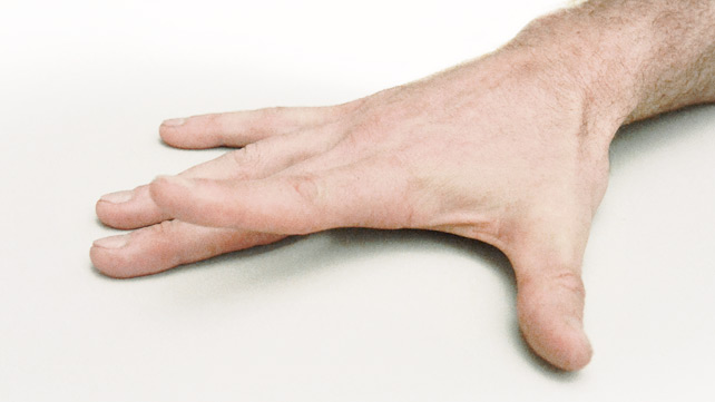
Finger lift:
- Keep your hand flat on a table.
- Gradually lift the finger off the table.
- Hold for 5 seconds.
Tennis ball squeeze:
- Take a soft tennis ball in your affected hand.
- Then release this position.
- Do this exercise 15 to 20 times and repeat it 2 to 3 times a day.

Summary:
A lumbrical plus finger is a paradoxical extension of the interphalangeal joints during the flexion of the fingers. In the case of a lumbrical plus finger secondary to a distal interphalangeal amputation, the proximal interphalangeal will extend upon attempted finger flexion.
The lumbrical plus finger phenomenon occurs most commonly in the middle finger. the origin of the lumbricals expects in to be very common in the index. treatment of lumbricals plus finger is lumbricals release and improve strengthening exercise of a finger.
FAQ:
Which fingers have lumbricals?
The first and second lumbricals originate from the radial side and palmer surface of the tendons of the first and second fingers.
What causes lumbrical plus?
We report a case of paradoxical extension phenomenon of the little finger, the so-called “lumbrical plus deformity” due to repetitive injury to the ulnar side of the palm.
What is the treatment for lumbrical?
Treatment of the lumbrical includes resting of muscle, ultrasound therapy, stretching of fingers, and strengthening exercises of fingers.
What nerve controls lumbricals?
First and second finger nerve control by the median nerve and the third and fourth finger nerve control by the ulnar nerve.
What are the signs of lumbrical injury?
Signs of lumbrical injury of pain in the palm of the hand, paradoxical finger extension, and Increased metacarpophalangeal flexion are noticed when attempting to grip an object or form a fist.



2 Comments