Lumbrical Muscles of the Foot
What are the Lumbrical muscles of the foot?
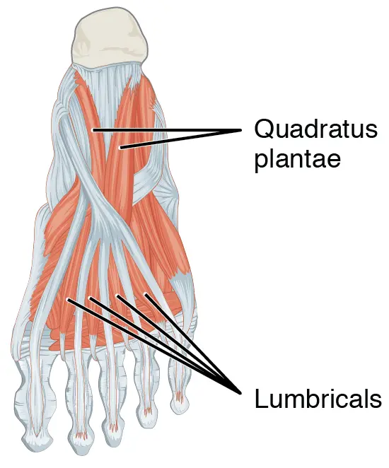
Lumbricals are the four small muscles located in the sole. Following the arrangement of plantar foot muscles into four layers (superficial to deep), the lumbricals and quadratus plantae include the second layer.
The lumbrical muscles extend at the interphalangeal joints and flex and adduct the lateral four toes at the metatarsophalangeal joints. The biomechanical balance of the foot while walking is contributed by these actions.
Origin and insertion
The lumbricals begin from the tendons of the flexor digitorum longus muscle, which is the reason they are incidentally referred to as the extra muscles of the flexor digitorum longus. They are labeled I-IV from the medial to the lateral side.
The first lumbrical muscle begins from the medial side of the first flexor digitorum longus tendon. The facing surfaces of two adjacent tendons of the flexor digitorum longus serve as the origin points for the remaining lumbricals.
Each lumbrical muscle travels anteriorly, plantar to the deep transverse metatarsal ligaments, from its point of origin. The belly of the muscle then twists in an obliquely upward direction to join the base of the proximal phalanges of the second through fifth digits and the extensor expansions thereon.
Relations
The lumbricals travel from the flexor to the extensor compartment of the foot after emerging from the tendons of the flexor digitorum longus muscle. The flexor digitorum longus tendons first cross the inferomedial and lateral surfaces of the lumbricals, where they serve as their starting points, before passing through the flexor sheaths of the second to the fifth toe.
Each lumbrical muscle inserts into the plantar surfaces of the deep transverse metatarsal ligaments, which connect the plantar ligaments of adjacent metatarsophalangeal joints via wide fibrous bands.
Innervation
The two terminal branches of the tibial nerve supply the lumbrical muscles;
The medial plantar nerve supplies the first lumbrical muscle (S1, S2).
The lateral plantar nerve (S2, S3) innervates the lateral three lumbricals.
Blood supply
The plantar arch is an anastomotic network that connects the lateral plantar and dorsalis pedis arteries and supplies the lumbrical muscles. The lateral plantar artery and four plantar metatarsal arteries, which supply the lumbrical muscles, are released by the convexity of the plantar arch.
Function
The lumbrical muscles flex and adduct the toes at the metatarsophalangeal (MTP) joints by pulling the medial base of the proximal phalanx. On the other hand, the lumbricals expand the toes at the interphalangeal (IP) joints by pulling on the phalanges’ extensor expansions.
The foot’s balancing function is provided by these lumbrical muscle actions. During the propulsive phase of gait, the MTP joints prevent hyperextension of the toes by opposing the extension of the long and short extensors. Because it opposes the flexion produced by the long and short flexors of the toes, the extension at the IP joints prevents clawing during gait.
Lumbrical muscle of the foot stretching
The lumbricals pedis muscles are stretched by extending toes 2-5 at the metatarsophalangeal joints or MTP joints and bending toes 2-5 at the proximal and distal interphalangeal joints.

Lumbrical muscle of the foot strengthening
Towel crunches
Put your foot on a towel and crunch the toes, keeping the towel with the toes. Finish three sets of ten repetitions. If this is simple, grab the pencil or pen with your toes instead of the towel.
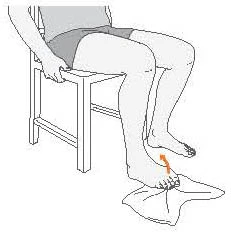
Towel toe pull
Sit on the bottom with your foot extended out in front. Keep the ends of the towel and twist the middle over the toes. Maintaining the knee straight with a bent foot (toes pointed upward). To bring the toes closer to the body, drag the towel’s finish. Maintain for twenty seconds. Repeat on the further side. Finish this stretch once a day, every other day.
FAQ
What do the lumbricals of the foot do?
The four lateral toes are extended at the interphalangeal joints by the foot’s lumbricals. Flex them at the joints of the metatarsophalangeal.
How many lumbricals are in a foot?
Four muscles called the lumbrical muscles of the foot come from the tendons of the flexor digitorum longus and pass dorsally before inserting into the free medial margins of the four lateral toe extensor hoods.
How long does a lumbrical injury take to heal?
The first step in recovering from a lumbrical strain is getting adequate rest from climbing: Grade I strains hope to rest for one to about fourteen days. From four to six weeks in Grade II. Grade III’s a month and a half or more.
What are the first and second Lumbricals of the foot?
On the medial sides of the four lesser toes, the muscles terminate in tendons that insert into the expansions of the tendons of the extensor digitorum longus muscle on the dorsal surfaces of the proximal phalanges.
What Innervates the first lumbrical of the foot?
medial plantar nerve
Innervation. The medial plantar nerve (S2 to S3) innervates the first lumbrical, while the lateral plantar nerve (S2 to S3) supplies the rest of the lumbricals. Both are terminal parts of the tibial nerve.