Muscle Tissue
Definition
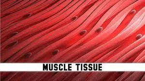
Muscle tissue is responsible for everything from the way you walk to the beat of your heart.
The cells that makeup muscle tissue are unique in that they may shorten or contract to cause movement of the body’s parts. The tissue has a high cell density and a good blood vascular supply. The cells are often grouped in bundles or layers encircled by connective tissue; because of their length and slenderness, they are occasionally referred to as muscle fibers. Muscle tissue includes the contractile proteins myosin and actin.
Skeletal muscle, smooth muscle, and cardiac muscle comprise the three types of muscular tissue.
Properties of muscle tissue
- Contractibility– The term refers to the capability of muscle cells to contract and shorten with great force.
- Extensibility–Muscles are capable of being stretched.
- Elasticity– The muscles have the ability to recoil back to their original length after being stretched.
- Excitability– The muscle tissue responds to a stimulus arrives from a motor neuron or hormone.
Structure of Muscular Tissue
- The epimysium, a thick connective structure that resembles cartilage, envelops the bundles of muscle tissues.
- The bundle of nerve cells that run in long fibers known as fascicles is encircled by the epimysium.
- The perimysium is a layer of protection that envelops the bundles. It permits blood and nerves to reach each individual fiber.
- The endomysium, another layer of defense, envelops the fibers.
- The contraction of various muscular sections is aided by these layers and muscles.
- As they compress, the many bundles slip past one another.
- The tendons that are connected to the periosteum, the connective tissue that envelops the bones, are connected to the epimysium.
- When the muscles contract, this facilitates the skeleton’s mobility.
- By forming connections with other connective tissues, the epimysium exerts a force on the organs, regulating everything from food processing to circulation.
Functions of muscle tissue
- Muscle tissue is generally associated with the same nerve bundles and acts as a single unit.
- The muscle contracts in response to a nerve impulse that travels from the brain or another external signal.
- The entire muscle contracts as a result of the nearly immediate transfer of the nerve impulse to every nerve cell in the muscle tissue.
- Every muscle cell comprises a combination of proteins that includes actin and myosin at the cellular level.
- The moment the signal to contract is received, these proteins move past each other.
- The filaments are attached to the ends of the cells, and the length of the cell decreases as they move past one another.
- When a muscle contracts, it shortens as a whole since a single cell can contract up to 70% of its length.
- Muscle tissue can be utilized to squeeze different organs, compress chambers, and move bones
Types of muscle tissue
- Skeletal
- Cardiac
- Smooth
Skeletal muscle tissue
- The contraction of skeletal muscle, which is affixed to the bones, enables voluntary movements of the body such as walking and expressing emotions on the face and in the posture.
- Skeletal muscle makes up forty percent of your body mass.
- By producing heat as a consequence of contraction, skeletal muscles contribute to thermal homeostasis.
- An involuntary contraction of the skeletal muscles occurs when the body temperature drops below average, causing shivering.
- The myoblasts that come from the mesoderm give rise to the muscle cell or myocyte.
- Throughout life, the number of myocytes is mostly constant.
- Connective tissue surrounds bundles of skeletal muscle tissue.
- Muscle cells seem striated and have many nuclei pressed along the membranes when viewed under a light microscope.
- The structural proteins that link the contractile proteins to connective tissues, as well as the contractile proteins actin and myosin, alternate regularly, resulting in striation.
- Each long muscle fiber is formed by the union of many myoblasts, which causes the cells to become multinucleated.
What Does Skeletal Muscle Do?
Your general health and daily activities depend on the function of your skeletal muscles.
Motion: Skeletal muscles contract and relax to produce movement. Your bone moves as a result of the contraction of these muscles, which shorten and pull on it. You can chew, swallow, urinate, or pass stool thanks to the skeletal muscles at these openings.
Stabilize the body: Skeletal muscles are also used to maintain posture and hold the body upright. These muscles constantly adjust slightly to keep your bones solid and prevent skeletal damage.
Keep your body temperature stable: Skeletal muscles assist in controlling body temperature. Your muscles use ATP, or energy, during contraction, which produces heat.
Consider how it would feel to shiver when you’re cold. This is a result of your body sensing a temperature drop below normal and causing your muscles to contract and relax in order to produce heat. Your temperature returns to normal as a result of the heat release.
Organ protection: Your skeletal muscles serve as a barrier to safeguard your organs, particularly the abdominal ones. They also aid in supporting your organs’ weight.
Storage: The building components of protein, amino acids, and glycogen, are stored in your muscles. When necessary, your body will use these amino acids to make proteins, and when you exercise or go without food, it will release glycogen to provide energy.
What Is Skeletal Muscle Made Of?
Skeletal muscle anatomy consists of numerous parts. On the other hand, muscle fibers, or myofibrils, are bundles of many proteins that make up skeletal muscle.
Sarcomeres are tiny protein structures found within each myofibril. These feature sections of light and dark that combine to form striation or patterns of red and white lines. Striated muscles are a common term for skeletal muscles. Your muscle shortens as a result of the sarcomeres enabling your muscle fibers to contract and slide across one another.
Additionally, blood arteries and nerve fibers in skeletal muscles transport waste materials and oxygen.
Every muscle is supported by three layers of connective tissue, which are as follows:
Epimysium: the outer layer of dense, irregular connective tissue surrounding the whole muscle
Perimysium: the middle layer of connective tissue surrounding bundles of fibers
Endomysium: the inner layer of connective tissue surrounding individual muscle fibers
Signs Something Might Be Wrong With the Skeletal Muscle
Weakness is the most typical indication of a muscle issue. While pain and twitching are common symptoms and indicators, they can also be expected side effects of activity or exercise.
During intense activity, your body may produce more lactic acid and other chemicals than it can eliminate, resulting in pain. These are common symptoms that normally go away in three to five days.
Signs and symptoms of more serious muscle problems may involve:
- Muscle Cramps
- Spasms
- Muscle Twitching
- Pain
- Weakness or trouble moving your limbs
- Muscle loss
- Loss of bladder or bowel control
- Trouble swallowing
- Balance problems
- Falling
- Tiredness
Cardia muscle
Muscle of heart
- The heart’s contractile walls are made up of cardiac muscle. Under a microscope, cardiomyocytes—the cells that comprise the heart muscle—also have striated appearances.
- Cardiomyocytes are solitary cells with a single nucleus positioned in the center, in contrast to skeletal muscle fibers.
- Cardiomyocytes’ ability to contract on their own intrinsic rhythm without outside stimulus is one of their main characteristics.
- Intercalated discs are specialized cell junctions that allow cardiomyocytes to adhere to one another.
- Gap junctions and anchoring junctions are both present in intercalated discs.
- Heart muscle fibers with attached cells branch out and function as a syncytium, enabling the cells to coordinate their movements.
- The heart muscle is an involuntary pump that circulates blood throughout the body.
How does cardiac muscle tissue function?
Additionally, certain cardiac tissue subtypes with “pacemaker” cell populations can be seen in the heart. In reaction to electrical impulses from the neurological system, they dilate and contract.
Action potentials, which are electrical impulses produced by pacemaker cells, instruct heart muscle cells to contract and relax. The pacemaker cells regulate heart rate and the heart’s rate of blood pumping.
How is it structured?
Heart muscle fibers, which are networks of interconnected heart muscle cells, provide cardiac muscle tissue its strength and suppleness.
While some heart muscle cells have two nuclei, most only have one. The genetic material of the cell is all contained in the nucleus.
Mitochondria are another feature of cardiac muscle cells that are sometimes referred to as “the powerhouses of the cell.” These are the organelles that produce adenosine triphosphate (ATP), which is energy, from glucose and oxygen.
Under a microscope, cardiac muscle cells have a striped or striated appearance. The alternating filaments of actin and myosin proteins are what cause these stripes. The dark stripes indicate the thick filaments made of myosin proteins, while the lighter filaments contain actin.
A cardiac muscle cell shrinks as a result of the myosin filament pulling the actin filaments near one another during contraction. This contraction is powered by ATP inside the cell.
Two actin filaments on either side are joined by a single myosin filament. This results in the formation of a sarcomere, a single unit of muscle tissue.
Heart muscle cells are connected by intercalated discs. Intercalated discs have gap junctions that allow electrical impulses to be sent from one cardiac muscle cell to another.
Other structures seen in intercalated discs are called desmosomes. These aid in the adhesion of heart muscle fibers.
What circumstances influence it?
A class of diseases known as cardiomyopathies harm the heart’s cardiac muscle tissue, making it more difficult for the heart to pump blood or rest correctly.
Among the typical signs of cardiomyopathy are:
breathing difficulties or dyspnea exhaustion
edema in the feet, ankles, and legs
discomfort in the neck or abdomen irregular heartbeat heart murmurs
feeling lightheaded or dizzy
The following variables may make someone more susceptible to cardiomyopathy:
- diabetes
- thyroid disease
- coronary heart disease
- heart attack
- chronic high blood pressure
- viral infections that affect the heart muscle
- valvular disease of the heart
- heavy alcohol consumption
- a family history of cardiomyopathy
- A heart attack because of a blocked artery may cut off the blood supply to a few areas of the heart. Over time, the muscle tissue in these particular areas of the heart will start to perish.
- The death of cardiac muscle tissue may also happen when the heart’s oxygen demand exceeds the oxygen supply. When the heart is damaged, it results in the release of cardiac proteins, like troponin, into the bloodstream.
Some examples of cardiomyopathy include:
Dilated cardiomyopathy
Dilated cardiomyopathy causes the heart’s left ventricle muscle tissue to stretch and the chamber to dilate.
Hypertrophic cardiomyopathy
Hypertrophic cardiomyopathy (HCM) is a genetic disorder characterized by the disorganized arrangement of cardiomyocytes. This condition can cause interruptions in blood flow out of the ventricles, abnormal electrical rhythms, and congestive heart failure.
Restrictive cardiomyopathy
Restrictive cardiomyopathy (RCM) is a condition where the heart’s ventricles become stiff, which prevents them from filling with enough blood.
Arrhythmogenic right ventricular dysplasia
This form of cardiomyopathy results in fatty infiltration in the right ventricular cardiac muscle tissue.
Transthyretin amyloid cardiomyopathy
Transthyretin amyloid cardiomyopathy (ATTR-CM) occurs when proteins known as amyloids accumulate and form deposits within the walls of the left ventricle. This results in the stiffening of the ventricle’s walls, making it difficult for the ventricle to fill with blood and reducing its ability to pump blood out of the heart. This type of condition is classified as a form of restrictive cardiomyopathy (RCM).
Smooth muscle
Internal organ motions that are not voluntary are caused by contractions of smooth muscular tissue. It creates the blood vessels, airways, and contractile elements of the reproductive, urinary, and digestive systems. Every cell has a single nucleus, a spindle form, and no discernible striations.
The structure
- The length and thickness of a smooth muscle cell range from 20 to 200 µm.
- Most of the myofilaments make up the homogeneously eosinophilic cytoplasm. During contraction, the center-positioned nucleus assumes the shape of a cigar.
- The cytoplasm is invaded by tiny pouch-like structures called caveolae, which are functionally similar to the skeletal muscle’s T-tubules.
- A basal lamina attaches the smooth muscle cells to the surrounding connective tissue.
- Branching bundles are the grouping of smooth muscle fibers.
- These bundles form a complicated system rather than a strictly parallel and orderly structure like skeletal muscle fibers do.
- As a result, the cells have far greater contractile power than striated muscle.
- Between dense bodies in the cytoplasm and attachment plaques at the cell membrane, the actin filaments are stretched.
- Actin filaments and myosin filaments are positioned between them. Furthermore, the cell structure is supported by intermediate filaments like vimentin and desmin.
Functions
- Smooth muscle can be found in practically every organ system, including the uterus, the eye, sphincters, hollow organs like the stomach and bladder, and tubular structures like arteries and bile ducts.
- It also has a significant function in the exocrine gland ducts.
- It performs a number of functions, including sealing orifices (such as the pylorus and uterine os) and moving chyme along the intestinal tube’s wave-like contractions.
- Smooth muscle cells are stronger, more maintained, and consume less energy than skeletal muscle cells, but they also contract more slowly.
- Myofibroblasts are a unique class of smooth muscle cells that also possess fibrocyte characteristics.
- They are also known as fixed (or stationary) connective tissue cells because they produce connective tissue proteins including collagen and elastin.
- Among other places, the lung’s alveolar septa and scar tissue contain myofibroblasts.
Innervation
Extremely intricate innervation exists inside the smooth muscle. It functions independently while also being influenced by the visceral nervous system.
Moreover, it is controlled by:
neurotransmitters, such as acetylcholine and norepinephrine;
hormones, such as oxytocin and estrogen;
tissue hormones, such as histamine and prostaglandins.
Stretching is one example of a local alteration that can be either calming or exciting. Smooth muscle is contracted involuntarily, in contrast to skeletal muscle.
One distinguishes between the multi-unit and single-unit categories functionally. The single-unit type smooth muscle cells contract uniformly and are electrically coupled by gap junctions. Internal organs and blood vessel walls (visceral smooth musculature) include this type of cell. Because the multi-unit smooth cells are independent of one another, they require separate innervation to provide more accurate muscle control. Among other places, they can be found in the iris and the muscles that control hair movement.
Comparison of structure and properties of muscle tissue types
| Muscle types | Structural elements | Function | Location |
| Skeletal | Long cylindrical fiber, striated, many peripherally located nuclei | Attached to bones and around entry & exit sites of the body (e.g., mouth, anus) | Short, spindle-shaped, with no evident striation, the single nucleus in each fiber |
| Short, spindle-shaped, no evident striation, the single nucleus in each fiber | Short, branched, striated, single central nucleus | Contracts to pump blood | Heart |
| Smooth | Voluntary movement produces heat, protects organs | Involuntary movement moves food, involuntary control of respiration, moves secretions, regulates the flow of blood in arteries by contraction | Walls of major organs and passageways |
FAQ
What functions do cells in muscles perform?
Virtually all voluntarily controllable movements are produced by skeletal muscle cells. Because of their extremely elongated structure, these cells—which may reach enormous lengths of 2-3 cm and diameters of 100 μm in an adult human—are frequently referred to as muscle fibers.
How do muscles function?
Body components are moved by the contraction and relaxation of muscles. Although muscles can pull bones, they are unable to push them back into place. Thus, they function as flexors and extensors in pairs. A limb can be bent at a joint by contracting the flexor.
What does a muscle do?
Soft tissues make up muscles. Your muscles consist of multiple elastic fibers. Your body contains more than 600 muscles. The functions of various muscle groups vary. Certain muscles are useful for running, jumping, and doing fine motor skills like threading a needle.
How does muscle grow?
Myoblast fusion, the process by which muscle fiber is created, requires a steady supply of nuclei. It is thought that an increase in satellite cell activity during postnatal growth is what causes the enlargement of muscle tissue. Satellite cells fuse to neighboring muscle fibers to increase their diameters.
What makes it a muscle?
Where did the term “muscle” come from? Its root word, musculus, is Latin and means “small mouse.” Hence the name originated from the belief that some muscles, most notably the biceps, moved and resembled mice.
References
- Muscle tissue | SEER training. (n.d.). https://training.seer.cancer.gov/anatomy/cells_tissues_membranes/tissues/muscle.html
- Admin. (2021, December 15). Muscular tissue – Structure, functions, and types of muscular tissue. BYJUS. https://byjus.com/biology/muscular-tissue/
- Biga, L. M. (2019b, September 26). 4.4 muscle tissue. Pressbooks. https://open.oregonstate.education/aandp/chapter/4-4-muscle-tissue/
- Eske, J. (2019, June 21). What to know about cardiac muscle tissue. https://www.medicalnewstoday.com/articles/325530#structure