Muscles Behind The Knee
Which are the Muscles Behind The Knee?
The anatomy of the back of the knee is complex. The thigh and calf muscles flow through this region, along with the knee joint positioned in the center. First, the big hamstring muscles link to the lower leg bones by passing over the knee from the pelvis. Furthermore, the huge Achilles tendon is formed by the vast calf muscles, which start at the bottom of the leg and cross the knee.
Ultimately, the lower leg and foot are supplied by essential blood vessels and nerves that pass through the back of the knee.
The back of the knee is also called The popliteal fossa or posterior knee. The popliteal fossa (also known as hough or it in analogy to the cubital fossa) is a shallow depression found at the rear of the knee joint. The bones that comprise the popliteal fossa are the femur and tibia.
A portion of the posterolateral corner of the knee, the popliteus is a small, thin, flat, triangular-shaped musculotendinous complex of the lower leg that includes the popliteofibular ligament and the popliteus muscle. It forms the floor of the popliteus fossa and is a deep knee muscle. It, along with the iliotibial band, makes up the lateral musculature of the knee joint. It is the only mono-articular muscle that does not affect the ankle joint in the posterior compartment of the lower leg, which is additionally used by the flexor digitorum longus, flexor hallucis longus, and tibialis posterior.
Because it is one of the principal posterolateral stabilizers of the knee joint, inducing both medial and lateral rotation of the knee, it plays a role in both the closed-chain and open-chain phases of the gait cycle. It acts similarly to a smaller stabilizer in terms of anterior translation, varus force, and internal rotation.
The back muscles of the knee are primarily composed of the hamstring group, along with a smaller, deeper muscle called the popliteus. Here’s a breakdown of each:
Hamstring Group:
- Biceps femoris: This large, three-headed muscle crosses both the hip and knee joints. It’s responsible for flexing the knee, extending the hip, and rotating the tibia medially
- semitendinosus. The semitendinosus is a long, thin muscle that originates from the ischial tuberosity, also known as the sit bone and ends at the medial tibia. It facilitates knee flexion and hip extension.
- semimembranosus. The semimembranosus muscle, which is situated on the medial side of the thigh, attaches to the medial tibia similarly to the ischial tuberosity. Its main purpose is to allow knee flexion, although it also helps with hip extension and tibia medial rotation.
Popliteus Muscle:
This tiny, triangular muscle can be found below the femur and above the tibia, deep within the knee joint. It is essential for maintaining knee stability, especially during flexion and rotation.
There are two types of muscles in the posterior knee: deep and superficial. This is an explanation:
The superficial layer:
- Gastrocnemius: This big, two-headed muscle connects the ankle and knee joints and forms the calf. It points to the foot and flexes the knee and ankle plantarflexed. the posterior knee gastrocnemius muscle
- Plantaris: A tiny, narrow muscle that acts similarly to the gastrocnemius but is frequently non-functional and vestigial in humans.
- Another big muscle located deep in the gastrocnemius is the soleus, which helps flex the knee in addition to plantarflexing the ankle.
Deep Layer:
Popliteus: Located deep within the knee joint, this triangle muscle is essential for rotating and stabilizing the tibia during flexion. Popliteus tendon posterior to the knee
Flexor Digitorum Longus: This muscle helps in plantar flexion and flexes the toes. It starts behind the knee and travels down the leg.
Tibialis Posterior: This inside side of the lower leg muscle helps with the bending of the knee and inverts and plantarflexes the foot.
Extra Information
- Strong muscles like the hamstrings are necessary for a variety of exercises like sprinting, jumping, and kicking.
- Athletes frequently sustain hamstring sprains or tears as a result of abrupt, strong contractions or overexertion.
- Regular hamstring strengthening and stretching can help reduce the risk of injury and enhance knee function in general.
Limits of the Muscles Behind The Knee
The popliteal fossa is a diamond-shaped region formed by the hamstring muscles, which are the muscles that collectively lie behind the knee. These three muscles define the limits of this fossa:
Superomedial border. The muscles of the semimembranosus and semitendinosus form the superomedial boundary. On the medial side of the thigh is a large, flat muscle called the semimembranosus. It originates from the ischial tuberosity and joins the semitendinosus in a shared tendon to attach to the medial tibia. The semitendinosus is a slender muscle located just lateral to the semimembranosus. It also arises from the ischial tuberosity and inserts onto the medial tibia through the common tendon.
Superolateral border: This border is formed by the biceps femoris muscle, specifically its long head. The biceps femoris is a large, two-headed muscle located on the posterior aspect of the thigh. The femur’s linea aspera is where the short head originates, while the ischial tuberosity is where the long head originates. Both heads join together to form a common tendon that inserts into the fibula head.
Inferomedial and inferolateral borders: These borders are formed by the medial and lateral heads of the gastrocnemius muscle, respectively. The gastrocnemius is a large, two-headed muscle that forms the calf. The medial head originates from the medial epicondyle of the femur and the posterior capsule of the knee joint. The lateral epicondyle of the femur is the source of the lateral head. Both heads join together to form a strong Achilles tendon that inserts into the calcaneus (heel bone).
Where is the popliteus muscle located?
The popliteus muscle is located deep within the back of the knee joint, situated beneath the femur (thigh bone) and above the tibia (shin bone). It forms part of the floor of the popliteal fossa, a diamond-shaped space behind the knee.
Origin
Through the strong tendon known as the popliteus tendon, the muscle fibers begin at the posterior horn of the lateral meniscus and the lateral condyle of the femur. Additionally, fibers from the styloid part of the fibular head have been observed in cadaveric dissections. These fibers then run obliquely and blend in with the main muscular structure.
It then proceeds inferiorly and mediolaterally in the direction of the Tibia. It is the deepest muscle in the posterior knee region and courses diagonally across the posterior upper tibia and a section of the joint capsule. The popliteal tendon does not penetrate the synovium; instead, it pierces the joint capsule. The biceps femoris tendon and the lateral collateral ligament (LCL) are passed beneath the popliteus tendon.
The lateral femoral condyle and the popliteal bursa are separated by this structure, which is often an extension of the synovial membrane. The popliteus muscle is a capsular structure that separates the lateral collateral ligament from the knee’s lateral meniscus, despite having extra-articular areas. In the lateral head of the gastrocnemius muscle, the sesamoid bone may give rise to a second popliteus head. The popliteus minor, an extra inconstant muscle that originates from the femur on the inside of the plantaris muscle and inserts into the knee joint’s posterior ligament, is occasionally observed.
The popliteus muscle is extra-articular, extra-synovial, and intra-capsular.
Insertion
It attaches to the tibia below the tibial condyles and is slightly proximal to the seal line.
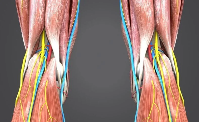
Nerve Supply to Muscles Behind The Knee
Nerve From spinal nerve roots L4 through S1, the tibial nerve supplies the popliteus muscle. There are roughly two to three parallel tibial nerve branches. The lateral distal margin of the muscle, which is located inferior to the fibular head, serves as the nerve’s entry point. From there, it divides into anterior, medial, and lateral distributions across the muscle.
The muscles behind the knee, also known as the popliteal fossa, are crucial for knee flexion and stability. Their nerve supply comes from a complex network, primarily originating from the sciatic nerve and its terminal branches:
Main Nerve Supply:
- Sciatic nerve: This large nerve originates from the lumbosacral plexus (L4-S3) and exits the pelvis through the greater sciatic foramen. It descends the posterior thigh and enters the popliteal fossa at the apex, where it typically divides into:
- Tibial nerve: Supplies the flexor muscles of the leg, the posterior compartment of the leg, and the sole. In the popliteal fossa, it gives off branches like the medial sural cutaneous nerve, medial collateral nerve, and tibial com
- Common fibular nerve: Divides into the peroneal nerve and the sural nerve in the popliteal fossa. The peroneal nerve supplies the anterior and lateral compartments of the leg, while the sural nerve supplies the skin of the posterior leg and lateral foot. Common fibular nerve popliteal fossa
The sciatic nerve is a major nerve that starts in the lumbosacral plexus (L4-S3) and leaves the pelvis via the larger sciatic foramen. It descends the posterior thigh and, at the apex, enters the popliteal fossa, where it normally separates into:
Tibial nerve: This nerve supplies the leg flexor muscles, the posterior compartment of the leg, and the sole. It gives out branches such as the medial sural cutaneous nerve, medial collateral nerve, and tibial communicating nerve in the popliteal fossa. Popliteal fossa tibial nerve
In the popliteal fossa, the common fibular nerve divides into the peroneal nerve and the sural nerve. The peroneal nerve provides sensation to the anterior and lateral compartments of the leg, whereas the sural nerve provides sensation to the skin of the posterior leg and lateral foot. Popliteal fossa of the common fibular nerve
Additional Contributors:
- Obturator nerve:
- The medial hamstring muscles and the hip joint capsule are supplied.To reach the popliteal fossa, it pierces the adductor magnus muscle.
- Sural communicating nerve: This nerve arises from the common fibular nerve and connects to the medial sural cutaneous nerve, providing sensory supply to the posterior leg.
Specific Muscle Innervation:
- Hamstring muscles:
- Biceps femoris (long head): Sciatic nerve
- Biceps femoris (short head): Common peroneal nerve
- Semimembranosus: Sciatic nerve
- Semitendinosus: Sciatic nerve
- Popliteus muscle: Tibial nerve
Understanding the nerve supply of these muscles is essential for:
- Diagnosing injuries or pain in the back of the knee
- Planning surgical procedures that may involve these nerves
- Developing rehabilitation programs for knee injuries
Blood supply
Artery Two arteries supply the popliteus muscle:
The posterior tibial artery’s muscular branch and the popliteal artery’s medial inferior genicular branch.
The muscles of the back of the knee, situated in the popliteal fossa, depend heavily on a dedicated network of arteries for their vital oxygen and nutrient supply. This intricate vascular web primarily originates from the popliteal artery, a continuation of the femoral artery that descends to the posterior thigh.
Main Arterial Supply:
- Popliteal artery: This large artery enters the popliteal fossa and generously branches out, nourishing the surrounding muscles, ligaments, and the knee joint capsule itself. Some key branches include:
- Superior genicular arteries (medial & lateral): These arteries climb towards the knee joint, supplying the powerful quadriceps muscles and the kneecap (patella).
- Inferior genicular arteries (medial & lateral): These arteries head down towards the ankle, providing blood flow to the calf muscles – gastrocnemius, soleus, and plantari.
Popliteal artery: This big artery enters the popliteal fossa and branches out liberally, supplying the surrounding muscles, ligaments, and the knee joint capsule. Among the most important branches are:
Superior genicular arteries (medial and lateral): These arteries nourish the strong quadriceps muscles as well as the kneecap (patella).
Inferior genicular arteries (medial and lateral): These arteries go down towards the ankle and supply blood to the calf muscles, including the gastrocnemius, soleus, and plantaris.
Understanding the arterial supply of these muscles is crucial for various reasons:
Diagnosis of vascular insufficiency or obstruction: If the blood supply to any of these muscles is disrupted, muscular weakening, discomfort, and even tissue death can occur.Identifying the damaged artery aids in determining the best therapy.
Planning surgical procedures: Knowing the precise position and path of these arteries is critical for surgeons working on the knee joint or adjacent tissues, reducing the chance of accidental arterial injury.
Understanding the arterial supply can guide treatment options aimed at boosting blood flow and relieving symptoms for illnesses such as muscular cramps or compartment syndrome (increased pressure inside a muscle compartment)
Function of the Muscles Behind The Knee
As a primary internal rotator of the tibia in a non-weight-bearing position, the popliteus muscle aids in knee flexion, and its function is determined by the lower extremity’s position, i.e., weight-bearing or non-weight-bearing.
It rotates the tibia medially in open-chain kinematics.
It laterally rotates the femur in the first phase of knee flexion in close-chain kinematics.
‘Locking’ of the knee happens with extension during weight bearing. When the femur medially rotates on the tibia, complete extension is possible without using any muscles. Popliteus muscle contraction is required to ‘unlock’ the knee, resulting in flexion and lateral rotation of the femur on the tibia; this gives the muscle the term ‘key’ to the locked knee.
During knee flexion: The popliteus muscle retracts the lateral meniscus posteriorly during knee flexion to prevent it from becoming trapped between the tibia and femur.
In knee stability: The Popliteus tendon is often attached to the lateral capsule, suggesting that the muscle may play a role in postero-lateral knee stability. Even if all other postero-lateral ligaments were severed, Krudwig et al. view the popliteus as an important component that prevents excessive external tibial rotation and preserves neutral tibial rotation.
The popliteus muscle, despite its compact size, packs a powerful punch when it comes to knee function. Nestled deep within the back of the knee, it plays a crucial role in two key actions:
1. Flexing the Knee:
- Consider yourself seated in a chair. The popliteus contracts as you bend your knee, dragging the tibia (shin bone) backward and lower. This rearward pull aids in the flexion of the knee joint, making it an important role in movements such as walking, jogging, and squatting.
2. Stabilizing the Knee:
- The popliteus acts like a silent guardian, stabilizing the knee joint throughout its range of motion. During knee flexion, it also rotates the tibia inwards (medially) on the femur. This inward rotation counteracts any outward displacement forces, preventing the knee from wobbling or buckling, especially crucial for movements like jumping and landing.
Here’s a quick recap of the popliteus muscle’s functions:
- Knee Flexion: Aids in the bending of the knee joint.
- Tibial Rotation: To maintain stability during knee flexion, the tibia is rotated inwards.
- Stabilization: Aids in preventing excessive tibial outward displacement, keeping the knee joint stable.
Remember, even though the popliteus is small, it’s mighty! Its contributions to knee flexion and stabilization are essential for various activities, from everyday movements to athletic endeavors
The functions of the muscles behind the knee, along with images for better understanding:
The Hamstrings: Powerhouse Behind the Knee
The muscles behind the knee are collectively known as the hamstrings. They’re a group of three powerful muscles located at the back of the thigh:
- The biceps femoris muscle has two heads and is located on the outside of the thigh.It is essential for all hamstring functions.
- Semitendinosus: Located on the inside thigh, this muscle largely aids in knee flexion and lower leg inward rotation.
- Semimembranosus: Similar to the semitendinosus in location and function, this muscle also aids in knee flexion and hip extension.
Mastering Knee Flexion
The hamstrings’ primary function is flexing the knee. They contract to pull the lower leg towards the thigh, bending the knee joint. This action is crucial for various activities like:
- Walking
- Running
- Kicking
- Climbing stairs
More Than Just Bending:
While knee flexion is their primary job, the hamstrings also play vital roles in other leg movements:
- Hip Extension: The hamstrings help to extend the hip joint, propelling you forward as you walk, run, or leap. The hamstring muscles extend the hip.
- Knee Stabilisation: The hamstrings, in collaboration with other muscles, stabilize the knee joint, limiting excessive hyperextension and providing support during weight-bearing exercises.
- Rotation: The hamstrings, particularly the biceps femoris, help rotate the lower leg inwards when the knee is flexed, assisting in motions such as kicking and changing direction.
Clinical significance
It frequently plays a role in knee posterolateral (PCL) corner injuries, which result from –
- A bent knee is subjected to a varus force.
- A direct strike to the knee (lateral to medial).
- Varus/hyperextension (from injuries sustained without contact as well).
- Knee dislocation.
Whatever the mechanism of injury to the PCL corner (as mentioned above), a rapid assessment of the patient’s limb’s neurovascular status is carried out. In a dislocated knee, the vascular status is evaluated first, then the knee joint is closed and the vascular status is evaluated once more.
In addition to trauma, bad posture, and movement habits can put a lot of strain on the popliteus muscle, making it vulnerable to damage and weakness. This muscle is frequently injured due to iatrogenic causes, which can have a negative functional prognosis. As a result, it is critical to treat this injury, particularly after knee reconstruction surgery. Extra care is also needed for anatomically smaller knees because there is a higher risk of popliteal injury.
Posterolateral knee pain is another possible symptom of popliteal tendinopathy. However, because other more prevalent causes of knee pain are nearby, it may be challenging to identify them specifically. This muscle can be pathologically overcome by excessive sprinting or running downhill, so these activities should be avoided or modified to run on flat surfaces like a treadmill. It also inhibits excessive tibial rotation by preventing significant anterior translation of the knee.
The popliteus muscle will weaken if the lateral hamstrings are stronger than the inner (medial) hamstrings. The popliteus will be under stress in the opposite direction when walking or running with excessive pronation or collapse of the inner foot.
Several EMG studies have demonstrated that popliteus muscle activity increases during knee extension and walking downhill, solidifying its function in regulating knee joint hyperextension.
Back of the knee pain is the pain pattern that is referred to when there is a popliteus trigger point.
The popliteus muscle, despite its small size, has significant clinical significance due to its crucial role in knee function and its susceptibility to various conditions. Here’s a breakdown of its clinical relevance:
Role in Knee Stability:
- The popliteus plays a vital role in stabilizing the knee joint, especially during flexion and rotation. It helps counterbalance outward forces on the tibia, preventing instability and potential ligament damage.
Clinical Implications:
- Knee Injuries: Popliteus muscle weakness or injury can lead to knee instability, discomfort, and trouble moving. This might be caused by ligament tears, meniscal tears, or overuse injuries.
- Diagnosis: Because of its deep placement and overlapping symptoms with other knee disorders, diagnosing popliteus involvement can be difficult. Physical examination, imaging tools such as MRI, and particular testing for unlocking the knee can all give useful information.
- Treatment for popliteus problems is dependent on the underlying reason. Physical treatment to strengthen the muscle, anti-inflammatory drugs, and, in certain circumstances, surgery for serious tears or instability are all options.
Specific Conditions:
Popliteus Tendinopathy is characterized by inflammation or degeneration of the popliteus tendon, resulting in discomfort and soreness behind the knee, especially during activities such as squatting or downhill jogging.
Popliteal Fossa Entrapment Syndromes: In rare situations, the popliteus muscle can compress nerves or blood vessels in the popliteal fossa, causing symptoms such as leg numbness, tingling, or weakness.
Additional Importance:
- The popliteus muscle can cause referred pain, which means that discomfort originating in the popliteus can be felt in other locations such as the calf or thigh.
- Understanding the function of the popliteus is critical for physiotherapists, sports medicine experts, and orthopedic surgeons when developing successful rehabilitation programs and treatment strategies for knee-related disorders.
Pathology popliteus muscle
The muscles behind the knee are collectively known as the hamstring muscles. They are a group of three muscles – biceps femoris, semimembranosus, and semitendinosus – that run down the back of the thigh and connect to the tibia and fibula bones in the lower leg. These muscles are in charge of extending the hip joint and flexing the knee joint.
Several pathological conditions can affect the hamstring muscles, including:
- Tears in the muscle fibers caused by overuse, abrupt stretching, or incorrect biomechanics are known as muscle strains. A hamstring strain manifests as discomfort, tightness, swelling, and weakening.
- Hamstring tendinitis: The tendons that join the hamstring muscles to the bones are inflamed in this situation. The causes are often overuse or repetitive strain. Pain, stiffness, and discomfort behind the knee are among the symptoms.
- Complete tears in the muscle or tendon are called hamstring tears.They are typically brought on by an abrupt, strong muscle contraction, like that which occurs during a sprint or jump. Severe pain, a popping sound, swelling, bruises, and a restriction in knee flexion are among the symptoms.
- Baker’s cyst: This is a fluid-filled sac that develops beneath the knee joint. It is frequently brought on by inflammation in the knee, which can result from torn ligaments or arthritis. Pain, stiffness, and edema are among the symptoms.
- Cysts in the popliteal fossa: These are sacs filled with fluid that develop in the area behind the knee joint. They can be brought on by some conditions, including tumors, arthritis, and trauma, but they are less prevalent than Baker’s cysts. Knee soreness, edema, and trouble bending the knee are among the symptoms.
Treatment for hamstring muscle pathology depends on the severity of the condition. Mild strains and tendinitis can usually be treated with rest, ice, compression, and elevation (RICE). More severe tears may require surgery. Baker’s cysts and popliteal fossa cysts may be treated with aspiration, medication, or surgery.
If you are experiencing any pain or other symptoms behind your knee, it is important to see a doctor to get a diagnosis and discuss treatment options. Early diagnosis and treatment can help prevent further injury and improve your chances of a full recovery.
The popliteus muscle is a small but important muscle located in the back of the knee. It plays a vital role in stabilizing the knee joint and aiding in flexion and rotation. Unfortunately, the popliteus muscle is also susceptible to various pathologies that can cause pain and dysfunction.
Here are some of the most common popliteus muscle pathologies:
1. Popliteus strain: This is a muscle fiber tear or overuse injury. It is frequently brought on by abrupt twisting or powerful knee motions, like those that occur during sports. A popliteus strain can cause pain at the back of the knee, as well as swelling, soreness, and trouble bending the knee.
2. Popliteus tendinopathy: This condition is an irritation or inflammation of the tendon that joins the tibia bone to the muscle. It may be brought on by the tendon experiencing repetitive stress from activities like running or leaping. Popliteus tendinopathy symptoms include a popping or snapping sensation in the knee and soreness behind the knee, particularly with weight-bearing exercises.
3. Popliteus entrapment: This is the result of surrounding structures, such as the popliteal artery or fibular head, compressing or pinching the popliteus tendon. Though it is a very uncommon condition, it can lead to leg and knee discomfort, weakness, and numbness.
The inflammation of the bursa, a fluid-filled sac that supports and cushions the popliteus tendon, is known as popliteal bursitis. Overuse, trauma, or other inflammatory disorders can be the reason. The back of the knee may hurt, swell, or feel tender and warm to the touch if you have popliteal bursitis.
Prevention of popliteus muscle pathologies:
- Warm up well before working out, and then cool down.
- Gain strength in the muscles that surround the knee, such as the quadriceps, hamstrings, and calves.
- Retain the appropriate amount of hamstring and calf flexibility.
- Steer clear of abrupt twisting or vigorous knee motions.
- Wear well-fitting, supportive shoes.
Diagnosis Muscles Behind The Knee
The muscles behind the knee are collectively known as the hamstrings and the popliteus. They play a crucial role in knee flexion, stability, and rotation.
Here’s a breakdown of the potential issues you might be experiencing, based on the location of your pain:
Hamstring pain:
- Location: Upper back of the knee, radiating up the thigh.
- Possible causes: Strain, tear, tendinitis, bursitis.
- Symptoms: Pain, tenderness, swelling, weakness, difficulty bending the knee.
Popliteus pain:
- Location: Deeper in the back of the knee joint, sometimes radiating down the calf.
- Possible causes: Strain, tendinitis, entrapment, bursitis.
- Symptoms: Pain, stiffness, catching or snapping sensation, difficulty bending or straightening the knee.
Diagnosing the cause of your pain:
To accurately diagnose the source of your pain, a doctor will typically perform a physical examination, inquire about your medical history and activity level, and possibly order imaging tests like MRI or ultrasound.
Evaluation
Due to its deep location, popliteus injuries are uncommon on their own but can occur in conjunction with other knee injuries like meniscus and ACL tears.
Swelling, soreness, edema, bleeding, and the patient maintaining the leg (tibia) in lateral rotation during knee flexion are some common signs of muscle injury.
To rule out injuries to the popliteus muscle, the following should be examined.
Tenderness: Only the terminal parts of the popliteus muscle can be palpated due to the numerous neurovascular structures that lie above it. A prone lying position is used to assess the tenderness of the proximal tendon, whereas tenderness over the posterolateral knee may indicate a lateral meniscus injury or a strain in the biceps femoris tendon.
In the Garrick test, the patient is seated upright with their hips and knees bent 90 degrees. There is resistance to the lower leg’s external rotation. Suffering while performing this action is regarded as a positive test.
Shoe removal manoeuvre. By internally rotating the afflicted leg to reach the heel of the opposing leg, the patient attempts to remove the contralateral shoe. A popliteus muscle injury is indicated by pain during this maneuver.
As previously mentioned, isolated popliteus muscle injuries are uncommon; however, popliteus tendon rupture was observed in isolated acute knee MRI studies.
In the stance phase, the popliteus muscle and PCL (posterior cruciate ligament) stabilize the femur over the fixed tibia, particularly when additional stability is required for activities like running downhill. Running downhill, particularly on a banked surface when hyperpronating, can therefore result in injuries to the popliteus muscle, such as tenosynovitis, tendinopathy, rupture, and strain.
Treatment
The management of popliteus muscle pathology has similarities to that of tendinopathy and other soft tissue and muscle injuries. Protection, rest, elevation, compression, elevation, and other anti-inflammatory medications are administered in RICE or PRICE therapy.
Depending on the pathology, related injuries, and patient condition, physiotherapy treatment is similar to that for other soft tissue and muscle injuries and includes mobility exercises, strengthening exercises, eccentric training, and many more rehab protocols.
Use your feet to sit. dorsiflexed to the point where the heel touches the floor. Turn your foot in an internal direction. Band tied to forefoot; can be done with or without resistance band.
Popliteal muscle release: Reposition a lacrosse ball behind a flexed knee and sit for a long time, feeling for sore spots. Increase pressure gradually. If the area is sensitive or numb, rotate the lower leg both internally and externally to increase movement.
according to isometry. When seated on a bar or table, rotate internally with your forefoot.
Chain-opening. complete range of motion in the knee with an external resistance band fastened to the forefoot to prevent internal rotation.
Close chain proprioception exercises on a bosu ball involving a cross lunge and the affected leg fixed on the bosu ball.
Jump forward with just one leg, bending the knee slightly.
Treatment for muscle issues behind the knee, primarily focusing on the hamstrings and popliteus, depends on the severity and specific cause of your pain. Here’s a breakdown of common approaches:
Rest:
- Take a break from activities that aggravate the pain, especially those involving repetitive bending or straightening of the knee. This allows for inflammation to subside and healing to begin.
Ice:
- Several times a day, apply cold packs to the affected region for 15 to 20 minutes at a time. Icing helps reduce inflammation and pain.
Compression:
- Wearing a knee brace or compression sleeve can provide support, and stability, and reduce swelling. Choose a brace that fits snugly but comfortably and allows for some movement.
Elevation:
- Keep the knee elevated above the heart whenever possible, especially when resting. This helps reduce swelling and promote drainage of fluids.
Pain medication:
- Acetaminophen or ibuprofen, two over-the-counter pain medications, can assist in controlling discomfort. Your doctor could occasionally recommend harsher painkillers.
Physical therapy:
- Once the initial inflammation subsides, physical therapy plays a crucial role in recovery. A physical therapist will design a personalized program of exercises to:
- Strengthen the hamstrings and popliteus, as well as surrounding muscles like the quadriceps and calves.
- Improve flexibility and range of motion in the knee joint.
- Address any muscle imbalances.
- Gradually return you to your desired activities.
Physical therapy exercises for knee muscles
Additional treatment options:
- In some cases, your doctor may recommend other treatment modalities like:
- Ultrasound therapy: This uses sound waves to promote healing and reduce pain.
- Electrical stimulation: This mild electrical current can help stimulate muscle healing and reduce pain.
- Massage therapy: Massage can help improve blood flow, reduce muscle tension, and promote relaxation
Risk factor
The muscles behind the knee, primarily the hamstrings and the popliteus, are susceptible to various risk factors that can increase the likelihood of injury or damage.
Anatomical Factors:
- Tight hamstrings or calves can restrict knee flexion and range of motion, putting additional pressure on the muscles and tendons during activity.
- Weak musculature: If the hamstrings or popliteus muscles are weak, the knee joint will be more prone to injury.
- Muscle imbalances: Asymmetry in hamstring and quadriceps strength can cause an imbalance in forces acting on the knee, increasing the risk of strains or tears.
Biomechanical Factors:
- Poor posture or misalignment of the knee joint can result in an unequal distribution of forces across the muscles, increasing the risk of injury.
- Flat feet: Pronated feet can have an impact on leg and knee biomechanics, perhaps putting more strain on the hamstring and popliteus muscles.
- Lack of flexibility in the hamstrings and calves can limit range of motion and increase the risk of muscle injuries, particularly during rapid movements.
- .
Activity-related Factors:
- Overuse: Repetitive activities requiring severe knee flexion, such as running or leaping, can overload the muscles and tendons, resulting in overuse problems.
- Sudden increases in activity level: Without sufficient preparation, rapidly increasing exercise intensity or length can place undue strain on the muscles and risk damage.
- Improper technique: Improper movement execution in sports or workouts can put unnecessary tension on individual muscles, increasing the risk of injury.
Other Factors:
- Age: As we become older, our muscular strength and flexibility decline, leaving the muscles behind the knee more vulnerable to injury.
- Previous injuries to the knee joint, muscles, or ligaments can weaken the tissues and increase the likelihood of repeat damage.
- Nutritional deficiencies: A lack of vital nutrients such as protein, vitamin D, and calcium can interfere with muscle repair and recovery, increasing the risk of injury.
- Medical problems, such as arthritis or neurological disorders, can impair muscle function and stability, increasing the risk of injury.
Remember:
- Identifying your personal risk factors can help you take preventative measures, such as strengthening exercises, stretching routines, and proper training techniques.
- Listening to your body and paying attention to pain or discomfort is crucial for preventing injuries.
- Consulting a healthcare professional for personalized advice and guidance is essential for managing risk factors and optimizing your training or activity plans.
By understanding these risk factors and implementing appropriate preventive strategies, you can significantly reduce your chances of encountering problems with the muscles behind your knee and keep them healthy and functional for a long time.
Preventing Popliteus Muscle Injuries
The popliteus muscle, though small, plays a crucial role in knee stability and flexion. Unfortunately, its deep location and involvement in various movements make it susceptible to injury. Luckily, several preventive measures can help keep your popliteus healthy and happy!
Here are some key strategies to prevent popliteus muscle injuries:
- Warm-up and cool-down periods:
Warm-up: Take 5-10 minutes before any exercise that requires knee flexion, such as jogging, leaping, or squatting, to warm up your legs with mild cardio and dynamic stretches.This boosts blood flow to the muscles, preparing them for exercise and lowering the likelihood of tears or strains. Warming up the legs before to workout
Cool-down: After your exercise, spend 5-10 minutes stretching to gradually lower your heart rate and increase muscular flexibility. This reduces muscular pain and tension, which can lead to injuries. After-exercise leg cooling
2. Build leg muscular strength:
Strong hamstrings and quadriceps support and stabilize the knee joint, relieving strain on the popliteus. To strengthen your knees, focus on activities like squats, lunges, leg presses, and hamstring curls.
- Maintain a high level of adaptability:
Tight muscles are more likely to be injured. Stretch your hamstrings, calves, and quadriceps regularly to enhance your range of motion and keep your muscles fluid. For focused flexibility training, consider including yoga or Pilates exercises in your workout program.
- Wear appropriate footwear:
Supportive shoes with excellent cushioning can absorb shock and keep the knee joint from being overworked. Choose running, walking, or sports shoes that are built for your unique activity.
- Pay attention to your body:
If you experience sudden pain, swelling, or weakness, stop immediately and seek medical attention. Ignoring discomfort can aggravate the damage and lengthen healing time.
- Use suitable techniques:
If you engage in sports or hobbies that require complicated motions, be sure you acquire and practice good technique. Improper form can place unnecessary strain on individual muscles, increasing the likelihood of injury.
7. Consider incorporating proprioceptive exercises:
Proprioceptive exercises help improve your body’s awareness of joint position and movement. Activities like balance boards, wobble cushions, and single-leg exercises can strengthen the small stabilizing muscles around the knee, including the popliteus.
Bonus tip: Stay hydrated! For the best possible muscular function and recovery, enough water is important. Ensure you drink plenty of water throughout the day, especially before, during, and after exercise.
By following these preventive tips and maintaining good overall fitness, you can significantly reduce your risk of popliteus muscle injuries and keep your knees healthy and happy
Complication
Muscle problems behind the knee can vary in severity from minor soreness to impairing pain and loss of function. The particular problems are contingent upon the injured muscle, the extent of the damage, and personal variables such as age and general health. Below is a summary of some typical issues:
Strains in the muscles:
Tendon inflammation, which links the muscles to the bones, can cause: Pain is typically exacerbated by exercise and gradually improves with rest.
Tenderness and swelling around the injured tendon.
The range of motion and flexibility are diminished.
Tears in the Muscle:
Tears can be partial or total, and more severe occurrences might result in serious problems. Among the symptoms could be:
Tendinitis:
- Inflammation of the tendons connecting the muscles to the bones can cause:
- Tendon inflammation, which connects the muscles to the bones, can result in:
- Pain is usually worse by exercise and progressively improves with rest.
- Tenderness and swelling in the area of the damaged tendon.
- Flexibility and range of motion are reduced.
Bursitis:
- Inflammation of the fluid-filled sacs that cushion the muscles and tendons can result in the following symptoms:
- Pain is frequently localized and exacerbated by certain motions.
- Tenderness and swelling in the area of the bursa.
- Knee range of motion is limited.
Popliteal Fossa Syndromes:
- Compression of nerves or blood vessels in the popliteal fossa behind the knee can cause foot or leg weakness, numbness, or tingling.
- Radiating pain down the leg.
- In extreme situations, there may be inadequate circulation and tissue damage.
Long-term Complications:
- Chronic discomfort, instability, and repeated injuries can result from untreated or badly managed injuries.
- Scarring and adhesions within the joint can restrict mobility.
- Complications such as arthritis or nerve damage might arise in extreme situations.
Remember:
- Early detection and treatment are critical for minimizing problems and ensuring a full recovery.
- Physical therapy is essential in rehabilitation since it helps to strengthen muscles, improve flexibility, and restore knee function.
- Surgery may be required in rare situations due to serious rips or recurrent instability.
Always consult a healthcare professional if you experience any pain, swelling, or difficulty with movement in your knee. They can accurately diagnose the problem, recommend appropriate treatment, and help prevent future complications.
In conclusion
All Things Considered:
Knowing how these muscles work is essential for:
Injury prevention: Strains and rips in the hamstrings or popliteus can be prevented by understanding appropriate form and movement mechanics.
Sports performance: Increasing the strength of these muscles can help with jumping ability, running speed, and general lower body explosiveness.
Overall health and mobility are enhanced by maintaining the flexibility and strength of the muscles located behind the knee. This is especially important for proper posture.
Keep in mind:
See a medical practitioner for an appropriate diagnosis and treatment if you feel any pain or discomfort in the back of your knee.
These muscles can be kept healthy and functional with regular stretching and strengthening workouts.
FAQs
What happens if the popliteus muscle is injured?
A damaged or weak popliteus muscle can cause some issues, such as:
Knee pain: Pain behind the knee, particularly when bending over, jogging downhill, or performing other activities requiring the flexion of the knee.
Knee instability: Due to a reduction in stabilization, the knee may feel shaky or buckle.
Movement difficulties: Bending or flexing the knee may be uncomfortable or difficult.
How is a popliteus muscle injury diagnosed?
Due to the popliteus muscle’s deep position, diagnosis might be difficult. Doctors could employ:
Physical examination: Checking for weakness and soreness, as well as using particular tests to determine popliteus function.
Imaging examinations: While MRIs can reveal inflammation or injuries in the muscle or tendon, X-rays are typically not useful..
How is a popliteus muscle injury treated?
Injuries to the popliteus muscle are usually treated with:
Rest and modify your activities: Steer clear of activities that exacerbate your pain so that your muscle can heal.
To lessen pain and inflammation, use ice and painkillers.
Physical therapy: Exercises to increase flexibility, strengthen the popliteus and surrounding muscles, and stabilize the body.
In rare instances: Severe tears or ongoing instability may require surgery. may be necessary for severe tears or persistent instability.
Can I prevent popliteus muscle injuries?
Yes, some precautions can be helpful:
Properly warming up before exercise: This improves blood flow and gets the muscles ready for action.
Building strength in the quadriceps and hamstrings: Robust leg muscles protect the knee joint and lower the chance of damage.
Sustaining a high degree of flexibilityRegular stretching increases the range of motion and maintains tendons and muscles flexible.
Donning appropriate footwear: Sturdy shoes offer support and lessen the risk of applying too much force on the knee.
Are there any other interesting facts about the popliteus muscle?
The popliteus muscle is sometimes called the “unlocking muscle” of the knee because it initiates flexion from a straight-leg position.
It is relatively small but surprisingly strong due to its unique location and leverage within the knee joint.
Injuries to the popliteus muscle can be mistaken for other knee problems, such as ligament tears or meniscal tears
What exercises are used to cure popliteus?
Step-ups: Step up onto a step or bench in front of you with your affected leg, then step back down. Leg press: Press against resistance with a leg press machine or a resistance band. Lunges: With your afflicted leg, take a step forward and lower your body until your thigh is parallel to the ground.
How long does it take for the popliteus to heal?
The majority of these injuries heal without accident or difficulty. After completing a functional evaluation, the patient can resume full physical activity without limitation. Recovery time can range from 3 to 16 weeks.
What causes discomfort in the popliteus?
What Is the Cause of Popliteus Tendinitis/Strain? A muscular strength imbalance in your hamstrings might cause the popliteus to weaken. The popliteus can be weakened by an unstable knee. Instability can arise for a variety of reasons, including a damaged ligament, a torn meniscus, or arthritis.
What are the four tendonitis symptoms?
Pain at the tendon’s location and in the surrounding region….
Joint stiffness or trouble moving your joints.
You might feel or hear something pop or crack when you move. Swelling is frequently accompanied by skin discoloration (red to purple or darker than your normal skin tone).
What is the Figure 4 popliteus test?
The popliteus tendon generally pushes the meniscus out of the joint when the knee is pulled into a figure-of-4 posture, which is how this test works. When the popliteomeniscal fascicles are ruptured, the meniscus returns to the joint and gets squeezed. This is especially frequent in wrestlers.
With a knee injury, what workouts should you avoid?
High-impact workouts might aggravate sore knees. Kickboxing, running, and leaping are all shocking exercises that one should stay clear of. Stay away from exercises that strain your knees excessively, such as deep squats and lunges. These can aggravate discomfort and, if not performed appropriately, might result in harm.
References
- Popliteus muscle. (2023, December 16). Wikipedia. https://en.wikipedia.org/wiki/Popliteus_muscle
- Popliteus Muscle. (n.d.). Physiopedia. https://www.physio-pedia.com/Popliteus_Muscle
- Wilson, C. (2021, September 28). Pain Behind The Knee: Causes & Treatment. Knee-Pain-Explained.com. https://www.knee-pain-explained.com/pain-behind-the-knee.html
- Digital, R. (2021, February 19). Pain Behind the Knee: Popliteus Strain and/or Tendinopathy. Burlington Sports Therapy. https://burlingtonsportstherapy.com/blog/pain-in-the-back-of-the-knee-popliteus-strain-and-or-tendinopathy/
- Popliteus Muscle. (n.d.). Physiopedia. https://www.physio-pedia.com/Popliteus_Muscle
- Wilson, C. (2021, December 1). Knee Muscles. Knee-Pain-Explained.com. https://www.knee-pain-explained.com/kneemuscles.html