
Protein
What Is Protein?
Large biomolecules and macromolecules made up of one or more extended chains of residues from amino acids are known as proteins.
Within animals, proteins carry out a wide range of tasks, such as triggering metabolic events, replicating DNA, reacting to stimuli, giving cells and organisms structure, and moving molecules from one place to another.
The nucleotide sequence of a protein’s genes dictates its amino acid sequence, which sets it apart from other proteins and typically causes it to fold into a certain three-dimensional shape that controls its function. A polypeptide is a linear chain of residues from an amino acid.
One lengthy polypeptide can be present in a protein. Peptides are short polypeptides with fewer than 20–30 residues that are frequently referred to as proteins. Peptide bonds and nearby amino acid residues hold the individual amino acid residues together.
The genetic code encodes the sequence of a gene, which determines the amino acid residues of a protein. The genetic code of most species specifies 20 normal amino acids, but in some, it can also contain pyrrolysine and selenocysteine in the case of some archaea.
Not long after Post-translational modification, which frequently occurs even during synthesis, modifies the chemical residues in proteins, changing their folding, stability, activity, and ultimately their physical and chemical characteristics.
Non-peptide groups, also referred to as prosthetic groups or cofactors, are connected to some proteins. Additionally, proteins can cooperate to carry out certain tasks, and they frequently join forces to create stable protein complexes.
Protein turnover is the process by which the machinery of the cell breaks down and recycles proteins after they have been created for a limited amount of time.
The half-life, which spans a broad range, is used to quantify the lifespan of proteins.
In mammalian cells, they have an average lifespan of 1-2 days and can persist for minutes or years. Proteins that are abnormal or misfolded break down more quickly because they are unstable or are intended for destruction.
Similar to other biological macromolecules like nucleic acids and polysaccharides, proteins are vital components of organisms and are involved in almost every cellular function. Numerous proteins are enzymes that are essential to metabolism and catalyze biological events.
Additionally, proteins can perform structural or mechanical tasks. For example, the proteins in the cytoskeleton and muscle’s actin and myosin work together to produce a scaffolding system that keeps cells in their proper shapes.
Cell adhesion, immunological responses, cell communication, and the cell cycle all depend on other proteins. For animals to get the essential amino acids that are non-synthesizable, their diet must include proteins. Proteins are broken down during digestion for utilization in metabolism.
Many methods, including ultracentrifugation, precipitation, electrophoresis, and chromatography, can be used to separate proteins from other components of the cell; the development of genetic engineering has enabled several ways to aid in this process.
Numerous techniques are frequently employed in the investigation of protein structure and function, including mass spectrometry, X-ray crystallography, site-directed mutagenesis, nuclear magnetic resonance, and immunohistochemistry.
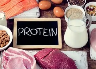
Protein is present in almost every bodily tissue and part, including muscle, bone, skin, and hair. It is the building block of hemoglobin, which delivers oxygen in your blood, and the enzymes that drive several chemical reactions.
You are made of a minimum of 10,000 unique proteins, which also maintain your unique identity. Amino acids are the more than twenty fundamental building blocks that makeup protein.
Our bodies produce amino acids from scratch or by modifying other amino acids since we cannot store them.
The nine amino acids that are considered essential are histidine, isoleucine, leucine, lysine, methionine, phenylalanine, threonine, tryptophan, and valine.
These amino acids must be obtained by diet.
History and Etymology
Antoine Fourcroy and others identified proteins as a unique class of biological molecules in the eighteenth century based on the molecules’ capacity to coagulate or flocculate when exposed to heat or acid.
Wheat gluten, blood serum albumin, fibrin, and albumin from egg whites were among the well-known examples at the time.
The Swedish chemist Jöns Jacob Berzelius termed proteins in 1838 after the Dutch chemist Gerardus Johannes Mulder had first described them. Almost all proteins shared the same empirical formula, C400H620N100O120P1S1, according to Mulder’s elemental analysis of common proteins.
He made the mistake of assuming that they were all one kind of (extremely massive) molecule. These molecules were first referred to as “proteins” by Mulder’s associate Berzelius; the word protein comes from the Greek word πρώτειoς (proteins), which means “primary,” “in the lead,” or “standing in front.”-in.
Mulder continued to identify the byproducts of protein degradation, determining the molecular weight of the amino acid leucine to be 131 Da, which is almost correct.
Other terms such as “albumins” or “albuminous materials” (Eiweisskörper, in German) were used before the term “protein”.
Because it was widely accepted that “flesh makes flesh,” early nutritional scientists like the German Carl von Voit thought that protein was the most crucial nutrient for preserving the body’s structure.
Karl Heinrich Ritthausen’s discovery of glutamic acid expanded the range of protein types that were previously recognized.
Thomas Burr Osborne of the Connecticut Agricultural Experiment Station put together a thorough analysis of the vegetable proteins.
Utilizing Liebig’s concept of the minimum in conjunction with Lafayette Mendel and laboratory rats as food sources, the biologically necessary amino acids were constituted. William Cumming Rose conveyed and carried on the job.
The work of Franz Hofmeister and Hermann Emil Fischer in 1902 helped to understand proteins as polypeptides.
It was not until James B. Sumner demonstrated in 1926 that the enzyme urease was in reality a protein that the crucial role that proteins play as enzymes in living organisms was clearly understood.
Proteins were extremely hard for early protein biochemists to examine because of the challenges associated with large-scale protein purification.
Because of this, early research concentrated on proteins that could be extracted in large amounts, such as those found in blood, egg whites, different types of toxins, and digestive and metabolic enzymes that were acquired from slaughterhouses.
The Armour Hot Dog Co. contributed to the widespread use of ribonuclease A as a prominent subject of biochemical research in the following decades by purifying one kilogram of pure bovine pancreatic ribonuclease A and making it freely available to scientists in the 1950s.
The effective prediction of regular protein secondary structures based on hydrogen bonding is attributed to Linus Pauling; William Astbury first proposed this theory in 1933.
Understanding of protein folding and structure mediated by hydrophobic interactions was aided by Walter Kauzmann’s later work on denaturation, which was partially based on earlier research by Kaj Linderstrøm-Lang.
In 1949, Frederick Sanger sequenced the first protein, insulin. With the accurate identification of the amino acid sequence of insulin, Sanger established without doubt that proteins were made up of linear polymers of amino acids as opposed to branching chains, colloids, or cyclols.
For this accomplishment, he was awarded the Nobel Prize in 1958.The development of X-ray crystallography made protein structure sequencing feasible.
Max Perutz’s hemoglobin and John Kendrew’s myoglobin protein structures were the first to be deciphered in 1958. The sequencing of complicated proteins was also made possible by the growing processing capacity and widespread use of computers.
In 1999, Roger Kornberg used high-intensity X-rays from synchrotrons to successfully sequence the incredibly complicated structure of RNA polymerase.
Large macromolecular assembly cryo-electron microscopy, or cryo-EM, has been developed since then. Instead of using crystals or X-rays, cryo-EM uses frozen protein samples and electron beams.
Because the sample is not as damaged, scientists are able to examine greater structures and gather more data.
Researchers are getting closer to the atomic-level resolution of protein structures with the aid of computational protein structure prediction of tiny protein domains.
There are currently more than 126,060 atomic-resolution protein structures in the Protein Data Bank as of 2017.
How Much Protein Do I Need?
Adults should consume no less than 0.8 grams of protein per kilogram of body weight, or a little over 7 grams for every 20 pounds of body weight, according to the National Academy of Medicine’s recommendations.
- That translates to around 50 grams of protein per day for a 140-pound person.
- That translates to around 70 grams of protein per day for a 200-pound person.
Additionally, the National Academy of Medicine establishes a broad range of permissible protein consumption, ranging from 10% to 35% of daily calories. Beyond that, there’s not a lot of reliable data regarding the optimal protein intake or the healthiest aim for calories from protein in the diet.
A Harvard study including over 130,000 men and women monitored for up to 32 years found no correlation between the percentage of calories from total protein intake and overall mortality or any particular cause of death.
But the source of the protein mattered. It’s crucial to remember that food instability prevents millions of people worldwide—especially small children—from getting adequate protein.
Malnutrition and protein shortage can have a range of serious consequences, from reduced immunity to weakening of the heart and respiratory system, and even death.
Other implications include growth failure and muscle loss. However, because there are so many foods high in protein, both plant- and animal-based, it is unusual for healthy adults in the United States and most other developed countries to have a shortfall.
Actually, a large portion of Americans consumes far too much protein, particularly from foods derived from animals.
Whether from plant or animal sources, “pure” protein most likely has identical health effects, yet the composition of amino acids can affect one’s health.
Certain foods include “complete” proteins, which are composed of all twenty-plus different types of amino acids required by the body to synthesize new proteins.
Some are insufficient because they lack one or more of the nine necessary amino acids, which our bodies are unable to produce on their own or from other amino acids.
Complete protein is typically found in meals derived from animals (meat, poultry, fish, eggs, and dairy products), whereas those derived from plants (fruits, foods including grains, seeds, nuts, and veggies) frequently lack one or more necessary amino acids.
In addition to consuming a range of plant foods high in protein that provide all the amino acids required to synthesize new protein, those who avoid animal products can also choose to include complete plant proteins such as chia seeds and quinoa.
Number of Proteins Encoded in Genomes
There may be a considerable number of genes that encode protein RNA, such as ribosomal RNAs, but overall, the number of genes and proteins encoded in a genome are nearly equal.
A few hundred to a few thousand proteins are usually encoded by viruses, a few hundred to a few thousand by bacteria and archaea, and several thousand to tens of thousands of proteins by eukaryotes (see genome size for a list of instances).
More than 150 genomic areas have been identified by genome-wide association studies (GWAS) as clearly holding variation predisposing to immune-mediated illness.
However, our capacity to identify the molecular pathways that these risk variants are disrupting will determine how we may infer disease biology from these results.
It has previously been noted that there is frequent physical interaction between genes carrying causative mutations for the same Mendelian disease.
Our objective was to assess the extent to which genes located within highly linked regions in complicated illnesses.
We construct protein–protein interaction (PPI) networks for genes within related loci using sets of loci established in GWAS for rheumatoid arthritis (RA) and Crohn’s disease (CD). We discover a wealth of physical connections between the protein products of associated genes.
We demonstrate that these networks are more densely connected than would be predicted by chance by using several permutation techniques.
In order to validate biological significance, we demonstrate that the constituents of the networks typically exhibit expression in analogous tissues pertinent to the symptoms.
In question, indicating that the network shows shared fundamental mechanisms that are impacted by risk loci.
Additionally, we demonstrate the predictive ability of the RA and CD networks by proving that proteins in these networks that are not encoded in the verified list of loci linked with the disease are significantly enriched for association with the phenotypes under investigation in extended GWAS analysis.
In order to determine whether our approach can be applied to complicated traits generally, we test it on three non-immune features.
We discover that genes at loci are linked to lipid and height Although vels organize form networks that are considerably coupled, the increased connection between Type 2 Diabetes (T2D) loci was not found to be more common than chance.
When combined, our findings provide proof that common genetic connections for many of the complex disorders under investigation involve areas encoding proteins that preferentially interact with one another physically, consistent with findings in Mendelian disease.
Classification
Although additional categories are frequently employed, sequence and structure are the primary means of classifying proteins.
The EC number system offers a functional classification scheme, particularly for enzymes. Likewise, genes and proteins are categorized by the gene ontology according to their intracellular location as well as their biological and biochemical functions.
Proteins are categorized according to their sequence similarity in terms of both evolutionary and functional similarity. Protein domains or entire proteins may be used in this, particularly in multi-domain proteins.
Protein domains, which can be joined in a variety of ways, enable the classification of proteins based on a combination of sequence, structure, and function.
About two-thirds of the 170,000 proteins in the initial study had at least one domain assigned to them, while larger proteins (those with more than 600 amino acids, for example, typically had more than five domains).
Biochemistry
The majority of proteins are composed of linear polymers composed of up to 20 distinct L-α-amino acid sequences. A common structural characteristic shared by all proteinogenic amino acids is an α-carbon that is linked to an amino group, a carboxyl group, and a variable side chain.
The only amino acid that deviates from this fundamental structure is proline, which has an odd ring attached to the N-end amine group that fixes the shape of the CO-NH amide moiety.
A protein’s three-dimensional structure and chemical reactivity are ultimately determined by the combined influence of all of its side chains, which are described in detail in the list of standard amino acids.
The side chains of the standard amino acids have a wide range of chemical structures and characteristics. Peptide bonds bind the amino acids together to form polypeptide chains.
A single amino acid is referred to as a residue once it has been connected to the protein chain; the linked sequence of carbon, nitrogen, and oxygen atoms is referred to as the main chain or protein backbone.
Two resonance forms of the peptide bond give some double-bond character and prevent rotation around its axis, resulting in approximately coplanar alpha carbons.
The local shape that the protein backbone adopts is determined by the other two dihedral angles in the peptide bond.
The protein’s sequence is written from the N-terminus to the C-terminus, from left to right. The end of the protein with a free amino group is called the N-terminus, or amino terminus, while the end of the protein with a free carboxyl group is called the C-terminus, or carboxy terminus.
There is some ambiguity and potential for meaning overlap when using the terms protein, polypeptide, and peptide.
While peptide is typically used to refer to short amino acid oligomers that frequently lack a solid 3D structure, protein is typically used to refer to the entire biological molecule in a stable shape.
However, the line that separates the two is usually not very clear and is anywhere between 20 and 30 residues.
Any single linear chain of amino acids, regardless of length, can be referred to as a polypeptide; nevertheless, the term frequently denotes the lack of a clearly defined shape.
Interactions
Proteins can interact with a wide range of substances, including DNA, lipids, carbohydrates, and other proteins.
Abundance in cells
An estimated 2 million proteins are present in each cell of an average-sized bacterium, such as Staphylococcus aureus and E. coli.
Lesser bacteria, including spirochetes and Mycoplasma, have between 50,000 and 1 million molecules in them. Eukaryotic cells, on the other hand, are larger and contain a lot more protein.
For example, it is estimated that human cells contain between one and three billion proteins, while yeast cells have roughly 50 million.
Individual protein copies can be found in quantities as low as a few molecules per cell or as high as 20 million.
Most cells do not express all of the genes that code for proteins, and the number of genes varies depending on factors including cell type and environmental stimulation.
For example, just 6,000 of the approximately 20,000 proteins encoded by the human genome are found in lymphoblastoid cells.
Synthesis
Biosynthesis
Amino acids are combined to build proteins utilizing information contained in DNA. The nucleotide sequence of the gene generating the protein specifies the specific amino acid sequence that each protein contains.
Each three-nucleotide combination in the genetic code, known as a codon, represents an amino acid. For instance, the code for methionine is AUG (adenine, uracil, and guanine).
There is some redundancy in the genetic code since there are 64 potential codons because DNA has four nucleotides.
Certain amino acids are designated by more than one codon. 1002–42 DNA-encoded genes are the first translated by proteins like RNA polymerase into pre-messenger RNA (mRNA).
The pre-mRNA also referred to as the primary transcript, is then processed by the majority of organisms through several types of post-transcriptional modification to create the mature mRNA, which the ribosome uses as a template for synthesizing proteins.
In prokaryotes, messenger RNA (mRNA) can be bound by a ribosome after it has separated from the nucleoid or used immediately upon production.
Eukaryotes, on the other hand, produce mRNA in the nucleus of the cell and move it into the cytoplasm, where protein synthesis occurs, across the nuclear membrane.
Prokaryotes can synthesize up to 20 amino acids per second at a rate higher than that of eukaryotes. Translation is the process of creating a protein from an mRNA template.
By matching each codon to its base pairing anticodon on a transfer RNA molecule, which carries the amino acid corresponding to the codon it recognizes, the mRNA is loaded onto the ribosome and read three nucleotides at a time.
Aminoacyl tRNA synthetase is the enzyme that “charges” the tRNA molecules with the appropriate amino acids.
A common phrase for the expanding polypeptide is the “nascent chain.” Proteins always undergo N-to-C-terminal biosynthesis. The total molecular mass of a produced protein can be determined by counting the amino acids it contains.
This mass is typically expressed in units of daltons, which are equivalent to atomic mass units, or in the derivative unit kilodalton (kDa).
Because higher species have more protein domains than archaea, bacteria, and eukaryotes (283, 311, 438 residues and 31, 34, 49 kDa, respectively), their average protein size increases with their evolutionary stage.
For example, yeast proteins have an average length of 466 amino acids and a mass of 53 kDa. The largest known proteins are called titins, and they are found in the muscular sarcomere.
They have a total length of approximately 27,000 amino acids and a molecular mass of nearly 3,000 kDa.
Chemical Synthesis
Peptide synthesis is a collection of techniques that can be used to chemically create short proteins.
These approaches rely on organic synthesis processes such as chemical ligation to yield high-yield peptides. Non-natural amino acids can be incorporated into polypeptide chains through chemical synthesis, for example, by attaching fluorescent probes to the side chains of amino acids.
These techniques are helpful in cell biology and laboratory biochemistry. yet usually not for use in business.
For polypeptides longer than roughly 300 amino acids, chemical synthesis is inefficient and the produced proteins might not easily adopt their natural tertiary structure.
The majority of chemical synthesis techniques work in the opposite direction of biological reactions, from the C-to-N-terminus.
Structure
The majority of proteins fold into distinct three-dimensional structures. A protein’s native conformation is the shape that it folds into when it is in its natural state.
While many proteins can fold into their native states on their own thanks to the chemical characteristics of their amino acids, others need the assistance of molecular chaperones.
The four unique components of a protein’s structure that biochemists frequently discuss are
- The sequence of amino acids is the primary structure. A polyamide is a protein.
- Secondary structure: local structures that repeat frequently and are held in place by hydrogen bonds. Turns, the β-sheet and the α-helix are the most prevalent examples. Multiple regions of distinct secondary structures can exist inside a single protein molecule due to the local nature of secondary structures.
- The overall form of a single protein molecule and the spatial arrangement of the secondary structures make up the tertiary structure. Nonlocal interactions, such as the creation of a hydrophobic core, but also salt bridges, hydrogen bonds, disulfide bonds, and even post-translational changes, are typically responsible for stabilizing tertiary structure. The terms “fold” and “tertiary structure” are frequently used interchangeably.
- The protein’s primary function is governed by its tertiary structure, Quaternary structure is made up of many protein molecules (polypeptide chains), which are referred to as protein subunits in this context and work together to produce a single protein complex.
- Quinary structure: the surface patterns of proteins that arrange the dense inside of cells. The quinary structure depends on brief but crucial interactions between macromolecules within live cells.
Proteins aren’t totally inflexible molecules. Apart from these structural levels, proteins can transition between multiple related structures during their functional actions.
These tertiary or quaternary forms are typically referred to as “conformations” in the context of these functional rearrangements, and transitions between them are known as conformational shifts.
The attachment of a substrate molecule to an enzyme’s active site, or the actual portion of a protein involved in chemical catalysis, frequently causes such alterations.
Proteins in solution can change structurally as a result of heat vibration and molecular collisions. Informally, proteins fall into three broad types: membrane, fibrous, and globular proteins.
These classes correspond to common tertiary structures. Numerous globular proteins are enzymes, and nearly all of them are soluble.
Like collagen, which makes up the majority of connective tissue, or keratin, which is a protein found in hair and nails, fibrous proteins are frequently structural.
Membrane proteins frequently function as channels or receptors that allow charged or polar substances to flow through the cell membrane.
Dehydrons are a particular type of intramolecular hydrogen bond seen in proteins that are poorly protected from water attack and so encourage dehydration.
Protein Domains
Numerous protein domains, or sections of a protein that fold into different structural units, make up a large number of proteins.
Additionally, domains typically serve as binding modules or have specialized roles, such as enzymatic activity (like kinase) or binding to proline-rich sequences in other proteins (like the SH3 domain).
A protein domain is a section of a protein’s polypeptide chain that folds independently of the rest and is self-stabilizing in molecular biology. Every domain creates a three-dimensional, compressed, folded structure.
A domain can be found in many different proteins, and many proteins are made up of many domains.
Domains are the building blocks of molecular evolution; they can be rearranged in various ways to form proteins with various functions. Generally speaking, domain lengths range from ranging in length from roughly 50 to 250 amino acids.
Metal ions or disulfide bridges stabilize the shortest domains, like zinc fingers. Domains frequently combine to form functional units, as in the case of calmodulin’s calcium-binding EF hand domain.
Domains can be “swapped” by genetic engineering between two proteins to create chimeric proteins since they are independently stable.
Sequence Motif
Proteins’ short amino acid sequences frequently serve as locations where other proteins can recognize one another.
As an example, SH3 domains generally bind to short PxxP motifs, which are composed of two prolines [P] separated by two unknown amino acids [x], albeit the precise binding specificity may depend on the surrounding amino acids.
An extensive collection of these motifs can be found in the Eukaryotic Linear Motif (ELM) database.
A sequence motif that shows up in an exon of a gene may encode the “structural motif” of the protein, which is a stereotyped component of the protein’s overall structure.
However, themes don’t always have to be connected to a unique secondary structure. “Noncoding” sequences do not result in protein translation, and nucleic acids containing these motifs do not have to conform to standard shapes (such as in the “B-form” DNA double helix).
There are motifs in the “junk”—such as satellite DNA—and regulatory sequence motifs outside of gene exons.
Some of them are thought to occasionally alter the form of nucleic acids (see RNA self-splicing, for example).
For instance, a large number of DNA binding proteins only bind DNA in its double-helical form when they have affinity for particular DNA binding sites.
By coming into contact with the major or minor groove of the double helix, they are able to identify patterns.
Short coding motifs include those that mark proteins for phosphorylation or to be delivered to certain regions of a cell; these motifs don’t seem to have any secondary structure.
Using computer-based sequence analysis techniques like BLAST, researchers look for themes within a sequence or database of sequences. These methods fall under the umbrella of bioinformatics. Also, see the consensus sequence.
Protein Topology
The arrangement of connections within the folded chain and the tangling of the backbone are referred to as a protein’s topology.
The characterization of protein topology has been done using two theoretical frameworks: circuit topology and knot theory.
Understanding protein topology broadens our understanding of diseases caused by misfolded proteins, including cancer and neuromuscular disorders, and provides new avenues for protein engineering and pharmaceutical development.
The sequence of secondary structure elements, together with their approximate orientations and relative spatial placements, make up the topology of a protein structure, which is a highly simplified representation of its fold.
A TOPS cartoon, which is a two-dimensional depiction of protein topology, can represent this information.
These cartoons are helpful for learning specific folds and establishing fold comparisons.
In this article, we provide a novel technique that produces TOPS cartoons with a far better success rate and greater robustness than earlier algorithms.
A database of protein topology cartoons that includes the majority of the database of known protein structures has been created using this approach.
Cellular Functions
The main players in the cell are thought to be proteins, which perform the functions dictated by the instructions contained in genes.
The majority of other biological molecules—aside from specific RNA subtypes—are comparatively inactive components that proteins interact with.
An Escherichia coli cell’s dry weight is composed primarily of proteins, with only 3% and 20% coming from other macromolecules like DNA and RNA, respectively.
A cell’s proteome is the collection of proteins that are expressed in that specific cell type. The primary property of proteins that also enables their wide range of functions is their capacity to firmly and precisely bind other molecules.
The binding site, which is commonly a “pocket” or depression on the molecular surface, is the area of the protein that binds another molecule.
The tertiary structure of the protein, which defines the binding site pocket, and the chemical characteristics of the side chains of the surrounding amino acids mediate this binding capacity. It is possible for protein binding to be incredibly precise and tight; for instance.
The protein known as the ribonuclease inhibitor shows sub-femtomolar dissociation constant (<10−15 M) when bound to human angiogenin, but no binding at all to its amphibian homolog increase (>1 M).
When an aminoacyl tRNA synthetase specific to the amino acid valine discriminates against the very similar side chain of the amino acid isoleucine, for example, binding can occasionally be almost completely eliminated by very small chemical changes, such as the addition of a single methyl group to a binding partner.
In addition to binding to small-molecule substrates, proteins can also interact with other proteins. Proteins can oligomerize to create fibrils when they bind selectively to other copies of the same molecule.
Structural proteins, which are made up of globular monomers that self-associate to form hard fibers, are examples of proteins that go through this process frequently.
Additionally, protein-protein interactions govern the course of the cell cycle, enzymatic activity, and the formation of massive protein complexes that perform a variety of closely related activities with a shared biological function.
Additionally, proteins have the ability to attach to and integrate into cell membranes. Binding partners’ capacity to cause conformational Protein alterations enables the creation of incredibly intricate signaling networks.
Studying the interactions between particular proteins is essential to comprehending key facets of cellular function and, eventually, the characteristics that set apart distinct cell types.
This is because interactions between proteins are reversible and largely depend on the availability of different groups of partner proteins to form aggregates that are capable of carrying out discrete sets of functions.
Enzymes
The most well-known function of proteins in a cell is as catalysts for chemical reactions, or enzymes.
Enzymes often only speed up one or a small number of chemical reactions and are quite specialized. The majority of metabolic reactions are completed by enzymes, which also work with DNA to manipulate it during activities like transcription, DNA replication, and DNA repair.
An action known as posttranslational modification occurs when certain enzymes interact with other proteins to introduce or remove chemical groups.
There are 4,000 known reactions that enzymes can catalyze.
In the instance of orotate decarboxylase, the rate acceleration provided by enzymatic catalysis can reach up to times faster than that of the uncatalyzed reaction (78 million years in the absence of the enzyme, 18 milliseconds in the presence of the enzyme).
Substrates are the molecules that enzymes bind to and work on. Even though enzymes can include hundreds of amino acids, only a small portion of the residues—usually three to four residues on average—come into contact with the substrate.
These residues are directly engaged in catalysis. The active site is the area of the enzyme that binds the substrate and houses the catalytic residues.
A class of proteins known as “dirigent proteins” controls the stereochemistry of a substance produced by another enzyme.
There are around 5,000 different types of biological reactions that enzymes can catalyze. Ribozymes, which are catalytic RNA molecules, are another type of biocatalyst.
An enzyme’s distinct three-dimensional structure determines its selectivity. Like other catalysts, enzymes work by reducing the activation energy of the reaction to speed it up.
Certain enzymes have the ability to convert substrate to product millions of times more quickly.
Orotidine 5′-phosphate decarboxylase is an extreme example; it can speed up a reaction that would normally take millions of years to complete in milliseconds.
Enzymes don’t change a reaction’s equilibrium or be consumed in chemical processes, just like any other catalyst does.
Enzymes are far more specific than the majority of other catalysts. Several substances can influence the activity of enzymes: Molecules classified as activators boost the activity of an enzyme whereas inhibitors reduce it.
Enzyme inhibitors are included in many medicinal medications and toxins. Outside of its ideal pH and temperature range, an enzyme’s activity sharply declines, and extreme heat can permanently denature many enzymes, causing them to lose their structure and catalytic capabilities.
Certain enzymes find economic application, such as in the synthesized creation of antibiotics.
Enzymes are used in certain home products to speed up chemical reactions: proteins in clothes stains caused by starch or fat are broken down by the enzymes in biological washing powders, and meat tenderizer breaks down proteins into tiny molecules that make the meat easier to chew.
Cell Signaling and Ligand Binding
The processes of cell signaling and signal transduction require several proteins. Extracellular proteins, like insulin, carry signals from the cell in which they are produced to other cells in different organs.
Others are membrane proteins that function as receptors and bind signaling molecules to trigger a cell’s metabolic reaction.
Many receptors contain an effector domain inside the cell that may undergo a conformational change that is picked up by other proteins in the cell, or they may have an exposed binding site on the cell surface.
The primary job of antibodies, which are protein components of the adaptive immune system, is to bind antigens—foreign substances—in the body and direct the body’s defenses against them.
Antibodies can be lodged in the membranes of specialized B cells called plasma cells or released into the extracellular milieu.
Antibodies do not have the same limitations on their binding affinity for substrates as do enzymes, which are constrained by the need to carry out their reaction.
The binding affinity of an antibody is incredibly strong for its target. Numerous ligand transport proteins bind specific tiny biomolecules and move them to different parts of a multicellular organism’s body.
When the ligand is present in the target tissues at low concentrations, these proteins must be able to release the ligand while maintaining a high binding affinity.
Haemoglobin, which carries oxygen from the lungs to various organs and tissues in all vertebrates and has close homologs in every biological kingdom, is the quintessential example of a ligand-binding protein.
Proteins called lectins bind sugar and are very selective to the sugar moieties they contain. Biological recognition processes involving cells and proteins are generally mediated by lectins.
Hormones and receptors are extremely selective binding proteins. Transmembrane proteins can also function as ligand transport proteins, changing the cell membrane’s permeability to ions and small molecules.
Polar or charged molecules cannot diffuse across the membrane’s hydrophobic core on their own. These chemicals can enter and exit cells through internal channels found in membrane proteins.
Potassium and sodium channels, for instance, frequently discriminate for just one of the two ions. Many ion channel proteins are designed to choose for only one specific ion.
Structural Proteins
Biological components that might otherwise be fluid are given stiffness and rigidity by structural proteins. The majority of structural proteins are fibrous proteins.
Hard or filamentous structures like hair, nails, feathers, hooves, and some animal shells contain keratin, whereas collagen and elastin are essential parts of connective tissue like cartilage.
Actin and tubulin, for instance, are globular and soluble as monomers but polymerize to create long, rigid fibers that make up the cytoskeleton, which helps the cell maintain its size and shape.
Some globular proteins can also serve structural roles. Motor proteins with the ability to produce mechanical forces, such as myosin, kinesin, and dynein, are additional proteins with structural roles.
These proteins are essential for the sperm of many multicellular organisms that reproduce sexually as well as the cellular motility of single-celled species.
They also serve crucial functions in intracellular transport and produce the forces that are applied by contracting muscles.
Protein Evolution
How proteins evolve—that is, how mutations, or more specifically, changes in amino acid sequence, can result in new structures and functions— remains a fundamental subject in molecular biology.
As many similar proteins across species demonstrate, most amino acids in proteins can be altered without affecting their activity or function (as collected in specialist databases for protein families, e.g. PFAM).
Before a gene can change freely, it may be duplicated to avoid the drastic effects of mutations. But it can also result in pseudo-genes or the total loss of gene function.
Changes to a single amino acid often have little effect, but occasionally, especially in enzymes, they can significantly alter how proteins operate.
For example, single or many mutations can alter the substrate specificity of many enzymes. Substrate promiscuity, or the ability of several enzymes to bind and process multiple substrates, facilitates changes in substrate specificity.
A mutation can cause an enzyme’s specificity to change, which can affect the enzyme’s enzymatic activity.
As a result, bacteria and other organisms are able to adapt to a variety of food sources, even artificial substrates like plastic.
It is possible to study protein structures and activity in silico, in vivo, and in vitro. To understand how a protein performs its function, in vitro.
Methods of Study
studies of purified proteins in controlled conditions are helpful.
Enzyme kinetics studies, for instance, investigate the chemical mechanism of an enzyme’s catalytic activity and its relative affinity for different potential substrate molecules.
On the other hand, data from in vivo tests can reveal details regarding the physiological function of a protein within the framework of a single cell or an entire organism.
Proteins are studied using computer techniques in silico studies.
Protein Purification
A protein needs to be separated from other cellular constituents in order to conduct an in vitro study.
Cell lysis, which involves rupturing a cell’s membrane and releasing its contents into a solution called a crude lysate, is typically the first step in this process.
Ultracentrifugation can be used to purify the resultant mixture by separating the different components of the cell into fractions that contain soluble proteins, membrane lipids and proteins, cellular organelles, and nucleic acids.
The proteins from this lysate can be concentrated by precipitation using a process called salting out.
Different kinds of proteins or proteins of interest are subsequently separated using chromatography according to characteristics including molecular weight, net charge, and binding affinity.
If the target protein’s molecular weight and isoelectric point are known, different forms of gel electrophoresis can be used to monitor the level of purification.
If the protein has distinct spectroscopic properties, spectroscopy can be used, and if the protein has enzymatic activity, enzyme tests can be used.
Furthermore, electrofocusing can be used to separate proteins based on their charge.
To get natural proteins sufficiently pure for use in lab settings, a number of purification processes could be required.
Genetic engineering is frequently used to give proteins chemical characteristics that make them easier to purify without changing their structure or activity in order to streamline this procedure.
Here, one protein terminus is joined to a “tag” made up of a particular amino acid sequence, which is frequently a string of histidine residues (a “His-tag”).
Because of this, the histidine residues in the lysate ligate the nickel in the chromatography column and adhere to it, allowing the untagged components of the lysate to flow through without obstruction.
Several tags have been created to assist scientists in removing particular proteins from intricate combinations.
The goal of protein purification is to separate one or a few proteins from a complicated mixture—typically cells, tissues, or entire organisms—by a number of procedures. To specify the role, structure, and interactions of the target protein, protein purification is essential.
Proteins and non-protein components of the mixture may be separated during the purification process, which will ultimately isolate the target protein from all other proteins.
Studying a protein of interest should ideally be kept apart from other cell components to prevent impurities from interfering with the analysis of the structure and function of the target protein.
The most difficult part of protein purification is usually separating one protein from the others. Disparities in protein size, physico-chemical characteristics, binding affinity, and biological activity are typically taken advantage of during separation phases.
One could refer to the pure outcome as a protein isolate. An essential first step in examining individual proteins and protein complexes and determining how they interact with other proteins, DNA, or RNA is protein purification.
Diverse protein purification techniques are available to fulfill specific requirements related to scale, throughput, and downstream uses.
Cellular Localization
The synthesis and location of the protein within the cell are frequently studied aspects of the research of proteins in vivo.
The cytoplasm is where many intracellular proteins are made, and the endoplasmic reticulum is where membrane-bound or secreted proteins are produced. However, it is frequently unclear how exactly proteins are targeted to particular organelles or cellular structures.
An effective method for Evaluating the localization of cells, genetic engineering is utilized to produce a chimera or fusion protein in a cell that combines a native protein of interest with a “reporter” like green fluorescent protein (GFP).
The location of the fused protein within the cell may then be clearly and effectively seen via microscopy, as illustrated in the figure across from this one.
The usage of recognized compartmental markers for areas like the ER, the Golgi, lysosomes or vacuoles, mitochondria, chloroplasts, plasma membrane, etc. Is necessary for other techniques to clarify the cellular location of proteins.
Utilizing fluorescently marked copies of Finding the location of a protein of interest becomes considerably easier with the use of these markers or antibodies to established markers.
For instance, fluorescence colocalization and location demonstration are made possible by indirect immunofluorescence.
For a similar reason, cellular compartments are labeled using fluorescent dyes. There are other options as well.
To obtain localization information, immunohistochemistry, for instance, often uses an antibody to one or more target proteins that are coupled to enzymes and produce luminous or chromogenic signals that may be compared between samples.
Cofractionation in gradients of sucrose (or other substances) using isopycnic centrifugation is another useful method.
Even though this method does not demonstrate that a protein of interest and a compartment of known density colocalize, it does raise the possibility and lends itself better to large-scale investigations.
Finally, immunoelectron microscopy is the gold standard for cellular localization. This method combines traditional electron microscopy methods with the use of an antibody against the target protein.
After the sample is ready for standard electron microscopy analysis, it is treated with an antibody against the target protein that has been coupled to a highly electro-dense substance, often gold.
This enables the localization of the target protein as well as ultrastructural features. Researchers can change the structure, cellular location, and regulatory susceptibility of proteins by using a different type of genetic engineering called site-directed mutagenesis.
Using modified tRNAs, this technology even permits the insertion of non-natural amino acids into proteins, and it may facilitate the logical design of new proteins with unique characteristics.
Eukaryotic organisms have complex cell divisions into membrane-bound compartments with diverse functions.
Extracellular space, plasma membrane, cytoplasm, nucleus, mitochondria, Golgi apparatus, endoplasmic reticulum (ER), peroxisome, vacuoles, cytoskeleton, nucleoplasm, nucleolus, nuclear matrix, and ribosomes are some of the main components of eukaryotic cells.
Additionally, when the cell is fractionated, the subcellular localizations of bacteria can be distinguished.
The cytoplasm, the cytoplasmic membrane (also known as the inner membrane in Gram-negative bacteria), the cell wall (which is often thicker in Gram-positive bacteria), and the external environment are the most frequently mentioned localizations.
The external environment is obviously not a subcellular location, but the cytoplasm, cytoplasmic membrane, and cell wall are.
Additionally, the majority of Gram-negative bacteria have periplasmic space and an outer membrane.
The majority of bacteria lack membrane-bound organelles, in contrast to eukaryotes, while there are a few exceptions (such as magnetosomes).
Proteomics
Proteomics, called after the related science of genomics, is the study of large-scale data sets containing all of the proteins that are present at any one time in a cell or cell type.
Important proteomics experimental methods include mass spectrometry, which enables quick, high-throughput protein identification, 2D electrophoresis, which separates many proteins, and peptide sequencing.
(most often after in-gel digestion), protein microarrays, which allow the detection of the relative levels of the various proteins present in a cell, and two-hybrid screening, which allows the systematic exploration of protein–protein interactions.
The total complement of biologically possible such interactions is known as the interactome. A systematic attempt to determine the structures of proteins representing every possible fold is known as structural genomics.
The extensive study of proteins is known as proteomics. Proteins have a variety of essential roles in the life of animals, including the synthesis and replication of DNA, the enzymatic digestion of food, and the development of the structural fibers of muscular tissue.
Other protein types include hormones, which carry vital messages throughout the body, and antibodies, which shield an organism from infection.
The whole collection of proteins that a system or creature produces or modifies is called its proteome. A growing amount of proteins can be identified thanks to proteomics.
This changes in response to different demands, or stresses, that an organism or cell encounters over time.
The interdisciplinary field of proteomics has benefited immensely from the genetic data of several genome studies, such as the Human Genome Project.
It is a crucial part of functional genomics and involves the investigation of proteomes from the general level of protein composition, structure, and activity.
While proteomics generally refers to the large-scale experimental investigation of proteins and proteomes, mass spectrometry and protein purification are frequently mentioned specifically.
In fact, mass spectrometry is the most effective technique for analyzing proteomes in single cells as well as massive samples made up of millions of cells.
Structure Determination
Finding a protein’s tertiary structure or the quaternary structure of its complexes might offer crucial hints about how the protein functions and can be influenced, for example in drug creation.
Proteins are too small to be viewed using a light microscope, thus structural information about them must be obtained through other means.
NMR spectroscopy and X-ray crystallography are two popular experimental techniques that can yield structural data at the atomic level.
Nonetheless, NMR studies can yield data from which a portion of the separations between pairs of The last potential conformations of a protein can be ascertained by solving a distance geometry problem, and atoms can be estimated.
A quantitative analytical technique for determining the overall protein shape as well as conformational changes brought on by contacts or other stimuli is dual polarization interferometry.
An additional laboratory method for figuring out the internal β-sheet/α-helical makeup of proteins is circular dichroism.
Lower-resolution structural data regarding very large protein complexes, including assembled viruses, can be produced by cryoelectron microscopy In some circumstances, electron crystallography can also yield high-resolution data, particularly for two-dimensional membrane protein crystals.
The Protein Data Bank (PDB), a publicly accessible repository that provides structural information about thousands of proteins in the form of Cartesian coordinates for each atom in the protein, often houses solved structures.
Protein architectures are much less known than gene sequences. Furthermore, the list of solved structures is skewed toward proteins that are easily exposed to the environmental requirements of X-ray crystallography, one of the primary techniques for determining the structure of molecules.
Specifically, globular proteins crystallize quite easily in advance of X-ray crystallography. On the other hand, membrane proteins and big protein complexes are challenging to crystallize and have a low PDB representation.
Initiatives in structural genomics have tried to address these shortcomings by methodically solving representative structures of significant fold classes.
Protein structure prediction techniques aim to give proteins whose structures have not been determined through experimentation a way to generate a believable structure.
Structure Prediction
Protein structure prediction, which is complementary to structural genomics, creates effective mathematical models of proteins in order to theoretically anticipate their molecular forms computationally rather than by observing their structures in a lab.
The existence of a “template” structure with sequence similarity to the protein being modeled is necessary for homology modeling, the most successful method of structure prediction.
The aim of structural genomics is to give enough solved structure representation to model the remaining ones.
It has been proposed that sequence alignment is the bottleneck in this process, as highly accurate models can be produced even when only distantly related template structures are provided, generated if a known “perfect” sequence alignment exists.
Numerous structure prediction techniques have been used to inform the rapidly developing subject of protein engineering, which has already resulted in the design of novel protein folds.
Additionally, ~33% of proteins in eukaryotes are categorized as intrinsically disordered because they contain extensive, unstructured portions that are still biologically functional.
Thus, predicting and analyzing protein disorder is a crucial component of characterizing protein structure. Peptide bonds hold strands of amino acids together, forming proteins.
The rotation of the main chain about the two torsion angles φ and ψ at the Cα atom allows for a wide range of conformations for this chain (see picture).
Proteins differ in their three-dimensional structure due to their conformational flexibility. Since the peptide bonds in the chain have partial charges—positive and negative charges that are separated—they are polar.
The NH group can function as a hydrogen bond donor, and the carbonyl group can function as an acceptor of hydrogen bonds.
As a result, these groups can interact inside the structure of proteins. Twenty distinct forms of L-α-amino acids, often known as proteinogenic amino acids, make up the majority of proteins.
These can be categorized based on the side chain’s chemistry, which is also crucial to the structure.
Glycine occupies a unique place in the protein structure because it has the smallest side chain—just one hydrogen atom—and can thus enhance the local flexibility of the structure.
However, cysteine has the ability to combine with another cysteine residue to produce a single cysteine, which creates a cross-link that stabilizes the entire structure.
Bioinformatics
Many computational techniques have been developed to study the evolution, structure, and function of proteins.
The abundance of genomic and proteomic data for a wide range of organisms, including the human genome, has fueled the development of such technologies.
Because it is just not practical to research every protein experimentally, only a small number are studied in lab settings, and comparable proteins are inferred using computational methods.
By aligning their sequences, homologous proteins can be effectively discovered in organisms that are distantly related. DNA and Many technologies are available to search gene sequences for specific attributes.
Restriction enzyme sites, open reading frames in nucleotide sequences, and secondary structure prediction are all possible with sequence profiling methods.
With specialized software such as ClustalW, evolutionary theories concerning the origins of current species and the genes they express may be generated, and phylogenetic trees can be built.
These days, the study of bioinformatics is essential to the analysis of proteins and genes.
In Silico Simulation of Dynamical Processes
Predicting intermolecular interactions, such as those in molecular docking, protein folding, protein–protein interaction, and chemical reactivity, is a more difficult computational task.
Molecular mechanics, and specifically molecular dynamics, are used in mathematical models to represent these dynamical processes.
Accordingly, the folding of tiny α-helical protein domains, such as the HIV accessory protein, and the villain headpiece, was found by in silico simulations, while the electronic states of rhodopsins were investigated by hybrid techniques that combined conventional molecular dynamics with quantum mechanical mathematics.
Quantum dynamics techniques go beyond classical molecular dynamics and enable realistic representation of quantum mechanical processes while simulating proteins with atomistic precision.
Examples are the hierarchical equations of motion (HEOM) methodology and the multi-layer multi-configuration time-dependent Hartree (MCTDH) method, which has been used for bacteria light-harvesting complexes and plant cryptochromes, respectively.
Because biological-scale system simulations using both quantum and classical mechanics are very computationally intensive, distributed computing projects (like the Folding@home project) help to make molecular modeling easier by taking advantage of advancements in GPU parallel processing and Monte Carlo methods.
Chemical Analysis
The amino groups found in proteins make up the majority of the overall nitrogen content of organic matter.
A common nitrogen measurement used in the examination of (waste) water, soil, food, feed, and organic matter in general is the Total Kjeldahl Nitrogen (TKN).
The Kjeldahl method is used, as its name implies. There are techniques that are more delicate.
Nutrition
All 20 of the normal amino acids can be biosynthesized by most plants and microbes, but animals, including humans, need to get some of their amino acid intake from food.
Essential amino acids are those that an organism is unable to synthesize on its own. Animals lack essential enzymes that catalyze the initial steps in the production of aspartate, lysine, methionine, and threonine, among other amino acids.
One example of such an enzyme is aspartokinase. Microorganisms can preserve energy by upregulating their biosynthetic pathways and absorbing amino acids from their surroundings if they are present in the environment.
Animals receive their amino acid intake from eating meals high in protein. After ingested, proteins are hydrolyzed by enzymes known as proteases and denatured by exposure to acid. This process breaks down ingested proteins into amino acids.
While some amino acids that are consumed are utilized in the production of proteins, others are fed into the citric acid cycle or gluconeogenesis, which produces glucose.
Because it enables the body to use its own proteins—particularly those contained in muscle—to sustain life, this utilization of protein as fuel is especially crucial during times of famine.
Protein helps animals like dogs and cats have healthy, high-quality skin by encouraging the formation of hair follicles and keratinization, which lowers the chance that skin issues may result in unpleasant odors.
In terms of gastrointestinal health, low-quality proteins also matter. When proteins enter the colon undigested, they ferment, releasing skatole, indole, and hydrogen sulfide gas.
This increases the risk of flatulence and other odorous chemicals in dogs. Animal proteins are better absorbed by dogs and cats than by plants, but inferior animal products—such as skin, feathers, and connective tissue—are not well absorbed.
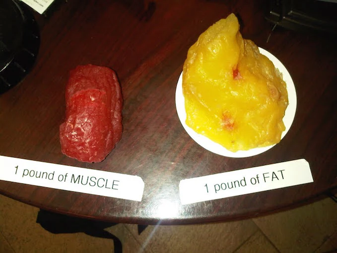
To know Best Protein To Build Muscle Click Here
FAQ
What is Protein?
Proteins are large, complex molecules that play many critical roles in the body. They do most of the work in cells and are required for the structure, function, and regulation of the body’s tissues and organs.
Which food is protein?
Animal-based foods (meat, poultry, fish, eggs, and dairy foods) tend to be good sources of complete protein, while plant-based foods (fruits, vegetables, grains, nuts, and seeds) often lack one or more essential amino acids.
What is the highest in protein?
Foods that are highest in protein typically include lean meat, poultry, and seafood. But you can also get protein from eggs, beans, nuts, seeds, and soy products.
Which food is 100% protein?
Complete List of High Protein Foods. Protein can come from both animal and plant sources. In general, foods such as beans, lentils, eggs, meats, poultry, nuts, seeds, seafood, soy products, dairy products, and whole grains are protein sources.
How much protein per day?
How much protein do I need? Most adults need around 0.75g of protein per kilo of body weight per day (for the average woman, this is 45g, or 55g for men). That’s about two portions of meat, fish, nuts, or tofu per day. As a guide, a protein portion should fit into the palm of your hand.
Do oats have protein?
Oat is considered to be a potential source of low-cost protein with good nutritional value. Oat has a unique protein composition along with a high protein content of 11–15 %.
References
Protein. (2023, December 15). In Wikipedia. https://en.wikipedia.org/wiki/Protein


159 Comments