
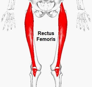
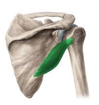
Rectus Femoris Muscle
The rectus femoris is one of the four quadriceps muscles located in the anterior (front) compartment of the thigh. It is a powerful extensor muscle that plays a crucial role in the movement of the knee and hip joints.
Introduction
The muscle closest to the skin and almost vertically oriented in the anterior thigh compartment is the rectus femoris. One of the most significant dynamic stabilizers of the knee is the quadriceps muscle complex, which includes this bipennate structure.
It is the sole quadriceps group muscle that crosses the hip, and it makes up the majority of the muscle found in the superior, anterior middle compartment of the thigh.
It is the superior and medial portion of the vastus lateralis and medialis muscles, as well as the vastus intermedius muscle.
From Latin, the word rectus means “straight.” The rectus femoris, which runs straight down the thigh, thus got its name.
Due to its two-way action, it helps the iliopsoas in hip flexion and contributes to 90° of knee flexion. This muscle spans over both the hip and the knee joint.
Anatomy
Origin
The muscle originates from two heads: the direct head, which arises from the anterior inferior iliac spine of the pelvis, while the superior acetabular ridge is the origin of the “indirect head.”
A common tendon is formed when the muscle heads join distally at an acute angle.
Insertion
Rectus Femoris together with vastus medialis, vastus lateralis, and vastus intermedius joins the quadriceps tendon to insert at the patella and tibial tuberosity (via patellar ligament).
Location
The rectus femoris is located in the middle of the anterior thigh.
Of the four quadriceps muscles, this is the only one that crosses the knee and hip joints.
Nerve supply
Rectus Femoris is innervated by the femoral nerve.
The corticospinal tract is the pathway via which cortical signals from the precentral gyrus apex descend. In an area known as the “pyramidal tract decussation,” at the level of the lower medulla, over 90% of the fibers in this nerve tract decussate. Up until they get to the lower motor neurons beneath the upper thoracic spinal cord, these fibers continue to descend on the contralateral side as the lateral corticospinal tract. Although they do not innervate structures below the upper thoracic spinal cord, the remaining 10% of the fibers that do not decussate continue as the ventral corticospinal tract.
Lower motor neurons in the lateral corticospinal tract connect with its fibers at the L4 level to reach the quadriceps femoris muscle. The lumbar plexus and the L4 anterior rami are the targets of the neuronal signal that travels from there.
Contributions from L2 and L3 fibers help build the femoral nerve, which originates from the L4 anterior rami. The rectus femoris is one of the four quadriceps femoris components that are innervated by the femoral nerve.
Bloody supply
Through the lateral femoral circumflex branch, blood from the femoral artery reaches the quadriceps muscle. The venous drainage of the muscle occurs via the femoral vein and its branches.
The superficial inguinal nodes receive the lymphatic veins from the anterior thigh. The arteries then rise to the lateral aortic and external iliac lymph nodes.
Function
Hip flexion
Hip flexion is caused by the Rectus Femoris and Iliopsoas, particularly when the knee is flexed.
It collaborates with the iliopsoas during the “toe off” phase of gait as a hip flexor.
Knee extension
- It helps with knee extension together with other muscles that are a part of the Quadriceps femoris.
- Rectus femoris is a muscle in the quadriceps group that functions as an extensor of the knee during the terminal swing phase. It produces the force required for loading (foot flat phase) during the stance phase.
- When moving from a position of knee extension and hip flexion, or when combining hip hyper-extension with knee flexion, the rectus femoris is more effective. booting a soccer ball, for instance.
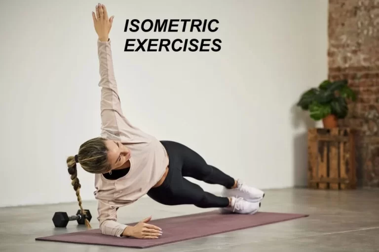
Exercise of Rectus Femoris Muscle
Strengthening Exercise
Exercises involving knee extension and hip flexion are useful for targeting and strengthening the rectus femoris muscle specifically. The following exercises target the rectus femoris:
Leg Extensions:
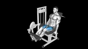
- Leg extensions are a popular machine-based workout in fitness centers. Position yourself on the leg extension machine, place your ankles beneath the roller pad, and push against the resistance to stretch your legs.
- Throughout the workout, make sure your movements are deliberate and free of sudden or jerky movements.
- Adapt the machine’s settings to your comfort level and degree of fitness.
Squats:
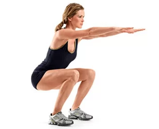
- Squats work the rectus femoris in addition to the glutes, hamstrings, and quadriceps collectively.
- Place your feet shoulder-width apart, bend your hips and knees to lower your body, and then take a step back up to your starting position.
- To guarantee optimal activation of the quadriceps, especially the rectus femoris, pay attention to your form.
Step-Ups:
- You can perform step-ups on a stable bench or platform. Elevate your body with a single footstep and then descend it again.
- With the other leg, repeat the motion.
- The rectus femoris is worked during this exercise when the knee is extended and the body is raised.
Stretching Exercise
Standing Quadriceps Stretch:
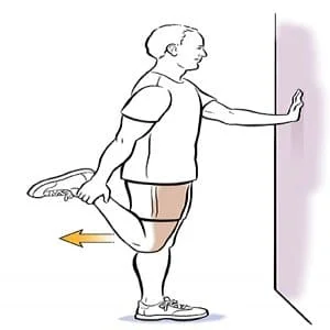
- While standing on one leg, turn your other heel toward the back end.
- Gently move your heel toward your buttocks while holding your ankle with your hand.
- Try for an erect posture and keep your knees close together.
- After 15 to 30 seconds of holding the stretch, switch legs.
Kneeling Hip Flexor Stretch:
- With one leg on the floor and the other foot in front with the knee bent at a 90-degree angle, begin in the lunge posture.
- Place your weight forward and notice a stretch in the back leg’s hip.
- After 15 to 30 seconds of holding the stretch, switch legs.
Embryology
The fifth week of development marks the emergence of the lower limb bud. The bud comprises the ectoderm, endoderm, and mesoderm, the three basic germ layers, and arises laterally from the L2 to S2 spinal segments. The mesoderm gives birth to the myotomic parts of somites, which in turn give rise to the rectus femoris muscle tissue.
The lateral plate somatic mesoderm is where the skeletal components that support the lower limb originate in the meantime. The knee is brought to the anterior aspect of the fetus during development when the lower limb rotates 90 degrees medially about the longitudinal axis.
Anatomical Variations
From the rectus femoris, accessory muscles can originate. One of these variations is a direct vastus lateralis inserted into the acetabulum via a muscle slip. With the acetabular origin absent and the lone tendon originating from the lower anterior iliac spine, the proximal origin of the rectus femoris muscle may also have just one head rather than two.
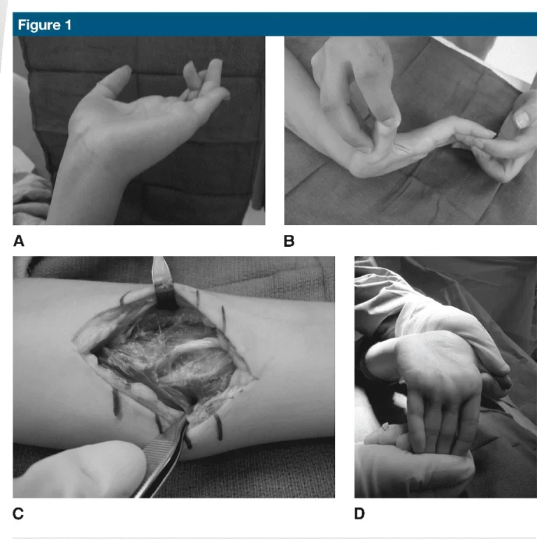
Surgical Importance
A tearing sensation and sudden onset of pain are the symptoms of acute rectus femoris muscle injury. Subacute injuries, on the other hand, typically start out as mild to moderate pain that gets worse when you run. This muscle’s injuries can be diagnosed and tracked with the use of MRIs and ultrasonography.
Although they are rare, proximal rectus femoris tears are linked to sports like soccer and other activities that need a lot of running and kicking. Involved parties are more frequently the direct head than the indirect head. Avulsions totale involve both. Although they are prone to recurrence, acute single-head proximal rectus femoris tears can be treated conservatively. Total avulsion and recurrent single-head tears should be treated surgically. Patients can recover to their pre-injury functional levels following surgery for both disorders, which have good prognoses.
Ruptures of the quadriceps tendon, affecting the entire quadriceps muscle, are more frequently linked to distal rectus femoris tears. Tears shed on their own are rare. Disabling quadriceps tendon injuries are typically treated surgically.
In order to repair an avulsed tendon, the tendon must be stitched into sutures that are fixed to the bone origin. If there is enough tissue to allow for surgical stitching, the broke tendon may be stitched to the tendon stump. In cases where tendon edges are not suited for repair or cannot be sufficiently mobilized, tendon allografts may be used for reconstruction.
Clinical significance
Rectus femoris strain
Hip flexor strain, also known as rectus femoris strain, is an injury that typically occurs in the muscle or at the tendon that connects to the patella. Usually a partial tear, however a full rip is possible. The injury is frequently sustained in sports like football or soccer and is brought on by a strong movement connected to running, jumping, or kicking.
The rectus femoris spans both the hip and the knee, making it vulnerable to damage. The incapacity to fully contract the rectus femoris with a tear is one of the symptoms, along with swelling and bruises, and an abrupt, acute pain in the front of the hip or in the groin.
Avulsion fracture
Avulsion fractures are caused by the Rectus femoris tendon, which can cause a portion of the anterior inferior iliac spine of the hip (AIIS) to avulse. This results from a muscular contraction that is so strong that it exerts more force than the force holding the bone together. Young athletes may be impacted by this well-known, atypical sports injury.
7 Comments