The Triceps Brachii muscle: Anatomy, Origin, Insertion, Function | Triceps muscle
The triceps muscle is the only muscle in the posterior compartment of the arm. The triceps brachii or more commonly known as triceps is a large, strong, fleshy muscle that kind of has a horseshoe experience when flaunted.
The muscle gets its name because of the presence of three heads. The word ceps mean head and tri means three. The three head includes a long head, a head on the outer aspect of the arm, and one on the inner aspect of the arm.
All three heads though arise from elsewhere, but end together at a common point.
Origin of the Triceps Muscle
All the three heads of the triceps brachii muscle have varied origins, namely,
- The long head arises from the infraglenoid tubercule of the humerus.
- The lateral head originates from an oblique ridge above the spiral groove on the upper part of the posterior surface of the shaft of the humerus.
- The medial head arises from the lower part of the shaft of the humerus, below the spinal groove.
The medial head of the triceps brachii muscle actually runs deeper than the long and lateral heads.
The term medial and lateral is decided with respect to the positions of the head to that of the radial groove.
Insertion of the Triceps Muscle
As mentioned earlier the triceps muscle might have a different origin, but the insertion is the same. All three heads collaborate and form a single tendon. This single tendon inserts itself into the upper portion of the ulna, in the bony prominence known as the olecranon process on the posterior side.
Nerve Supply of the Triceps Muscle
The triceps muscle is innervated by the radial nerve, which is the branch of brachial plexuses with the nerve roots C6, C7, and C8.
The recent studies on the cadaver found that the inner and outer head of the triceps muscle is also supplied by the ulnar nerve.
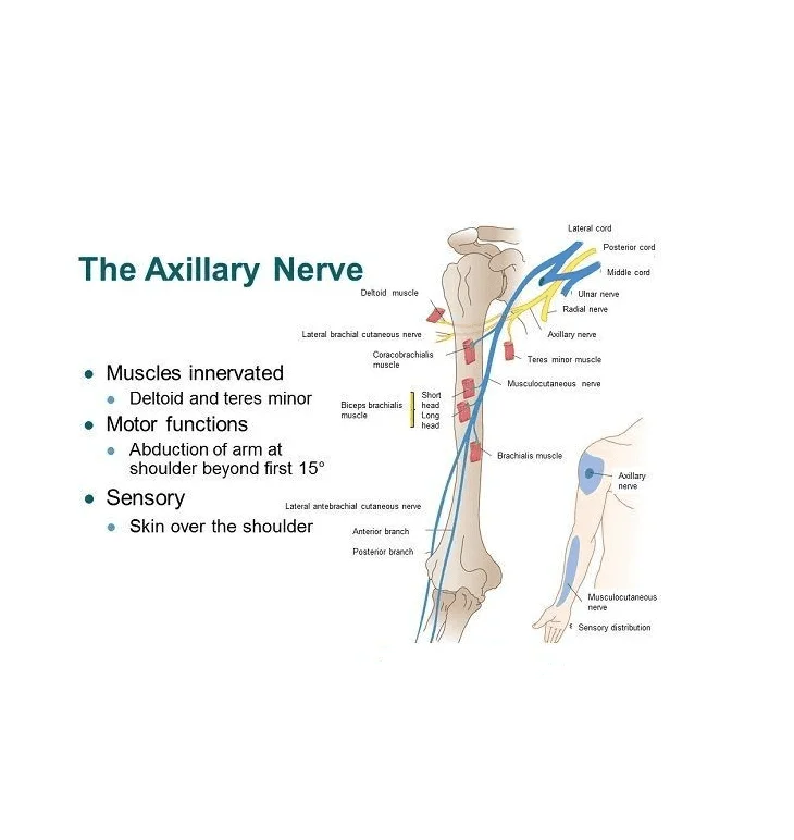
Also, the long head is supplied by the axillary nerve.
Blood Supply of the Triceps Muscle
The triceps muscle is supplied by the deep brachial artery along with the ulnar collateral arteries.
The venous drainage is done by the brachial vein which runs deeper along the brachial vein.
The function of the The Triceps Brachii muscle
- The chief function of the triceps is the extension of the forearm from the elbow joint.
- Along with this, the triceps also works as a stabilizer of the elbow joint while performing fine movements.
- Since the long head of the biceps originates from the infaglenoid tubercule, it plays a role in stabilizing the head of the humerus while the arm is outstretched sideways. It also plays a role in assisting with the extension and adduction of the arm.
- The medial head plays a role in keeping the forearm straight while rotated outwards or inwards.
- The lateral head is regarded as the strongest of all the heads and plays the same role as that of the medial head.
Palpation of the Triceps Muscle
To feel all the three heads of the triceps muscle, have the subject sitting comfortably on a high stool with the examiner standing facing the back of the subject.
Medial Head
To palpate the medial head, we use a landmark such as the medial epicondyle of the humerus. The examiner places their middle, ring and little fingers just above the medial epicondyle of the humerus. The subject is then asked to straighten the elbow while pushing the seat downwards as if they are pulling themselves up. The contraction of the muscle which you feel under your fingers while performing this movement is the medial head.
Long Head
To Palpate the long head, the examiner moves the fingers backwards from the medial condyle of the humerus to the axilla. Once the finger is in position, the examiner asks the subject to repeat the same motion. The muscle exertion felt known is that of the long head of the triceps.
Lateral Head
To palpate the lateral head, the examiner places place their fingers posteriorly and in the outward direction in the middle of the shaft of the humerus. The subject once again is asked to repeat the same motion and know the strength of the lateral head is felt.
Clinical Importance of the Triceps Muscle
The triceps is a relatively strong muscles. It is used in everyday activities in conjugation with the muscle of the anterior compartment of the arm to throw things, to straighten out the elbow while doing household chores or daily day-to-day activities, while pulling yourself up from a short sitting or long sitting position, etc.
The Triceps muscle is also used to test the triceps jerk or triceps reflex, which ultimately gives the scorecard on the C6 and C7 nerve roots, majorly on C7.
How to test the Triceps Reflex
- To test the triceps reflex, have the patient either sit or stand comfortably.
- If sitting, the patient should be in high sitting with no back support.
- The therapist or medical practitioner stands sideways to the patient, facing the arm to be tested.
- The patient is asked to raise their arm outwards perpendicular to the ground at a 90-degree angle.
- The forearm is asked to be left loose, making the elbow slightly bent.
- The examiner then, without the knowledge of the patient taps the triceps tendon with a reflex hammer.
- A positive result would be the extension of the elbow due to involuntary contraction of the triceps brachii muscle.
The reflex is graded on a scale from 0-4, with,
- 0 – No reaction
- 1 – Diminished reaction
- 2 – Normal reaction
- 3 – Exaggerated reaction
- 4 – Highly exaggerated reaction
The first two are seen in case of damage to the radial nerve either due to peripheral nerve injuries or trauma.
The last two are seen in the case of spasticity or rigidity which usually follows in the case of stroke or head trauma.
Associated Conditions of the Triceps Muscle
Triceps Tendinitis
- Triceps tendinitis is a soft tissue injury which may occur due to an overload on the muscle.
- The common signs and symptoms are a long time of pain behind the elbow joint which worsens while trying to straighten out the elbow.
- Is commonly found in males between 30-40 years of age.
- Is predominant in athletes involved in professional weight lifting, in games requiring repetitive throwing and catching, football players, motorcycle riders, Bunge jumpers, rugby players, etc.
- It can also occur when a person sustains an injury when falling on the outstretched hand or in people with less flexibility and strength who lifts heavy objects suddenly.
- The condition is generally treated with conservative measures such as resting the muscle, applying ice over the tender point to reduce pain and swelling, elevating the arm in order to dissipate the swelling and over the counter painkillers to reduce the pain little bit.
Tendon Ruptures
- Since the triceps is such a strong muscle, it is very rare for it to undergo tendon rupture.
- If so, they are more common in anabolic steroid users.
- Though uncommon distal triceps tendon rupture may occur when a person falls on an outstretched hand or someone directly hits the triceps tendon.
- The rupture occurs at the tendon-bone junction usually when the elbow is being extended out as in eccentric contraction.
- The typical patient presentation for triceps tendon rupture includes a painful popping sensation followed by pain and swelling over the back of the elbow. Along with the incapability of straightening the elbow against force.
- In case of acute partial rupture, a conservative method with minimally invasive techniques might be enough. But in case of an acute complete rupture, a full-on surgical invasion followed by 2-3 weeks of rehabilitation phase might be required.
- In case of chronic rupture, the surgery is often followed by remodeling techniques before completing with rehabilitation period.
Triceps muscle tightness:
In the triceps muscles tightness, you may feel heaviness or stiffness at the backside of your arm during elbow movement. The triceps muscle tightness refers to a tight stiff feeling in the back of your arm muscles and also you feel discomfort while bending or extending elbow joints.
Treatment Options for the Triceps Muscle
Triceps tendinopathies and partial tendon tears with sufficient strength are taken care of conservatively with rest, ice, immobilization, nonsteroidal anti-inflammatory drugs (NSAIDs), and physiotherapy treatment.
The physiotherapy treatment includes a progression from gravity independent pain-free exercises to gravity-dependent pain-free exercises, followed by a period of strength training. The strength training includes isometric contractions followed by closed chain exercises and then onto the open chain exercises. Once the patient is able to do open chain exercises, weighted exercise with dumbbells or bodyweight is started. It is advised to the patient to carry out the strength training for a minimum period of three months and then incorporate it into their lifestyle at least twice or thrice a week.
The surgery is considered, if,
- The conservative treatment approach fails even after religiously being followed for 6-months,
- The patient comes in with a neurovascular deficit or strength deficit at the time of injury.

15 Comments