
Azygos vein
The azygos vein is positioned on the back right side of your chest. It aids in transporting blood from your chest and abdomen to your heart, where it is reoxygenated. The azygos vein is a section of the azygos venous system.
These veins collaborate with other veins in your body to circulate blood and provide oxygen and nutrients to tissues.
Overview
The azygos vein is a vein that drains into the superior vena cava from the right side of the thoracic spinal column. It joins the superior and inferior vena cava systems and can provide an alternate conduit for blood to the right atrium when either vena cava is blocked.
Structure
The azygos vein transfers deoxygenated blood from the thorax and abdomen’s posterior walls to the superior vena cava.
It is formed at the level of the 12th thoracic vertebra by the union of the ascending lumbar veins with the right subcostal veins, ascending to the right of the descending aorta and thoracic duct, passing behind the right crus of the diaphragm, anterior to the vertebral bodies of T12 to T5, and right posterior intercostal arteries.
It joins the superior vena cava at the level of the T4 vertebrae by arching over the root of the right lung from behind to front. The trachea and esophagus are positioned medially to the azygous vein arch.
The “arch of the azygos vein” (arcus venae azygos) is an anatomic landmark. The arch can be shifted laterally in 1-2% of the population, resulting in a pleural septum separating an azygos lobe from the upper lobe of the right lung.
The azygos vein’s origin and anatomical path are highly variable. There is usually a single azygos vein on the right side of the body. However, the azygos vein may be found in the midline on occasion, or two independent veins may be present, as in early embryonic development. It drains thoracic, bronchial, and gonadal veins in some rare variants. The vein is named for the fact that there is no proportionally corresponding vein on the left side of the body.
Function
The azygos vein transports blood to your heart from the back of your chest and belly. This vein delivers blood upward via your diaphragm and the mediastinum, the region between the membranes that protect your lungs (pleural sacs).
The function of the azygos vein is to discharge deoxygenated blood into one of your body’s major veins (the superior vena cava). The superior vena cava transports blood to your heart’s right upper chamber (atrium) to be reoxygenated.
What is the azygos vein system?
The azygos venous system is a network of veins that assist blood circulation. This system directs blood flow between your two major veins, the inferior and superior vena cava. If the inferior or superior vena cava becomes obstructed, this channel (anastomosis) allows blood to return to the heart.
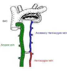
The azygos venous system consists of two smaller veins, or tributaries, to the azygos vein:
The accessory hemiazygos vein begins in the spaces between your middle ribs (intercostal spaces). It drains into the azygos vein from the top left side of your spine. The accessory hemiazygos vein is fed by the left bronchial vein and a vein in the digestive tract (esophagus).
Hemiazygos vein: This vein starts where tributaries of other veins meet. Mediastinal veins, lumbar veins in your spine, and subcostal veins that run down the bottom of your ribs are examples of tributaries. The hemiazygos vein drains many intercostal veins and the left subcostal vein before joining the azygos vein.
Etymology
The Greek root zyg means “a pair.” ‘A-‘ denotes no. As a result, azygos signifies “unpaired.” There is only one azygos vein in the body, which is predominantly on the right side. While the hemiazygos vein and its accessory are located on the left side of the body, they are regarded as tributaries of the azygos vein rather than its left-side counterpart.
Furthermore, azygos vein injury can occur during thoracic procedures (surgeries performed inside the chest cavity). Implanted device complications, such as pacemakers, might also damage your azygos vein.
Can you explain the differences between the azygos vein and other veins?
Veins typically form in pairs. There is normally a vein on the left and a vein on the right side of your body. However, the structure of the azygos vein differs. The majority of people only have one azygos vein on the right side of their body. During fetal development, two azygos veins may arise.
Anatomy
Location
The azygos venous system drains the viscera within the mediastinum, as well as the back and thoracoabdominal walls, and is positioned on either side of the vertebral column. The azygos vein and its two major tributaries, the hemiazygos vein and the auxiliary hemiazygos vein, comprise this system.
The azygos vein typically develops from the lumbar azygos vein or from the posterior side of the inferior vena cava near the level of the renal veins. It flows through the diaphragm, into the mediastinum, and then into the superior vena cava. There are numerous anastomoses between the azygos venous system and the inferior vena cava and spinal venous plexuses. This makes it a crucial link between the two venae cavae, allowing for an additional drainage route in the event of inferior vena cava occlusion.
Anatomy course
The azygos vein normally arises from the inferior vena cava’s posterior portion, near the level of the renal veins. It ascends to the level of T4 within the posterior mediastinum before arching above the right pulmonary hilum. Just before it pierces the pericardium, it drains into the superior vena cava.
Anatomical relations
The azygos vein’s connections to other structures in the mediastinum can be significant in therapeutic practice. The azygos vein is followed by the following structures:
The anterior longitudinal ligament is a type of ligament.
arteries of the right posterior intercostal space
T4-T12 body parts
The azygos vein is located in the posterior thorax, posterior to the recess of the right pleural sac and the oesophagus.
Medial to the vein can be found:
Thoracic duct Aorta Oesophageal Trachea Right vagus nerve
Laterally to the azygos vein are the following structures:
The larger splanchnic nerve on the right side
Pleura of the Lung
The azygos vein is likewise near to the descending thoracic aorta’s right posterolateral side. Pulsations in the aorta may enhance venous return in the azygos and hemizygous veins.
Tributaries
The hemiazygos vein and the supplementary hemiazygos vein are two tributaries of the azygos vein.
The hemiazygos vein is frequently linked to the left renal vein. The oesophageal and mediastinal tributaries, the common trunk of the left ascending lumbar vein and left subcostal vein, and the lowest three posterior intercostal veins all contribute to its formation. At T8, it ascends anterior to the vertebral column before passing posterior to the aorta, esophagus, and thoracic duct.
Veins from the fourth to eighth intercostal spaces, as well as the left bronchial veins, comprise the supplementary hemiazygos vein. It falls to the left of the spinal column before connecting with the azygos vein.
When it connects with the hemiazygos vein, their shared trunk drains into the azygos vein.
Anatomical variations
The azygos vein’s origin and anatomical path are highly variable. There is usually a single azygos vein on the right side of the body. However, the azygos vein may be found in the midline on occasion, or two independent veins may be present, as in early embryonic development.
The azygos vein usually begins at the lumbar vertebrae, but it can begin further up. The azygos vein can ascend anterior to the lumbar vertebrae and pass behind the right crus of the diaphragm, or it can cross the aortic hiatus to the right of the cisterna chyli, a dilated lymph sac.
The azygos vein joins the common trunk of the right ascending lumbar vein and the right subcostal vein prior to the body of T12. If the lumbar segment is not present, this trunk may create the azygos vein.
Clinical Importance
Diseases and disorders affecting the azygos vein can have major consequences due to its location in the body and function as part of the circulatory system. Everything from endemic illnesses to circulation or cardiac problems to physical damage might affect this vein.
Laceration
The azygos vein can be severed or burst as a result of a fall or a car accident. This can result in a pneumothorax, which is a collection of blood in the pleural space (the gap between the membranes that cover the lungs). These are spotted by X-ray and necessitate a thoracotomy, a surgical operation in which blood is evacuated through a chest incision.
Aneurysm
Heart failure, internal bleeding, excessive blood pressure in the portal vein, and blockage in the inferior vena cava can all cause aneurysms, which are weakening and bulging of the arterial walls. Although this is usually asymptomatic, surgery may be required if there is a risk of rupture or blood clots reaching the lungs (pulmonary embolism).
Syndrome of the Superior Vena Cava
When blood flow from the superior vena cava to the right atrium of the heart is impeded, not enough blood is drained from the head and neck. This can cause blood flow to reverse, or travel away from the heart, resulting in breathing difficulties, lightheadedness, and edema.
To treat superior vena cava syndrome, computerized tomography (CT) imaging is utilized for diagnosis, and surgery or medicines are used.
Fibrosing Mediastinitis
Scar tissue forms in the mediastinum of this extremely rare illness, blocking blood flow. Fibrosing mediastinitis is most usually caused by a fungal or bacterial infection, although it is also linked to autoimmune illnesses such as Behcet’s disease and other conditions.3
X-ray imaging can reveal fibrosing mediastinitis growth. Treatments for this largely asymptomatic illness include, among other things, surgery to remove scar tissue and medicines.
Syndrome of the Inferior Vena Cava
When the inferior vena cava becomes clogged, new vessels emerge and the azygos can enlarge. As a result, lesions can form in the vein, allowing inadequate blood to return to the heart. Symptoms include breathing difficulties, edema, cognitive impairments, and heart arrhythmia.
After imaging to determine the cause of the blockages, surgery or blood-thinning drugs may be utilized to treat the disease.
Complications of Medical Devices
Because the azygos vein may be a location of catheter injection (as in cardiac catheterization) or as a result of pacemakers put near the heart, issues such as pneumothorax or azygos vein laceration might occur. Implanted devices or medical equipment left in the area can also result in abnormal tissue growths (fistulas) and blood clotting.
The key to treatment, as with other illnesses, is to eliminate the cause of the problem. X-rays or CT scans may be used, and surgery, among other options, may be required.
What are the most common conditions that affect the azygos vein?
Changes in the form of your azygos vein might occur as a result of changes in blood flow caused by high blood pressure or something blocking the vein (obstruction). Your azygos vein may engorge or grow larger.
Injury can potentially cause damage to the azygos vein. Car accidents or other severe contacts with your chest, such as a fall or a heavy impact during sports, can cause cuts (lacerations) or ruptures. These alterations may cause your azygos vein blood pressure to become too high or too low.
What tests do doctors perform to check for azygos vein problems?
Your doctor may advise you to get an imaging test to assess the function of your azygos veins:
Arteriogram or venogram, in which a dye is injected and X-rays are taken to assess blood flow.
X-ray of the chest.
The CT scan.
MRI.
How do medical professionals treat azygos vein damage?
When a vein is injured or damaged, healthcare personnel may perform operations to release pressure and allow the vein to drain. To assist blood drain from the space between your lungs (pleural cavity), providers may make an incision in your chest (thoracotomy).
When your azygos vein does not form properly before birth, you may have a vein abnormality (congenital defect). Some people with azygos vein congenital abnormalities may be more vulnerable to:
- Conditions affecting your spleen.
- Congenital cardiac disease.
What should I do to maintain the health of my veins?
These lifestyle choices can assist in maintaining the health of your circulatory system, particularly your veins:
- Consume nutritious foods.
- Exercise on a regular basis.
- Drink plenty of water.
- Control your blood pressure.
- Stop smoking.
FAQ
What are the azygos vein’s relationships?
The azygos vein is a vein that drains into the superior vena cava from the right side of the thoracic spinal column. It joins the superior and inferior vena cava systems and can provide an alternate conduit for blood to the right atrium when either vena cava is blocked.
What are the two azygos vein tributaries?
The hemiazygos vein and the supplementary hemiazygos vein are two tributaries of the azygos vein. The hemiazygos vein is frequently linked to the left renal vein.
What is the meaning of azygos vein?
“Azygos” means “without a pair” in Greek, which explains the absence of a similar vein on the left side of the spinal column in the thorax.
What distinguishes the azygos vein?
The Azygos vein drains venous blood from the top portion of the posterior abdominal wall and the thoracic wall. It is particularly essential since it connects the superior vena cava to the inferior vena cava and has few or no valves.
What is the azygos vein absence?
The absence of the azygos vein is a very rare congenital venous defect, however we should consider it when the chest radiograph displays the “aortic nipple” on the lateral side of the aortic arch instead of the azygos arch shadow.
References
- Gurarie, M. (2021, September 24). The anatomy of the Azygos vein. Verywell Health. https://www.verywellhealth.com/azygos-vein-anatomy-function-5191070
- Azygos vein – wikidoc. (n.d.). https://www.wikidoc.org/index.php/Azygos_vein
- MBChB, C. O. B. (2023, October 30). Azygos vein. Kenhub. https://www.kenhub.com/en/library/anatomy/azygos-vein
- Professional, C. C. M. (n.d.-a). Azygos vein. Cleveland Clinic. https://my.clevelandclinic.org/health/body/24036-azygos-vein
- Wikipedia contributors. (2023a, September 20). Azygos vein. Wikipedia. https://en.wikipedia.org/wiki/Azygos_vein
One Comment