
Biceps Femoris Muscle
Biceps Femoris Muscle Anatomy
Biceps femoris is a long muscle of the posterior aspect of the thigh. Together with the semitendinosus and semimembranosus muscles, it makes the group of muscles commonly known as the hamstrings.
Biceps femoris muscle runs from the ischial tuberosity, all the way to the proximal part of the fibula. In doing so the muscle crosses two joints; the hip joint and the knee joint. Acting simultaneously on these joints, biceps femoris has many important functions; flexion and external rotation at the knee joint, extension and external rotation in the hip joint.
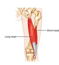
As its name suggests, this muscle consists of two heads, one lying deep to the other. Each head has a different origin and innervation but they share the same insertion.
Origin and insertion:
Biceps femoris is the most lateral hamstring muscle located in the posterior thigh. As the name suggests, this muscle has two heads; long and short. These have different origins but share one common insertion.
The long head of biceps femoris muscle originates from the medial facet (inferomedial impression) of ischial tuberosity, medial to the origin of semimembranosus and superior to the origin of adductor magnus muscle. It is important to highlight that this is a shared tendon with both the semitendinosus muscle and sacrotuberous ligament. The tendons of biceps femoris and semitendinosus run together for a distance before separating into two distinct muscles.
The short head originates quite distally from the long head, arising from the lateral lip of the inferior third of the linea aspera and supracondylar ridge of femur. This origin lies medially to vastus lateralis muscle and laterally to adductor magnus muscle.
Near the muscle’s insertion, the long head of biceps femoris continues as an aponeurosis. The muscle fibers from the short head join the aponeurotic sheet, comprising the round common tendon that inserts to the lateral aspect of the head of the fibula. Just prior to insertion, the tendon splits into two slips, passing on either side of the fibular collateral ligament. A few fibers attach to the ligament, a few others spread to the adjacent tibial condyle. When the knee is flexed, the biceps femoris tendon can be easily palpated in the posterolateral aspect of the knee.
Relations:
For its largest part, the biceps femoris runs superficially in the posterolateral thigh, sitting deep only to skin, fat and fascial layers. The exception to this is at its superior aspect, where it is only to skin, fat and fascial layers. The exception to this is at its superior aspect, where it is covered by the gluteus maximus muscle. While descending from the pelvis into the posterior thigh region, biceps femoris passes on top of semimembranosus muscle, adductor magnus muscle, and the lateral head of gastrocnemius muscle.
Along the way, it is also located superficial to the sciatic nerve, providing protection for it. The sciatic nerve gives its terminal branch (common fibular nerve) near the insertion of the biceps femoris. The nerve travels briefly along the medial border of biceps femoris, adhering to the tendon. This is an important clinical relation when considering injury or performing surgery procedures in this region.
Blood supply:
The majority of the blood supply for biceps femoris comes from branches of the deep femoral artery (perforating arteries and medial circumflex femoral artery). Additional supply comes from the inferior gluteal and superior lateral genicular arteries.
Function:
In general, the biceps femoris muscle acts on both the knee and hip joints. Although, due to its attachments, the short head of this muscle acts only on the knee joint while the long head acts on both.
When acting on the hip joint, biceps femoris produces the movement of hip extension. This action is the strongest when the trunk is bent forward and is to be brought in an upright position. The biceps femoris is also sometimes described as assisting with external rotation (when the hip joint is in an extended position). When acting at the knee joint, the most prominent action of the biceps femoris muscle is flexion of the leg. This occurs when the lower limb is in an anatomical position. In contrast, when the knee is semiflexed, biceps femoris acts to produce external rotation of the leg at the knee.
Together with other hamstring muscles, biceps femoris stabilizes pelvis, especially during the forward flexion of the trunk occurs. Therefore it has a major role in the gait cycle. Its most important antagonist is the quadriceps femoris muscle which is nearly three times stronger than the hamstrings.
To expand your knowledge check out our learning materials about the muscles of the hip and thigh. You can expand your knowledge with the video tutorials and actively test yourself with our integrated quizzes.
Palpation:
Position the client in prone lying with the knee in slight flexion
Starting distally locate the lateral proximal border of the popliteal fossa to locate the insertion of the tendon
Palpate the hamstrings laterally to locate the biceps femoris
Move palm toward the ischial tuberosity (proximally) to locate the muscle belly of the biceps femoris
Length Tension Testing / Stretching
With one hand, palpate the patient’s ASIS and iliac crest with your thumb and index finger
With the other hand, support the patient’s leg just above the ankle
Raise the patient’s leg into hip flexion keeping the knee extended, and add internal rotation of the tibia to bias the biceps femoris
Flexibility of the biceps femoris is exhausted when the innominate starts to rotate posteriorly
Trigger Point Referral Pattern
Pain referred from TrPs in the lower half of the biceps femoris (long or short head) focuses on the back of the knee and may extend up the posterolateral area of the thigh as far as the crease of the buttock.
Treatment:
Stretching:
Position of patient is side lying and support is provided to lower leg with hip and knee flexed
Hold the upper leg and bring into abduction
During abduction, try to support the leg against your body, so that the patient can extend the knee whilst pressing the hip into flexion
With one hand, hold the upper leg and with thenar of your other hand try to provide stretch to the muscle away from the site of insertion.
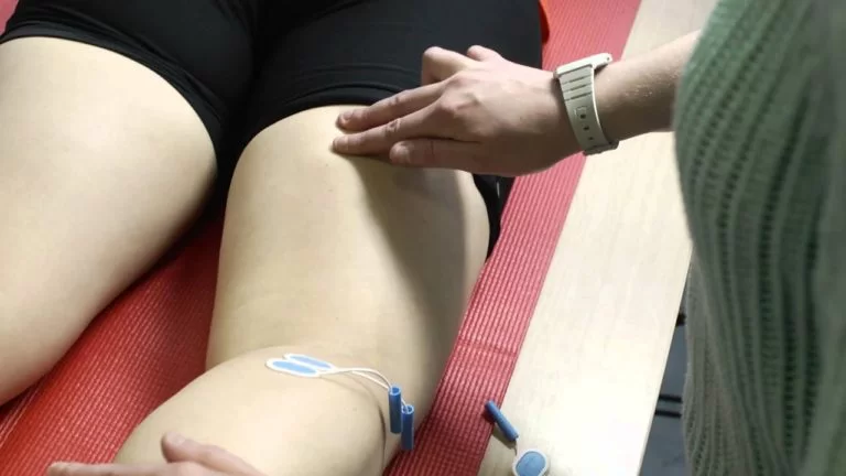
Quadriceps foam rolling:
The present study offers findings that foam rolling to quadriceps alone is effective in decreasing the activation of biceps femoris muscle. Foam rolling on patients muscle can lead to alteration in range of motion, performance and muscular co-activation around the particular joint. According to the researcher, when there is simultaneous contraction of the agonist and antagonist muscle, it helps in providing stability to the knee joint. However, if any disturbance occurs to the stability of the joint, the knee particularly goes for injury. Furthermore, the study portrays about ACL injury, which is more common due to alteration in muscular co-activation, and further research is required to identify the efficacy of foam rolling on activation of muscle around the knee joint.
Soft tissue mobilization:
Performing soft tissue mobilization via IASTM to gluteals, IT band, Quadriceps, adductors, and hamstrings, twice per week for 3 weeks helps in improving the knee extension range of motion among patients suffering from a decrease in flexibility or decrease movement range due to soft tissue limitations.

5 Comments