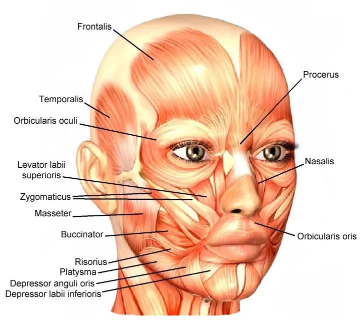
Median Nerve: Anatomy, Origin, Course, Function, Clinical Importance
| | |

Median Nerve: Anatomy, Origin, Course, Function, Clinical Importance

The Median Nerve is a nerve in humans and other animals in the upper limb. It is one of the five main nerves originating from the brachial plexus. Anatomy of Median Nerve: The median nerve originates from the lateral and medial cords of the brachial plexus and has contributions from ventral roots of C5-C7 (lateral…

Calf Muscle Anatomy: Anatomy and Exercise
| | |

Calf Muscle Anatomy: Anatomy and Exercise

Anatomy of the Calf muscle The calf muscle is located on the back of the lower leg and is actually made up of two muscles: The medial and lateral heads of the gastrocnemius muscle, which is the most superficial, are present. The gastrocnemius muscle belly is formed by the convergence of its two heads. The…

Hamstring Muscle: Anatomy And Exercise
| | |

Hamstring Muscle: Anatomy And Exercise

Hamstring Muscle Anatomy The hamstrings are a group of muscles and their tendons at the rear of the upper leg. They include the biceps femoris, semitendinosus, and semimembranosus. The hamstrings flex the knee joint and extend the thigh to the backside of the body. They are used in walking, running, and many other physical activities….

Coarctaction of Aorta
| | | |

Coarctaction of Aorta

Coarctation (ko-ahrk-TAY-shun) of the aorta — or aortic coarctation — is a narrowing of the aorta, the large blood vessel that branches off your heart and delivers oxygen-rich blood to your body. When this occurs, your heart must pump harder to force blood through the narrowed part of your aorta. Coarctation of the aorta is…

PATENT DUCTUS ARTERIOSUS
| | |

PATENT DUCTUS ARTERIOSUS

PATENT DUCTUS ARTERIOSUS PDA is a heart problem that is frequently noted in the first few weeks or months after birth. It is characterized by the persistence of a normal fetal connection between the aorta and the pulmonary artery which allows oxygen-rich (red) blood that should go to the body to recirculate through the lungs….

ATRIAL SEPTAL DEFECT
| | |

ATRIAL SEPTAL DEFECT

OVERVIEW An atrial septal defect (ASD) is a hole in the wall between the two upper chambers of your heart (atria). The condition is present at birth (congenital). Small defects may never cause a problem and may be found incidentally. It’s also possible that small atrial septal defects may close on their own during infancy…