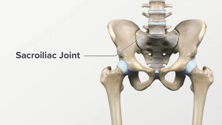
SI JOINT (Sacroiliac Joint) : Anatomy, Movement, Dysfunction, Exercise
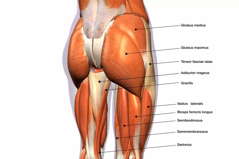
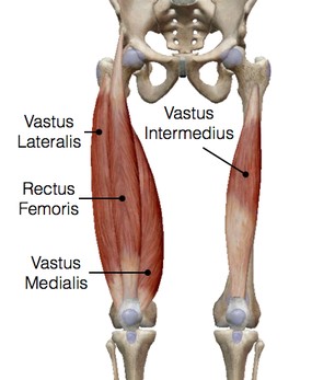
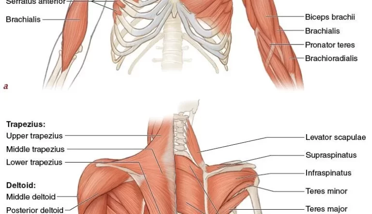
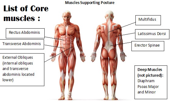
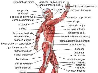
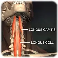
Introduction : Anatomy of the Sacroiliac Joint : Joint Capsule : Ligaments : Nerves : Muscles : Movement of the SI joint : Nutation & Counternutation : Nutation occurs: Counternutation occurs : Torsion: Primary mechanisms of to this joint dysfunction include to the : Causes & Risk Factors of this Joint Dysfunction : Symptoms of…