

Ulnar Nerve
The ulnar nerve is a major peripheral nerve of the upper limb. The ulnar nerve is located in the medial side of the forearm. It runs from the large cubital tunnel in the elbow down to the fourth and fifth fingers. The ulnar nerve supplies sensation to both sides of the hand and contributes muscles to the wrist and fingers. Injury to the ulnar nerve can cause weakness in the hand and fingers with possible loss of sensation.
Anatomy of the Ulnar Nerve:
Ulnar nerve: The ulnar nerve originates in the medial cord of the brachial plexus and extends from the medial epicondyle of the humerus to the medial border of the ulnar bone. It receives filaments from the medial and posterior divisions of the brachial plexus and passes through the compartment of the cubital fossa. The nerve supplies motor function to flexor digitorum profundus and flexor pollicis longus; it also supplies sensory innervation to the skin of the medial side of the forearm and hand and part of the medial palm.
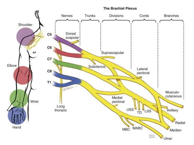
The ulnar nerve arises from the brachial plexus. It is a continuation of the medial cord, containing fibres from spinal roots C8 and T1.
After arising from the brachial plexus, the ulnar nerve descends down the medial aspect of the upper arm.
At the elbow, it passes posterior to the medial epicondyle of the humerus and gives rise to an articular branch that supplies the elbow joint.
The ulnar nerve is palpable and vulnerable to injury at the medial epicondyle.
In the forearm, the ulnar nerve pierces the two heads of the flexor carpi ulnaris, and travels deep to the muscle, alongside the ulna.
Three main branches arise in the forearm:
- Muscular branch – innervates two muscles in the anterior compartment of the forearm.
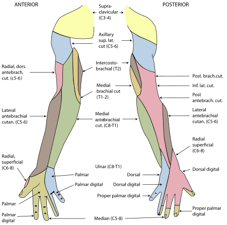
- Palmar cutaneous branch – innervates the medial half of the palm.
- Dorsal cutaneous branch – innervates the dorsal surface of the medial one and a half fingers, and the associated dorsal hand area.
At the wrist, the ulnar nerve travels superficially to the flexor retinaculum, and is medial to the ulnar artery.
It enters the hand via the ulnar canal (Guyon’s canal). In the hand, the nerve terminates by giving rise to superficial and deep branches.
Description:
The ulnar nerve originates from C8-T1 nerve roots, which form the medial cord of the brachial plexus.
The ulnar nerve runs down the hand where it passes behind the medial epicondyle of the humerus at the elbow.
The ulnar nerve doesn’t give branches in the axilla or in the upper arm. It starts giving muscular and cutaneous branches in the upper forearm and hand.
After the ulnar nerve passes behind the medial epicondyle, it enters the forearm between the two heads of the flexor carpi ulnaris muscle.
The ulnar nerve may become compressed or irritated as it passes behind the medial epicondyle.
The ulnar nerve travels through the cubital tunnel that runs under the medial epicondyle.
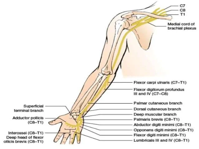
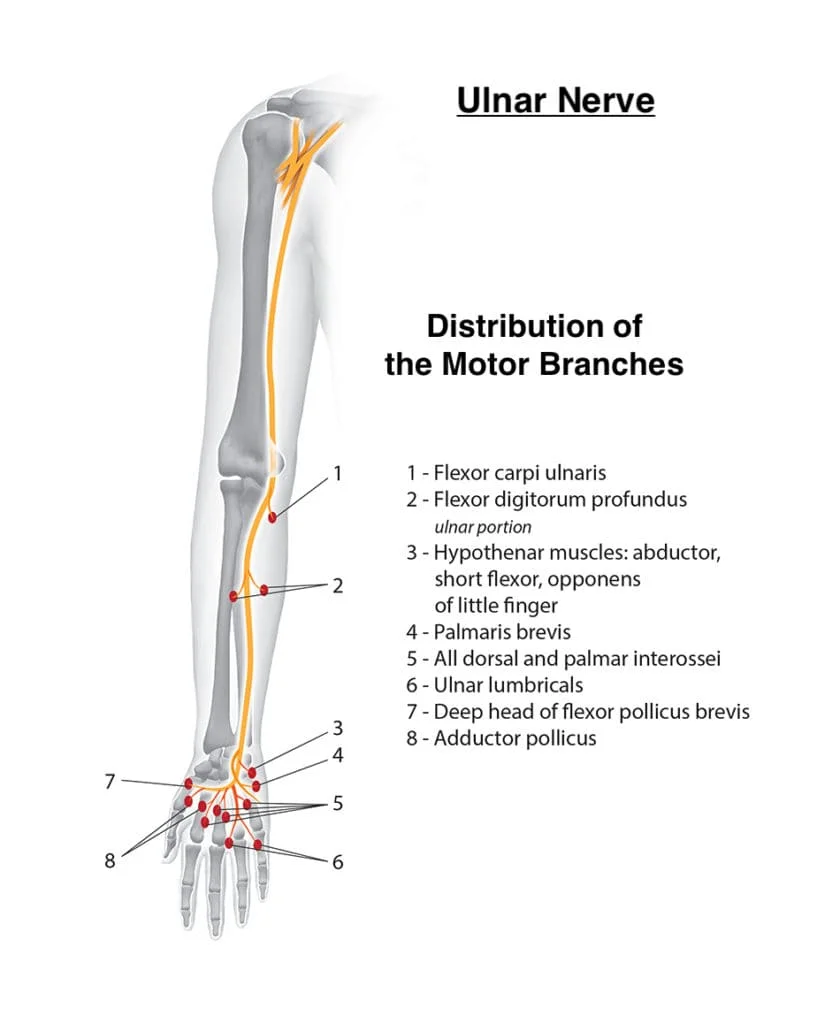
The nerve gives branches to the flexor carpi ulnaris and medial half of flexor digitorum profundus.
The ulnar nerve then travels alongside the ulnar bone of the forearm into the wrist.
As the nerve descends into the forearm, it stays medially above the flexor digitorium profundus and under the flexor carpi ulnaris, giving branches to these muscles.
In the lower part of the forearm, the ulnar nerve lies lateral to the flexor carpi ulnaris muscle and medial to the ulnar artery.
The ulnar nerve enters the palm of the hand through the Guyon canal. The nerve and artery pass superficial to the flexor retinaculum.
At the wrist, the ulnar nerve lies just lateral to the pisiform bone.
The superficial branch of the ulnar nerve supplies and passes under the Palmaris brevis muscles and divides into palmar digital nerves.
The deep branch of the ulnar nerve innervates the three hypothenar muscles, the two medial lumbricals, the seven interrosei, the adductor pollicis and the deep head of the flexor pollicis brevis.
It supplies all intrinsic hand muscles which lie medial to the flexor pollicis except the last lateral two lumbricals.
Nerve Root:
- C8, T1
- From the Medial cord of the brachial plexus.
Branches of Ulnar Nerve:
- Muscular branch
- Palmar cutaneous branch
- Dorsal cutaneous branch
Structure of the ulnar nerve:
Arm:
The ulnar nerve originates from the C8-T1 nerve roots (and occasionally carries C7 fibres which arise from the lateral cord ) which then form part of the medial cord of the brachial plexus, and descends medial to the brachial artery, up until the insertion point of coracobrachialis muscle (middle 5 cm over the medial border of the humerus).
Then, it pierces the medial intermuscular septum and enters the posterior compartment of the arm, accompanied by superior ulnar collateral vessels. It runs at the posteromedial aspects of the humerus, passing behind the medial epicondyle (in the cubital tunnel) at the elbow, where it can be palpated by hand.
Forearm:
The ulnar nerve is not a content of the cubital fossa. It enters the anterior (flexor) compartment of the forearm between the two heads of flexor carpi ulnaris, lies along the lateral border of the flexor carpi ulnaris.
The ulnar nerve runs between the flexor digitorum superficialis (laterally) and flexor digitorum profundus medially. Near the wrist, it courses superficial to the flexor retinaculum of the hand, but covered by the volar carpal ligament to enter the hand.
In the forearm it gives off the following branches:
- Muscular branches of ulnar nerve – supplies one and a half muscles (flexor carpi ulnaris and the medial half of flexor digitorum profundus)
- Palmar branch of ulnar nerve – arises from the middle part of the forearm and supplies the skin over the hypothenar eminence.
- Dorsal branch of ulnar nerve – arises from 7.5 cm above the wrist, winds backwards to supply the skin of the proximal part of the ulnar one and a half fingers and the adjoining area between the fingers.
Articular branches are given off to the elbow joint.
Hand:
Ulnar nerve enters the palm of the hand via the Guyon’s canal, superficial to the flexor retinaculum and lateral to the pisiform bone.
Here it gives off the following branches:
- Superficial branch of ulnar nerve – supplies the palmaris brevis and gives digital branches to the medial one and a half fingers.
- Deep branch of ulnar nerve – It accompanies the deep branch of the ulnar artery. It passes backwards between the abductor digiti minimi, flexor digiti minimi, and opponens digiti minimi, supplying all three muscles, and lying on the hook of the hamate bone.
- It then turns laterally, supplying the 3rd and 4th lumbricals and all the palmar interossei muscles and dorsal interossei of the hand. It terminates by supplying the adductor pollicis.
- Articular branches to the wrist.
Function of Ulnar Nerve:
Ulnar nerve is also known as “musician’s nerve” as it controls the fine movements of the fingers.
Sensory:
- Cutaneous innervation of the right upper extremity. Areas innervated by the ulnar nerve are the areas on the hand colored in light blue.
- The ulnar nerve also provides sensory innervation to the fifth digit and the medial half of the fourth digit, and the corresponding part of the palm.
- Palmar branch of ulnar nerve – supplies cutaneous innervation to the anterior skin and nails.
- Dorsal cutaneous branch of ulnar nerve – supplies cutaneous innervation to the dorsal medial hand and the dorsum of the medial 1.5 fingers.
Motor:
- The ulnar nerve and its branches innervate the following muscles in the forearm and hand:
- An articular branch that passes to the elbow joint while the ulnar nerve is passing between the olecranon and medial epicondyle of the humerus.
In the forearm, via the muscular branches of ulnar nerve:
- Flexor carpi ulnaris
- Flexor digitorum profundus (medial half)
In the hand, via the deep branch of the ulnar nerve:
Hypothenar muscles:
- Opponens digiti minimi
- Abductor digiti minimi
- Flexor digiti minimi brevis
- The third and fourth lumbrical muscles
- Dorsal interossei
- Palmar interossei
- Adductor Pollicis
- Flexor pollicis brevis (deep head)
In the hand, via the superficial branch of the ulnar nerve:
- Palmaris brevis
Clinical Relevance:
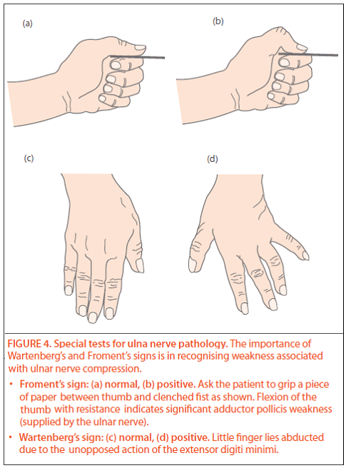
Froment’s Sign: Froment’s sign is a test for ulnar nerve palsy – specifically paralysis of the adductor pollicis:
The patient is asked to hold a piece of paper between the thumb and index finger, as the paper is pulled away.
They should be able to hold the paper there with no difficulty (via adduction of the thumb).
A positive test is when the patient is unable to adduct the thumb. Instead, they flex the thumb at the interphalangeal joint to try to maintain a hold on the paper.
Ulnar Nerve Palsy:
Damage at the Elbow:
Mechanism of injury: Trauma at the level of the medial epicondyle (e.g. isolated medial epicondyle fracture, supracondylar fracture). It can also be compressed in the cubital tunnel.
Motor functions:
All the muscles innervated by the ulnar nerve are affected.
- Flexion of the wrist can still occur, but is accompanied by abduction (due to paralysis of flexor carpi ulnaris and medial half of flexor digitorum profundus).
- Abduction and adduction of the fingers cannot occur (due to paralysis of the interossei).
- Movement of the 4th and 5th digits is impaired (due to paralysis of the medial two lumbricals and hypothenar muscles).
- Adduction of the thumb is impaired, and the patient will have a positive Froment’s sign (due to paralysis of adductor pollicis).
Sensory functions:
- All sensory branches are affected, so there will be a loss of sensation over the areas that the ulnar nerve innervates.
- Characteristic signs: Patient cannot grip paper placed between fingers, positive Froment’s sign, wasting of hypothenar eminence.
Damage at the Wrist:
Mechanism of injury: penetrating wounds, Guyon canal cyst
- The claw hand deformity is more prominent with injury at the wrist as opposed to a lesion higher up in the arm, for instance, at the elbow, as the ulnar half of the flexor digitorum profundus is not affected. This pulls the distal interphalangeal joints of the 4th and 5th digit into a more flexed position, producing a more deformed ‘claw’. This is known as the ulnar paradox.
Motor functions:
Only the intrinsic muscles of the hand are affected.
- Abduction and adduction of the fingers cannot occur (due to paralysis of the interossei).
- Movement of the 4th and 5th digits is impaired (due to paralysis of the medial two lumbricals and hypothenar muscles).
- Adduction of the thumb is impaired, and the patient will have a positive Froment’s sign (due to paralysis of adductor pollicis).
Sensory functions:
- The palmar branch and superficial branch are usually severed, but the dorsal branch is unaffected. This results in sensory loss over the palmar side of the medial one and a half fingers only.
- Characteristic signs: Patient cannot grip paper placed between fingers, positive Froment’s sign, wasting of hypothenar eminence.

24 Comments