
Cervicogenic Headache:
What is a Cervicogenic Headache?
Cervicogenic Headache presents as unilateral pain which starts in the neck. A common chronic and recurrent headache usually begin after neck movement. It is usually associated with decreased range of motion (ROM) of the neck. The cervicogenic headache was first introduced by Sjaastad et al.(1983). It is a chronic headache that strat from the atlantooccipital and upper cervical joints and is perceived in one or more regions of the head and face.
Definition:
Referred pain perceived in any region of the head is caused by a primary nociceptive source in the musculoskeletal tissue which is innervated by the cervical nerves. (Albc,1999). Sources of cervicogenic headache pain lie in the structures innervated by the C1-C3 spinal nerves. Cervicogenic headache because of chronic headache.
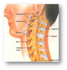
Clinical relevant anatomy of the cervical spine
Anatomy of cervicogenic headache
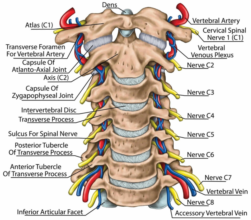
The cervical spine is an important one anatomically, clinically, and therapeutically.
It is the region where the nerves of arms arise through the brachial plexus and where the cervical plexus forms providing innervation to the diaphragm surrounded by other structures.
It also allows the opening of important vascular structures to reach the brain and allows attachment area for muscles that move the head, neck, and shoulder girdle.
To understand this complicated region, we will consider the bony structures first, and then discuss the ligaments, nerves, and musculature that are associated with this region of the cervical spinal column, come to the conclusion with some clinical implications of damage to some of these structures.
The cervical spine includes 7 vertebrae, C1 to C7, the cervical nerves from C1 to C8, muscles, and ligaments.
The first two vertebrae C1 and C2 have unique shapes and functions. They form the upper cervical spine.
Upper Cervical Spine
The cervical spine is composed of 7 vertebrae, C1 to C7.
In that, the first two vertebrae C1 and C2 form the upper cervical spine.C1 and C2 have unique shapes and functions.
Nerves
Many important nerves arise from the cervical spinal cord.
Most of these nerves originate in one of two plexuses: The cervical plexus or The brachial plexus
C1 (Atlas) vertebrae support the skull. It articulates superiorly with the occiput which is known as the atlanto-occipital joint.
The atlanto-occipital joint is responsible for 33% of flexion and extension. The structure of the atlas allows forward and backward movement of the head.
Below the C1 (atlas) is the C2 (axis) that allows rotation. This joint (The atlantoaxial joint) is responsible for 60% of all cervical rotation.
C3-C7 cervical vertebrae that make up the lower cervical spine, are similar to each other but very different from C1 and C2.
SPINAL NERVES
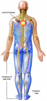
Certain spinal nerves are involved in cervicogenic headaches. Spinal nerves are signal transmitters that allow communication between the brain and the body via the spinal cord. At each level of the cervical spine is a set of spinal nerves; one on the left side and another one on the right side of the spine. C1, C2, and/or C3 may be involved in the development of cervicogenic headaches because these nerves allow function (movement) and sensation of the head and neck. Nerve compression can also cause inflammation and pain.
Pathophysiology
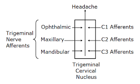
The C1-C3 nerves receive pain signals to the nociceptive nucleus of the head and neck(the trigeminocervical nucleus). Aseptic inflammation and neurotransmission within the C-fibers because of cervical disc pathology are thought to originate and worsen the pain in a cervicogenic headache.
The nociceptive nucleus of the head and neck(the trigeminocervical nucleus) receives afferents from the trigeminal nerve as well as the upper three cervical spinal nerves. Neck trauma, whiplash injury, strain, or chronic spasm of the scalp, neck, or shoulder muscles can increase the sensitivity of the area which is similar to the allodynia(Pain due to a stimulus that does not normally provoke pain) that is seen in late chronic migraines. A lower pain threshold makes patients more prone to more severe pain. Because of that early diagnosis and therapeutic intervention are very important.
Epidemiology
It is a rare chronic headache in people who are 30 to 45 years old.
According to NCBI(national center for biotechnology information), a prevalence of 4.1% was found. In 41 cases with the highest number of cervicogenic headache criteria, there was a male prevalence (F/M: 0.71). While cervicogenic traits (mechanical precipitation etc.) were frequently present in cervicogenic headaches, ‘migraine traits’, like nausea, vomiting, and throbbing seemed to be rarely present. In 97% of the cases, pain exacerbations began in the neck and occipital region.
Which are the Causes of Cervicogenic Headache?
Many things can cause a cervicogenic headache.
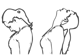
Cervicogenic headaches can come from problems with your cervical vertebrae, joints, or neck muscles. e.g.people in certain jobs, like carpenters, truck drivers, and hair stylists can get cervicogenic headaches from the way they hold their head and neck when they work. Some people who hold their heads out in front of their bodies are also caused by cervicogenic headaches. This movement is called “forward head movement,” and it puts extra stress on your neck and upper back.
It is thought to be referred pain arising from irritation caused by cervical structures innervated by the upper three cervical spinal nerves C1, C2, and C3; therefore, any structure innervated by the C1 to C3 spinal nerves could be the source of a cervicogenic headache. The cause of a cervicogenic headache is often related to excessive tension in the neck. The headache may result from cervical spondylosis, a damaged disc, or whiplash-type movement that irritates a cervical nerve or sometimes over-activity of neck muscles also cause cervicogenic headache because of spasm of para-spinal neck muscles.
The facet joints of cervical vertebrae (neck bony structure) and neck muscles also contribute to the development of a cervicogenic headache.
Medical conditions which can cause cervicogenic headaches are :
Poor Posture
Whiplash or another injury to the neck
Cervical Disc Prolapse
Fractures
Infections
Tumors in the Cervical region
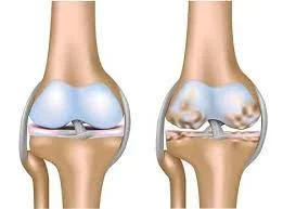
Cervical spondylosis and osteochondritis are not accepted as a cause of cervicogenic headaches.
Symptoms of Cervicogenic Headache
CH (cervicogenic headache) is pain that comes from a sudden movement of your neck.
Another is that you get headaches when your neck remains in the same position for some time.
Symptoms of a Cervicogenic headache may include:
- a decreased range of motion (ROM) in the neck
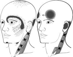
- pain on one side of the face
- pain and stiffness of the neck c
- pain around the eyes
- Spasm of Paraspinal Neck Muscles
- Pain radiate from the Upper trapezius muscle to the shoulder and sometimes arm region.
- pain in the neck, shoulder, or arm on the affected side
- pain while coughing or sneezing
- Headache which is triggered by certain neck movements or postures
- throbbing head pain
- sensitivity to light and noise
- Weakness in the deep neck flexors
- Tenderness in the Upper Cervical area
- nausea
- blurred vision
- Poor Posture
Other signs may include:
- Pain on one side of your face
- Steady pain
- When you cough, sneeze or take a deep breath cause headache
- A cervicogenic headache pain that can last for hours or days
- Stiff neck (you can’t move your neck normally)
- Pain that stays in one spot, like the back, front, or one side of your head
Even though cervicogenic headache and migraine are different but some of the symptoms can be similar.
For example, you may:
Feel sick to your stomach
Throw up
Have pain in your shoulder or arm
Feel uncomfortable in bright light
Feel uncomfortable with loud noise
Have blurry vision
Some people get cervicogenic headaches and a migraine at the same time.
Risk Factors of Cervicogenic Headache:
Risk factors that may be aggravated cervicogenic headaches include:
- Fatigue
- Muscular stress
- Cervical disc problems
- Current or prior neck injuries
- Poor posture (High cell phone use)
- Sleep difficulties
Differential Diagnosis:
Differentiate from serious pathology such as:
- Cervical Instability
- Cervical Myelopathy
- Occipital neuralgia
- Vascular Pathologies of the Neck
- Intracranial Pathology
How do doctors diagnose cervicogenic headaches?
Diagnosis of cervicogenic headache involves ruling out other conditions.
Diagnosis also involves testing for movement and ruling out the affected nerves, bones, and muscles.
For diagnosis, your doctor will start by asking some questions regarding your symptoms.
Then, they’ll run through a set of some special tests.
All of this helps your doctor to rule out if your nerves and spinal cord are under too much pressure.
Your doctor will perform a physical examination to find the cause of your neck pain or other symptoms.
Physical examination:
Pain localized in the neck and occiput, which can spread to other areas in the head, such as the forehead, orbital region, temples, vertex, or ears, usually affected side. Pain is the trigger or aggravated by specific neck movements or sustained postures.
At least one of the following:
- Limitation of passive neck movements
- Changes in neck muscle texture, tone, shape, or reaction to active and passive stretching and contraction
- Abnormal tenderness of neck muscle.
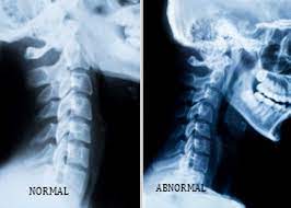
Radiological examination:
Radiological examination discloses at least one of the following:
- Movement abnormalities (flexion/extension)
- Abnormal posture
- Fractures
- Congenital abnormalities
- Bone tumors
- Rheumatoid arthritis
- Other distinct pathology (not spondylosis or osteochondrosis)
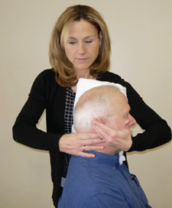
Flexion rotation test
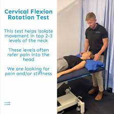
The inverse relationship between headache severity of CGH (cervicogenic headache) and ROM (range of motion)towards the most restricted side for the Cervical Flexion rotation test (FRT) was statistically significant for all patients with cervicogenic headaches
(Sn = 0.91, Sp = 0.90).The patient must feel no pain at the time of the FR test. During the test, the neck of the patient is passively held in end-range flexion. The therapist rotates the neck to both sides until they feel resistance or until the patient says they are in pain. At this endpoint, the therapist makes a visual estimate of the rotation range and says on which side the FRT was positive or negative. The test was positive when the approx range was decreased by more than 10° from the anticipated normal range (44°).
Other tests are:
- X-ray: X-rays can help to know about degenerative changes to the spine (cervical spine) and problems like bone spurs.
- Computed tomography (CT): A CT scan is an imaging technology that uses multiple X-ray images to see the neck and spine in different “slices.”
- Magnetic resonance imaging (MRI): An MRI scan is used with powerful magnetic and radio waves to create highly detailed images of bone and soft tissues, including those of the spinal cord.
- Blood tests: A complete blood count (CBC), erythrocyte sedimentation rate (ESR), and C-reactive protein (CRP) test can help to determine if inflammation or infection is present.
- In a myelogram, a dye injection highlights certain areas of your spine. CT scans or X-rays are used to provide more detailed images of affected areas.
- . A nerve conduction study checks the speed and strength of the signals sent by nerves. This is done by placing electrodes on your skin where the nerve is innervated in the muscle.
RED FLAGS
- Sudden onset of a severe headache;
- An aggravate pattern of a pre-existing headache in the absence of obvious predisposing factors
- If the headache is associated with fever, neck stiffness, skin rash, and a history of cancer, HIV, or other systemic illness
- If the headache is associated with focal neurologic signs other than a typical aura
- Moderate or severe headache aggravated by cough, exertion, or bearing down
- New onset of a headache during the pregnancy.
Patients with one or more red flags must be referred for an immediate medical consultation and further investigation.
Treatment of Cervicogenic Headache
Medical Treatment:
There are many ways to reduce the pain, or get rid of it completely:
Medicine:
Non-steroidal anti-inflammatory (NSAIDs) such as ibuprofen, muscle relaxers, and other pain relievers may ease the pain.
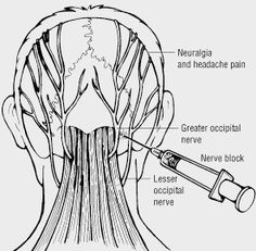
Nerve block:
This may temporarily relieve your pain and help you better work with physiotherapy.
Spinal manipulation:
This is the coordination of massage, physiotherapy, and joint movement.
It should only be done by a chiropractor, a physiotherapist, or an osteopath.
Other options:
Non-surgical ways to cure the pain include relaxation techniques, such as deep breathing, yoga, and acupuncture.
Physiotherapy treatment of Cervicogenic Headache:
Your doctor might refer you to a physical therapist for treatment.
Physiotherapy treatment can help you to stretch your neck muscles and also help to relieve neck pain.
You can try a couple of things at home if your condition is mild:
- Rest
- Take an over-the-counter(OTC)pain reliever, such as acetaminophen (Tylenol) or non-steroidal anti-inflammatory drugs (NSAID), which include ibuprofen and naproxen sodium.
- Use a hot pack or a cold pack on your neck for 7-10 minutes which helps to relieve pain.
- Exercise regularly to help you recuperate faster.
- Wearing a cervical collar to limit movement and provide support
- In moderate conditions, you might prefer a physiotherapist.
Pain management:
Treating CGHs with Electrotherapy.
Some people respond to treatments like OTC (over-the-counter) pain medications or physiotherapy, some people may need different solutions for dealing with their pain.
Electrotherapy can effective for pain management.
What is Electrotherapy?
Electrotherapy uses mild electrical pulses to stop the pain in a point area of the body. Generally, all of these therapies use a device with electrodes to supply a low-voltage electrical current to pain areas.
The main goal of electrotherapy is pain management.
Uses of electrotherapy :
It can help improve blood circulation, which activates the body’s healing process.
This may also help to strengthen muscles, stimulate bone growth, and repair damaged tissues.
Moreover, electrotherapy may increase movement and function in an affected area.
These are low-voltage electrical currents, a person usually only senses a tingling sensation.
Some people even find using electrotherapy machines to be relaxing purpose.
The side effects of electrotherapy are generally mild.
The most common side effect is patients complaining of skin irritation because of the adhesive on the electrodes. Sometimes burning sensation on the surface of the skin because of not using the device properly.
Here are common types of electrotherapy that your physiotherapist may recommend:
TENS (Transcutaneous Electrical Nerve Stimulation)
One of the most common electrotherapy devices use for pain relief. Small and sticky pads with electrodes are placed over or near the area where you sense pain. Your physiotherapist will decide the area of placement for electrodes. A series of low-voltage electrical currents send by the battery-operated unit to the area. You will increase or decrease the electrical current as well as the frequency and patterns of stimulation by the controller.
PENS(Percutaneous Electrical Nerve Stimulation)
An alternative to TENS, PENS (percutaneous electrical nerve stimulation) uses needles on behalf of pads to supply electrical currents. It works as acupuncture with electrical stimulation. The benefit of using needles rather than pads means the pulse of the electrical current is closer to the pinched nerves and muscles. This can help to relieve pain immediately in some patients.
Generally, this therapy is used as an outpatient procedure. You can also use a PENS device at your home.
PEMF (Pulsed Electromagnetic Field Therapy)
It takes a different process to use electricity for pain relief. PEMF tries to heal at the cellular level rather than treating nerves or muscles. Specialized coils create an electromagnetic field around part or all of the body throughout the treatment. These electromagnetic waves stimulate the cell’s electrons and assist the healing process and stimulate bone growth.
PEMF treatments can recover the cells and make them more active in healing, rebuilding, and fighting disease. Treatment is performed in a therapist’s clinic. Some PEMF products can be purchased for home use.
Cervical traction:
Cervical traction help to relieve pain. It involves using weights to increase space between the cervical joints and also relieve the pressure on the cervical discs and nerve roots.
Chiropractic manipulation can help to control severe pain
Other forms of physiotherapy, include the application of heat and cold therapy, traction, or exercise.
Exercises for cervicogenic headache

- Cervical spine mobilization
- Strengthening exercises for neck muscles including deep neck flexors and upper quarter muscles
- Thoracic spine thrust manipulation and exercise
- C1-C2 Self-sustained Natural Apophyseal Glide (SNAG) has shown to be effective for lessen the cervicogenic headache symptoms
- active mobilization exercises
- Upper cervical spine mobilizations
- Passive mobilization with movement
- Work-related ergonomic training
- active mobilization with movement
- Thoracic Manipulation
- Deep Neck Flexor Exercise
Trigger Point Therapy :
This is composed of different manual approaches, e.g., compression, stretching, or transverse friction massage. Pressure release over the sternocleidomastoid muscle. Pressure is applied progressively to increase over the trigger point until the finger encountered an increase in tissue resistance (tissue barrier). This pressure is maintained until the therapist senses relief from the stressed band. At that time, the pressure is increased again until the next increase in tissue resistance. Stretching of the tight band muscle fibers is also important. Trigger point therapy is effective for lengthening the trigger point in the muscle and the associated connective tissue. The physiotherapist applies moderate slow pressure over the trigger point and slides the fingers in the opposite directions.
This manual therapy is applied slowly and is performed without inducing pain in the patients.
Co-contraction of the Neck Flexor and Extensor :
It is facilitated with neck rotation and neck exercises are introduced once the patient can activate the deep muscles.
The patient does a self-resisted isometric rotation in a correct sitting posture. They look into the palm and allowing that the resistance to facilitate the muscles and perform the consecutive rhythmic stabilization exercises with an emphasis on the slow onset and slow-release holding contractions, using resistance to match about a 10–20% effort. Retrain the strength of the superficial and deep flexor synergy.
Extensors of the Craniocervical Spine:
The patient exercises eccentric control of the head into flexion followed by concentric control of the back to the neutral position in a 4-point kneeling position to train the coordination of the deep and superficial cervical extensors. These exercises are incorporated with re-education of the scapular muscles in these positions and are begin in the program. The exercise is progressed by performing alternating small ranges of craniocervical extension and flexion while maintaining the cervical spine in a normal position.
Retrain the cervical flexors for anti-gravity function in a sitting position:
This exercise is a controlled eccentric action of the neck flexors into the cervical extension range followed by a concentric action of these muscles to return the head to the normal upright position. The return to the correct position must be initiated by CCF (craniocervical flexor), rather than a dominant action of SCM (sternocleidomastoid).
The exercise is progressed by slowly increasing the range to which the head is moved into extension as control improves, and by introducing isometric holds through the range.
Retrain the Scapular Muscles :
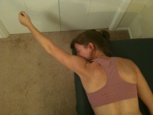
Retrain scapular muscle which is oriented in posture correction strategy is to have the patient move the coracoid upward and the acromion backward, which results in a slight retraction and external rotation of the scapula. The goal is to facilitate the coordinated action of all parts of the trapezius and serratus anterior muscles, allowing the lower trapezius to slightly depress the medial border of the scapula, consequently relaxing the levator scapulae.
Once the patient practices correct scapular orientation, Train the endurance capacity of the scapular stabilizers repeated repetitions of 10-second holds of the corrected scapular position encourages early endurance retraining. The endurance of the middle and lower trapezius muscles is also trained by accomplishing an exercise in the prone lying position against the effects of gravity.
Retrain scapular control with arm movement and load. This is important when activities such as computer work or deskwork trigger the pain. The patient is inspired to maintain their newly learned scapular position while performing small range (+/- 60 degrees) arm movements, for example, working at a computer. Scapular control in cooperation with control of cervicothoracic postural position is also educated for functional activities such as lifting and carrying.
Upper Quarter Strengthening Exercises
Middle Trapezius Strengthening
Lower Trapezius Strengthening
Adding upper quarter exercises in treatment for patients with cervical dysfunction is important to integrate ‘global’ muscles that have connections to the cervical spine through anatomical chains.
Reeducate the Strength of the Superficial and Deep Flexors Synergy :
The head lift must go first with CCF (craniocervical flexors) followed by cervical flexion to just lift the head from the bed. Gravity and head weight provides resistance. Care must be taken that high load exercise is not introduced too early, as it may be evocative of symptoms.
Re-education of Posture :
Posture is an attitude assumed by the body and an indirect measure of the functional status of the neuromuscular system. The postural position is trained in a sitting position and is corrected from the pelvis. Another aspect of re-education of postural position is the correction of scapular position. Maintenance of a correct scapular position with appropriate muscle coordination has the added benefit of inducing reciprocal relaxation in muscles.
Sensory-motor Training :
Because CGHs(cervicogenic headache) is thought to be a dysfunction of the sensorimotor system. Sensorimotor training includes progressive exercise on unstable surfaces to promote reflexive stabilization and postural stability. Unstable surfaces such as exercise balls can be used to add challenge to the cervical spine as well as the whole body for stabilization exercises. These final stages of the rehabilitation program for cervicogenic headache patients can be progressed toward functional activities to return the patient to full participation.
Mobility, Strength, Stability, and Postural Exercises :
Neck Strengthening with thera-band
These technics were primarily posted isometric relaxation procedures (A) myofascial mobilization, and (B) selected elements of McKenzie therapy. Exercises were mainly applied to the muscles with Trigger points, showing a pathological increase in EMG amplitude. When relaxation of painful, tensed muscles is achieved, the next step of treatment includes strengthening exercises of the neck muscles. All of the exercises are supervised by a physiotherapist. Exercise intensity shall not increase the pain sensation in the cervical spine, or shoulder girdle muscles. They aren’t supposed to provoke the headache.
There are isometric exercises with a slowly loading increase and dynamic exercises
.Elastic thera bands are most commonly used during these exercises. Additionally, the self-control exercises of the correct body posture are carried out in front of the mirror relying on visual feedback.
Prevention Measure:
Maintaining a good posture while sitting or driving ( sitting tall with shoulders back and without protruding the head forward)
Using a supporting travel pillow on the plane or bus to avoid excessive side-bending or flexion
Avoid poor postures or movements that evoke pain to start
Using a neck brace (soft collar) can be helpful while sitting upright or sleeping in a chair
Avoid tummy-sleeping posture
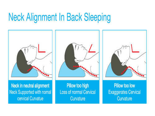
Find the correct pillow to prevent the head from being too high or too low when sleeping.
Prevention while sleeping:
How to select the proper pillow for cervicogenic headaches?
The best pillow for neck and shoulder pain is firm enough to hold the head at a correct angle but soft enough to alleviate pressure points. Most people find success with either memory foam, latex, buckwheat, or feather pillow, as these materials provide the best balance of support and pressure relief.
1. Cervical pillows for neck pain relief
Cervical pillows especially provide neck support and keep your spine in proper posture while you’re asleep. It’s elevated where your neck is more compressed where your head lies. Your pillow works ergonomically to support your neck. Cervical pillows come in different shapes and materials according to your comfort.
If you’re changeover from a regular pillow to a cervical pillow, you may feel discomfort for some days. Using it every day for taking a short rest during work can adapt easier.
2. Pillows that mold to the neck for back sleepers
Choosing the right pillow for your neck depends on how you sleep. If you sleep on your back, try to choose a pillow that molds to your head and neck so that your neck is enough to support. Memory foam pillows or water pillows are good choices for back sleepers. They keep their shape but adjust according to your body.
3. Firm pillows for side sleepers
If you sleep on your side, then choose a firm pillow. Place the bulk of the pillow under your neck rather than your head, which provides proper support to the spine.
It helps if the pillow has elasticated. A pillow with a elasticate is one with extra material in it, with the join into rectangular panels on all four sides of the pillow so that the cushioning adjusts when you move your head. A pillow without a elasticate has only two pieces, a top and a bottom that are stitched together. Side sleepers also need to put a pillow between their legs to help maintain spine posture.
4. Thin pillows for stomach sleepers
When you’re sleeping on your stomach you put stress on your back. So, you have to use a thin pillow or no pillow to avoid stress on your back. Also, use a thin pillow under the abdominal area that can help to decrease the stress on your back.
5. Horseshoe pillows when traveling
When you’re traveling by car, plane, or train, a horseshoe pillow is great support for your neck.
If you have neck pain, don’t let your head bend to the side when you sleep during traveling. Make sure your neck is supported
5 Comments