
Longus colli muscle
Introduction
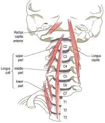
Longus colli is a paired muscle found on the anterior aspect of the vertebral column. As such, it is usually referred to as an anterior prevertebral muscle concurrently with longus capitis, rectus capitis anterior, and also scalenus anterior muscles. The longus colli muscle (the ‘long muscle of the neck) is also known as longus cervicis since it transits the entire cervical spine and the first three thoracic vertebrae. By acting on the cervical vertebrae, longus colli is accountable for forward and lateral flexion of the neck, as well as rotation of the neck.
The Longus colli muscle, and all prevertebral muscles, are found deep in the prevertebral fascia, which is a layer of the deep cervical fascia of the neck. The muscle seats in the groove are formed by the junction of the transverse process with the vertebral body, posterior to the retropharyngeal space. Therefore, the esophagus, trachea, thyroid gland, and muscles of the hyoid (supra-, infra-) all sit anterior to it. Longus colli is connected medially by the cervical plexus, brachial plexus, and subclavian artery.
Origin of Longus colli muscle
Longus colli drives the entire length of the neck; between the atlas (first cervical vertebra) and T3 (third thoracic vertebra). It is limited at its superior and inferior ends and has a broad central section. The muscle consists of three components which are attached to the vertebral column via tendinous slips:
Anterior tubercles and the anterior surfaces of bodies of C3 to T3. The superior Oblique portion originates from anterior tubercles of the transverse processes of the third, fourth, and fifth cervical vertebræ. The inferior Oblique portion originates from the front of the bodies of the first two or three thoracic vertebræ. The vertical portion originates from the front of the bodies of the upper three thoracic and lower three cervical vertebræ
Insertion of Longus colli muscle
Anterior arch of the atlas, anterior tubercles of C5-6, and the anterior surfaces of bodies of vertebrae C2-4.The superior Oblique portion is inserted into the tubercle on the anterior arch of the atlas. Inferior Oblique portion into the anterior tubercles of the transverse processes of the fifth and sixth cervical vertebræ.Verticle part into the front of the bodies of the second, third, and fourth cervical vertebræ.
The medial border of the scalenus anterior muscles fibs close to the lateral border of the longus colli. Here, a pyramidal space is formed, with the first section of the subclavian artery developing its base. The thoracic duct, cervical sympathetic trunk, and the first portion of the vertebral artery (V1) all lie within this pyramidal space, passing between the longus colli and scalenus anterior muscles. The cervicothoracic ganglion also fibs lateral to the longus colli muscle at the level of the seventh cervical vertebra.
Posterior to the longus colli muscle, one can find the carotid artery (including the carotid sheath) and phrenic nerve. These lie anterosuperiorly to the tubercle of the transverse process at the sixth cervical vertebrae. In addition, the inferior thyroid artery pursues a medial course inferior to this tubercle and then descends along the longus colli muscle towards the thyroid gland.
The origins of the vertical and inferior portions of the longus colli represent the deepest structures of the superior mediastinum.
Blood supply
The blood supply to the longus colli arrives from the muscular branches of the following three arteries: The vertebral artery, the Inferior thyroid artery, and also Ascending pharyngeal artery.
Nerve supply
The Longus colli muscles are innervated by the branch of the anterior rami of the second to sixth cervical spinal nerves (C2-C6).
Functions of Longus colli muscle
Bilateral contraction of the muscle induces flexion of the neck (i.e. forward movement). Unilateral contraction, especially of the inferior oblique portion, also results in weak lateral flexion (ipsilaterally) and also contralateral rotation of the neck. Nevertheless, some references doubt the unilateral action of longus colli due to its almost mid-sagittal position. These movements have important stabilizing functions for the vertebral column, aligning the head and neck for an upright posture.
Clinical significance
It is generally injured by rear-end whiplash injuries, usually resulting from a car crash. This muscle is in front of the spine and is thought by some scientists that it may cause some whiplash patients to have an abnormal lack of curvature in the patient’s neck.
As well as working with the other cervical flexors to produce neck flexion, Longus Colli has been shown to have a postural function on cervical curvature, counteracting the lordosis increment connected to the weight of the head and also the contraction of the posterior cervical muscles. Impaired strength and endurance of the deep neck flexors muscles are a feature of cervicogenic headaches.
Acute calcific tendinitis of the longus colli muscle can happen. This suggests the acute onset of neck pain, stiffness, dysphagia, and odynophagia, and must be distinguished from the retropharyngeal abscess and other sinister conditions. Imaging diagnosis is by CT or MRI, indicating calcification in the muscle in addition to retropharyngeal edema. Treatment is supported, also with non-steroidal anti-inflammatory drugs.
Exercise of Longus colli muscle
Strengthening of a weakened longus colli muscle is possible through performing these exercises.
The chin tuck exercise
Begin by lying on the floor with neck support. Without turning the neck, tuck the chin in and hold for five seconds. Now push the head into the floor without turning the neck, and hold for 5 seconds. Replicate this exercise twelve times for two sets. Execute this exercise three times weekly.
Head nods
In the same position as a chin tuck, without the pillow, and tuck the chin in. Keep the head straight; raise the head off the floor approximately three or four inches. Hold the position for twenty seconds. Replicate this exercise by completing two sets of five repetitions. Execute this exercise three times a week.
FAQ
Where are the longus colli muscles found?
The left and right longus colli muscles are located along the anterior part of the cervical vertebral bodies. Each of these muscles consists of three regions: vertical, inferior oblique, and superior oblique. Together the three elements of this muscle flex the neck.
What is the action of longus colli?
The longus colli muscle (the ‘long muscle of the neck) is also known as longus cervicis since it travels the entire cervical spine and the first three thoracic vertebrae. By working on the cervical vertebrae, longus colli is responsible for forward and lateral flexion of the neck, as well as rotation of the neck.
What are the symptoms of longus colli?
Tendinitis of the longus colli muscle is an aseptic inflammatory procedure leading to acute posterior neck pain, neck stiffness, and dysphagia or odynophagia. We present a patient exhibiting an irregular symptom, vertigo.
How do I activate my longus colli?
Begin by lying on the floor with neck asset. Without bending the neck, tuck the chin in and keep it for five seconds. Now push the head into the floor without turning the neck, and hold for 5 seconds. Replicate this exercise twelve times for two sets.
What is the calcification of the longus colli muscle?
Acute calcific tendinitis of the longus colli muscle—also known as retropharyngeal calcific tendinitis—is an occasional and benign condition caused by basic calcium phosphate deposition in the tendons of the longus colli muscle that is accompanied by an aseptic inflammatory procedure.

3 Comments