Iliacus muscle
What is the Iliacus muscle?
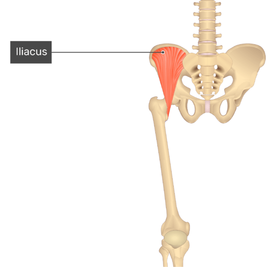
The iliacus muscle is a large muscle with a triangular shape in the pelvic area. It is part of the group of muscles known as the hip flexors, which are responsible for lifting the thigh toward the torso.
The iliopsoas muscle is made up of the iliac triangular muscle, the iliacus, and the psoas major. The iliacus muscle has a spot with the get-together of the inward hip muscles, alongside the psoas minor, psoas major, obturator internus, obturator externus, inferior gemellus, superior gemellus, piriformis, quadratus femoris muscles.
The inner hip muscles work together to affect the hip joint. The work of the iliacus muscle specifically is specified along with that of the psoas major, for example from the part of the iliopsoas muscle. Having said that, the hip joint’s primary flexor is the iliopsoas. Additionally, it aids in the trunk’s lateral flexion. However, the psoas major muscle alone is responsible for the latter movement, which does not involve the iliacus muscle.
Origin of Iliacus muscle
Upper 2/3 of the iliac fossa of the ilium, the internal lip of the iliac crest, the ventral sacroiliac ligament, the lateral part of the sacrum, and the lower region of the iliolumbar ligament
Insertion
Femur’s lesser trochanter. Its fibers are in many cases inserted before those of the psoas major and broaden distally over the lesser trochanter.
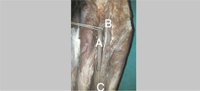
Relations
The iliac fascia separates the pelvic portion of the iliacus muscle from the peritoneum by covering its anterior surface. The lateral femoral cutaneous nerve, the cecum on the right side, and the iliac part of the descending colon on the left side also cross this iliacus surface. The iliacus muscle’s medial margin and the psoas major muscle’s lateral margin are connected to the femoral nerve.
The fascia lata, rectus femoris, and sartorius muscles are posterior to the anterior surface of the iliacus muscle’s thigh portion. The deep femoral artery (profunda femoris) also passes through it. A large subtendinous iliac bursa separates the posterior surface of the iliacus from the hip joint.
Innervation
The femoral nerve (L2-L4) innervates the iliacus muscle.
Blood supply
The iliacus is primarily supplied by the iliolumbar artery, just like the psoas major muscle. It also receives blood from the femoral, obturator, and deep circumflex iliac arteries.
Function of Iliacus muscle
To move the hip joint, the iliacus muscle and the psoas major muscle work together. At the point when its proximal connection is fixed, the muscle adds to the flexion of the thigh. The muscle, on the other hand, works against resistance to move the trunk forward because its distal attachment is fixed. These activities are fundamental for lower limb work like walking, running, and jumping.
Clinical relations
Pain in the iliacus muscle can occur due to a variety of reasons, including overuse, strain, or injury. Symptoms of iliacus muscle pain may include pain in the groin or hip area, difficulty with hip flexion, and stiffness or soreness in the area.
Treatment for iliacus muscle pain may include rest, ice or heat therapy, stretching, physical therapy, and in severe cases, medication or surgery. It is important to seek medical attention if the pain is severe or if it persists for an extended period of time. A healthcare professional can diagnose the underlying cause of the pain and recommend the appropriate treatment.
Shortening of the iliacus muscle
People who work at a desk or who exercise a lot without stretching tend to shorten the iliacus muscle. The shortening of the iliacus muscle brings about its dysfunction and considerable reduction of hip joint motions.
The following symptoms are associated with this kind of injury to the iliacus:
The aggravation of the iliacus muscle is the most extreme of the known trigger points, which are the tight or firm marks of the muscle
Reduced movement on the hip joint and impaired gait
The most widely recognized treatment is an exercise-based therapy that advances extending of the iliacus muscle.
It can become shorter if the muscle is used too much, especially if a person works at a desk every day. This muscle can likewise become more limited through lots of activity without stretching. Injuries and dysfunction in the hip and pelvic areas of the body can begin as this muscle shortens, including:
Trigger points – tight regions or bunches in the iliacus muscle that is painful, stiff, and tender.
Iliacus ischemia is a condition in which the muscle receives less blood.
Problems with the hips, knees, and lower back: A person may walk with imbalanced hips if the iliacus on one side of the hip is shorter than the iliacus on the other side. This can affect a person’s normal gait. ( Try walking with one hip lower than the other; if you do this, you will probably start to feel pain and irritation in your hips, knees, and lower back.) Pain is the most common iliacus dysfunction symptom. The hips, groin, lower back, and upper thigh are all possible locations for this pain. Different side effects include:
- Tenderness
- decreased range of motion
- stiffness
Traumatic iliacus muscle injury is infrequent. it is typically brought about by injury or extraordinary activity including pelvic support; It can cause a hematoma with femoral nerve neuropathy. Patients with coagulation disorders frequently develop spontaneous muscle hematomas. It causes severe pain in the right buttock, groin, and iliac fossa, and inadequate exercise (supine double leg lifts) prevents hip flexion and ambulation. Upper thigh ecchymoses may be present.
Iliacus compartment syndrome
Rare retroperitoneal compartment neuropathy known as iliacus compartment syndrome is characterized by sensory and motor deficits caused by compression of the femoral nerve and bleeding within the iliacus muscle. Iliac hematomas are uncommon and can occur as a result of traumatic or non-traumatic injury, and anticoagulant therapy side effects can exacerbate the condition.
The hematoma’s surgical evacuation can cause a long-term or permanent disability. Be that as it may, there are different reports depicting great healing with non-operative treatment. If radiological studies do not explicitly confirm the presence of a discrete hematoma compressing the femoral nerve, unless the progression of symptoms increases, non-surgical intervention is recommended.
Assessment of Iliacus muscle
Palpation
Place a pillow under the client’s knees and have them lie down. Place the palpating hand on the anterior iliac crest and palpate into the iliac fossa with your fingertips while flexing the knee slightly laterally. The majority of the iliacus cannot be felt. The client should actively flex their thigh at the hip joint, and they should feel the iliacus contracting.
Tightness
The client should stand with their heels (or feet) apart from the wall and their shoulders and head touching the wall. The ability to posterior tilt to touch the small of the back against the wall is typical. The restriction could be shortened iliacus if the client is unable to posterior tilt by flattening their back against the wall with their feet apart and their hips and knees straight, but can do so with their knees bent and their hips flexed.
Iliacus muscle stretching
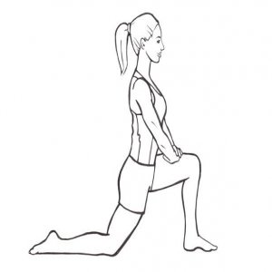
Kneeling Hip Flexor Stretch
With the left leg approximately two feet in front of the right leg, assume a half-kneeling position.
The left knee ought to be bent at 90 degrees. You want to involve a mat for padding.
Put your hands on your left side knee, and keep an erect posture.
Next, on your right side, incline slightly forward until you feel a stretch in the front of your hip, groin, and thigh.
Stand firm on this stooping footing for 20 to 30 seconds. An individual shouldn’t feel any low back aggravation. Release the stretch if you do.
Switch sides and slowly get back to where you started.
On each side, perform the kneeling hip flexor stretch twice to three times.
Iliacus muscle strengthening exercise
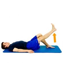
Lying Leg Raises
Leg raises boost the iliopsoas. On a mat, begin lying on your back. To stabilize your pelvis, place your hands under your buttocks. Pull your knees toward your chest without allowing your lower back to lift, then return to the starting position by straightening your legs. To raise the resistance, attempt the activity with straight legs. Leg raises on an incline board make the exercise harder. The exercise will be more difficult the steeper the board angle.
Hanging Leg Raises
The iliacus and psoas major are also targeted by hanging leg raises. Hanging from a pull-up bar with an overhand grip is the first step. Your hands ought to be about the same width as your shoulders. Twist your knees, pulling them up to the hip level or higher, then stretch out your legs down to get back to vertical. To expand the opposition, keep your legs straight as you raise them. A variety of this exercise should be possible by utilizing stomach muscle straps to help your arms.
Situps
The abdominal muscles, including the iliacus and psoas major, are the focus of situps. Lie on your back on a mat. Put your feet on the bottom and flex your knees. Put your fingers together behind your head. Twist your chest area up from the mat, then lower down. Hook your feet under support or have a partner hold them down if they lift off the ground. Situps on a board that is inclined will make the exercise harder.
Scissor Kicks
Your iliopsoas is challenged by scissor kicks with a limited range of motion. On a mat, lie on your back to begin. To stabilize your pelvis, place your hands under your buttocks. While keeping your knee straight, raise one leg off the ground. Lift the opposite leg a little bit off the ground. Continue alternating legs in a scissor-kicking motion after changing positions. To make the activity more straightforward, let your heels, on the other hand, contact the floor with every count.

To make the activity more straightforward, let your heels, on the other hand, contact the floor with every count. To make the activity more straightforward, let your heels, on the other hand, contact the floor with every count.
FAQ
Where are you experiencing iliac pain?
You will see the ache in the hip/groin/low back region, particularly with kicking and pivoting your hip. You might try and notice a snapping sound or feeling.
How can I make my iliacus stronger?
Maintaining the foot flexed, engaged core, and neutral spine and hips, slowly lift one knee at a time in a marching motion. As you raise the leg, try not to lean too much to one side or the other. Instead, engage your core and feel the stability provided by your hip musculature. For each side, repeat five times.
How much time will it take for the iliacus muscle to heal?
Recovery from mild conditions typically takes one to three weeks of rest and treatment. Then again, more serious cases can take around four to about a month and a half or longer.
When the iliacus is tight, what happens?
A tight iliacus will externally rotate your thigh bone, bringing about your foot turning outward. The psoas and the iliacus both add to hip flexion movement, however, their essential occupation is to keep intact the hip, pelvis, and lower back region.
Can back pain be caused by a tight iliacus?
Together, they aid in balancing the tension around the pelvis. If a short, contracted iliopsoas overwhelms a more fragile gluteus maximus on one or the two sides of the pelvis, this can bring about a shortened region of the low back and potentially ache.