
Ulna Bone
Definition
The Ulna and Radius are the two main bones that make up a human’s forearms. Each arm has a single ulna bone. It is a long bone that plays a key role in the formation of the elbow and wrist joints.
The two major bones that jointly make form the forearm are the radius and the ulna. The location where the interosseous membrane, which connects the two forearm bones, attaches. It creates the elbow joint with the Humerus and also connects with the radius both proximally and distally.
When your arm is in the anatomical position, it is situated in the medial forearm. Of the two forearm bones, it is the bigger one. The ulna aids in the forearm and hand’s pronation and supination.
Overview
In the forearm lies a lengthy bone called the ulna. The second forearm bone, the radius, is located medially and parallel to it. The radius pivots to create mobility, while the ulna serves as the Stabilising bone.
The elbow joint is where the ulna and Humerus articulate most closely. The distal radio-ulnar joint is formed when the ulna and radius articulate distally.
This article will examine the ulna’s osteology and bony markers. We will also take into account its clinical relationships.
What is Ulna Bone?
One of the bones in your forearm is the ulna. It facilitates hand, wrist, and arm movement. If your bones get weaker due to osteoporosis, you might not even be aware that you have a higher risk of fractures.
Of the two bones in your forearm, the ulna is the longer one. It facilitates hand, wrist, and arm movement. Your ulna also supports dozens of essential muscles, tendons, ligaments, and blood vessels.
In order to restore your bone structure and restore your range of motion, physical therapy and surgery may be necessary if you suffer a fractured ulna.
Like all bones, your ulna may be impacted by osteoporosis.
Since the ulna is linked to so many other muscles and nerves, damage to one can frequently have an impact on the others.
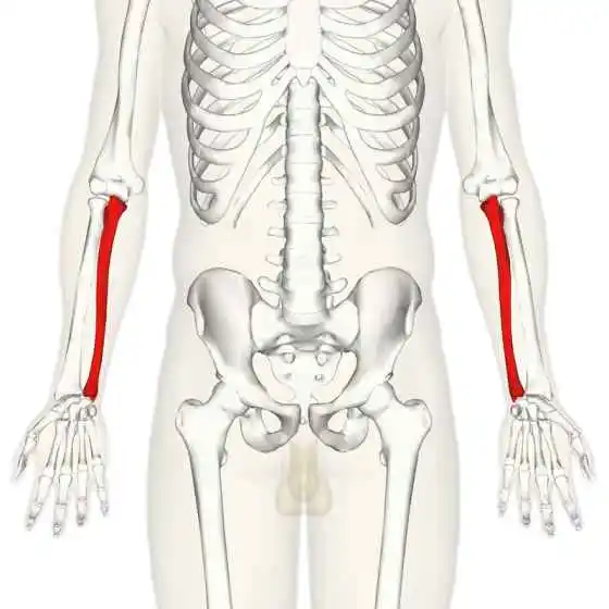
The long bone in the forearm that extends from the elbow to the wrist is called the ulna or ulna bone (plural: ulnae or ulnas). In its anatomical position, it is located on the medial side of the forearm.
That is, the ulna is on the exact same side of the forearm with the little finger. It runs in line with the other long bone in the forearm, the radius. The radius is shorter and the ulna is longer, but the ulna is thinner and the radius is broader.
Thus, of the two bones in the lower arm, the ulna is regarded as the smaller bone. The fibula is the equivalent bone in the lower leg.
Where is The Ulna located?
One of the two bones in your forearm is the ulna. Your radius is the other. The medial (pinkie) side of your forearm is home to the ulna.
It is the forearm’s medial bone, which runs from the wrist area to the elbow and is situated on the side of the little finger, opposite the thumb.
Put another way, the ulna runs parallel to the other lower arm bone, the radius, and is located between the proximal carpal row and the upper arm bone, the humerus.
Anatomy
It is the forearm’s medial bone, which runs from the wrist area to the elbow and is situated on the side of the little finger, opposite the thumb.
In other words, the ulna can be found between the proximal carpal row and the upper arm bone humerus, traveling parallel to the other lower arm bone radius.
The ulna is a long bone in the forearm that extends from the elbow to the wrist. It is located on the medial side of the forearm in its anatomical position. It gets narrower as it gets closer to the wrist and gets broader around the elbow.
The ulna has a notched end where it connects your humerus (upper arm bone), a long shaft in the middle that’s somewhat bent, and a narrow end that meets your wrist. It surpasses the radius by a small amount.
Your ulna is a single long bone, but it is actually composed of several pieces. Among them are:
The proximal part of the Ulna
The trochlea of the humerus articulates with the hook-shaped proximal ulna to produce the hinge joint of the elbow and The ulna has a unique anatomy with bony prominences for muscle attachment that permits movement at the elbow joint.
The coronoid process and olecranon combine to generate the articulation.
Your ulna’s higher (proximal) end joins with your humerus. It is also known as the head of the ulna. It shapes your elbow’s bony tip.

The Olecranon, Coronoid process, Trochlear notch, Radial notch, and Tuberosity of Ulna are significant landmarks of the proximal ulna.
Olecranon
a sizable proximal bone protrusion that is a component of the trochlear notch. It feels like the “tip” of the elbow when touched. Its superior surface is where the triceps brachii muscle attaches.
When the elbow extends, this sizable, curved bony protrusion is accepted into the olecranon fossa on the humerus.
When the elbow bends and extends, the olecranon articulates with the humeral trochlea to form the upper portion of the large, smooth semi-lunar notch.
The olecranon is a broad, thick protrusion that curves and is located in the back and upper portion of the ulna. Its tip is curved forward to create a noticeable lip that extends the forearm and is received into the olecranon fossa of the humerus.
Where it connects the body and the narrowest portion of the upper end of the ulna, its base is constricted. Its rear surface is smooth, subcutaneous, triangular, and covered in a bursa while facing backward.
Its superior surface has a quadrilateral shape, with a rough insertion for the triceps brachii noted in the back and a minor transverse groove near the edge in front that serves as an attachment point for a portion of the elbow joint’s posterior ligament.
Its smooth, concave anterior surface makes up the upper portion of the semilunar notch.
Its boundaries are extensions of the groove on the superior surface edge, and they act as attachment points for the posterior ligament laterally and the rear portion of the ulnar collateral ligament medially.
A portion of the flexor carpi ulnaris comes from the medial border, while the anconeus is linked to the lateral border.
The coronoid process
The coronoid process forms a horizontal, bony protrusion that hooks directly onto the ulnar shaft. It is received by the coronoid fossa of the humerus in elbow flexion. Additionally, the semi-lunar notch’s lower portion is formed by the coronoid process.
On the lateral side of the coronoid process is the radial notch where the head of the radius resides.
This bone ridge, which is a portion of the trochlear notch, protrudes anteriorly.
A triangular protrusion extending forward from the front and upper regions of the ulna is known as the coronoid process. Contingent with the bone’s body, its base possesses significant strength.
Its apex is sharp and somewhat upwardly curved, and it enters the humerus’ coronoid fossa during forearm flexion. Its smooth, concave upper surface creates the bottom portion of the semilunar notch.
Its concave antero-inferior surface is distinguished by a rough imprint where the brachialis inserts. The oblique cord is joined to the lateral border of the tuberosity of the ulna, a rough eminence that affords insertion to a portion of the brachialis, near the junction of this surface and the front of the body.
The radial notch is a thin, oblong, articular depression that is visible on its lateral surface.
Its prominent, free margin on the medial surface allows a portion of the ulnar collateral ligament to connect to it.
A small, rounded eminence at the front of this surface marks the origin of one head of the flexor digitorum superficialis; a depression behind the eminence corresponds to a portion of the flexor digitorum profundus origin; a ridge that descends from the eminence marks the origin of one head of the pronator teres.
A spherical bundle of muscle fibers often originates the flexor pollicis longus from the lower portion of the coronoid process.
The trochlear notch
The coronoid and olecranon processes combine to generate it. It articulates with the humerus’s trochlea and has the shape of a wrench.
The olecranon and coronoid process combined to form the semilunar notch, a sizable depression that functions as an articulation point with the humeral trochlea.
The point where the olecranon and coronoid process converge is marked by an indentation that somewhat shrinks it at the middle of either side of the notch.
The notch is split into a medial and lateral section by a smooth ridge that extends from the tip of the coronoid process to the summit of the olecranon. It is concave from above downward.
The radial notch
It articulates with the head of the radius and is situated on the lateral surface of the trochlear notch.
The circumferential articular surface of the radius head is received by the radial notch, a thin, oblong articular depression located on the lateral side of the coronoid process.
Its large extremities serve as the annular ligament’s attachment point, and it is concave from the front to the back.
Ulnar tuberosity
Immediate distal to the coronoid process, there is a roughening. The brachialis muscle attaches there.
Shaft of Ulna
The ulna shaft has three surfaces and three edges, giving it a triangle form. The width decreases as it approaches the distal end.
The ulna’s body has three main parts: a straight center section, a rounded, smooth lower part, and a curving, prismatic upper part that is convex behind and lateralward. It has three borders and three surfaces, and it tapers progressively from top to bottom.
Surfaces
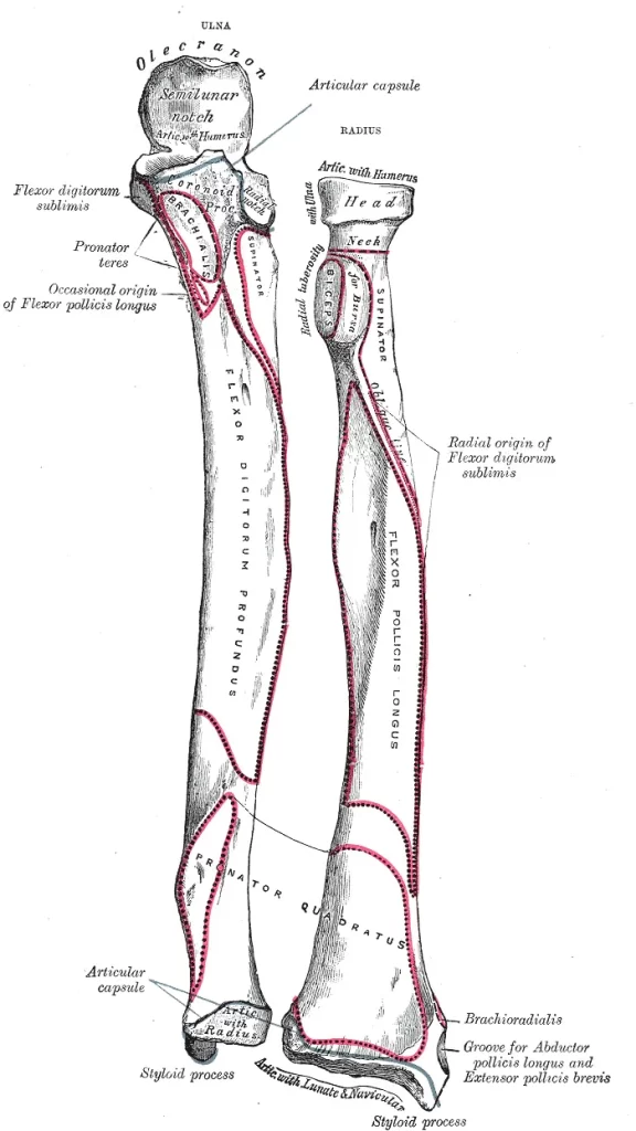
Anterior surface
The quadratus pronator muscle’s distal attachment point.
The flexor digitorum profundus originates from the volar surface (facies volaris; anterior surface), which is much broader above than below. Its lower fourth, which is likewise concave, is covered by the pronator quadratus.
The pronator quadratus’s origin is marked by a ridge that runs obliquely downward and medially, dividing the bottom fourth from the remaining section. The nutrient canal is located at the obliquely upward junction of the middle and upper thirds of the bone.
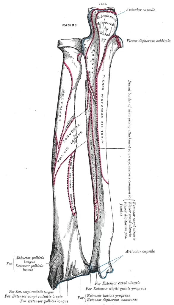
Posterior surface
Numerous muscles attach to the posterior region.
Directed backward and lateralward, the dorsal surface (facies dorsalis; posterior surface) is broad and concave above, convex and considerably narrower in the center, and thin, smooth, and rounded below.
An oblique ridge on its upper portion extends from the dorsal end of the radial notch down to the dorsal border; the Anconaeus inserts itself on the triangular surface above this ridge, and the supinator can be attached to the upper portion of the ridge.
Below this, the surface is split into two sections by a longitudinal ridge that is sometimes referred to as the perpendicular line: the lateral portion, which is wider and rougher, provides the Supinator, the abductor pollicis longus, the extensor pollicis longus, and the extensor indicis proprius with their origin from above downward.
The medial portion is smooth and covered by the extensor carpi ulnaris.
Medial surface
The interior surface, also known as the medial surface (facies medialis), is narrow and convex below and broad and concave above.
The Flexor digitorum profundus originates from its top three-fourths, whereas the lower third is subcutaneous.
Boundaries
Interosseous Border
The location where the interosseous membrane, which connects the two forearm bones, attaches.
The junction of two lines that converge from the extremities of the radial notch and create a triangular space for the origin of a portion of the Supinator between them forms the interosseous crest (crista interossei; also known as the external or interosseous border).
It finishes at the head of the ulna below.
Its lowest fourth is smooth and rounded, while its top section is spiky. This crest divides the volar from the dorsal surface and provides a connection to the interosseous membrane.
Anterior Border
The volar border, also known as the margo volaris or anterior border, starts at the coronoid process’s conspicuous medial angle on top and finishes in front of the styloid process on the bottom.
The pronator quadratus originates from its lower region, whereas the flexor digitorum profundus originates from its well-defined upper part and smooth, rounded middle portion. The medial surface and volar are divided by this boundary.
Posterior Border
Palpable all the way along the forearm’s posterior portion.
The dorsal border, also known as the margo dorsalis or posterior border, is well-marked in the upper three-fourths and attaches to an aponeurosis that provides a common origin for the extensor carpi ulnaris, flexor digitorum profundus, and flexor carpi ulnaris.
Its lower fourth is smooth and rounded. It starts above the apex of the triangular subcutaneous surface at the back part of the olecranon and ends below at the back of the styloid process. The dorsal surface and medial surface are divided by this boundary.
The proximal part of the Ulna
The lateral side of the head of the Ulna features a convex articular surface that allows it to articulate with the radius’s ulnar notch. The distal radio-ulnar joint is formed by it.
Compared to the proximal end, the distal end of the ulna has a substantially lower diameter. It ends in a rounded head and has the ulnar styloid process as its distal projection. Other than that, it is very nondescript. The styloid process is linked to the ulnar collateral ligament.
The head articulates with the ulnar notch of the radius to produce the distal radio-ulnar joint.
The articular surface of the ulna is composed of two parts: the remaining portion, which is directed lateralward and narrow and convex, is received into the ulnar notch of the radius.
The oval or semilunar portion of the head articulates with the upper surface of the triangular articular disc, which separates it from the wrist joint.
The ulnar, which is located close to the wrist, has two eminences: the styloid process, a non-articular eminence, is located on the medial side and is narrower and more projecting than the lateral, articular, rounded eminence known as the head of the ulna.
- There is an Articular surface on the head. A portion of it is oval or semilunar in shape, oriented downward, and articulates with the upper surface of the triangular articular disc that divides it from the wrist joint. The remaining portion is narrow and convex, oriented laterally, and is received into the radius’s ulnar notch.
- The rounded end of The styloid process, which protrudes from the medial and rear portion of the bone and drops slightly beyond the skull, allows it to be attached to the ulnar collateral ligament of the wrist joint.
A shallow groove for the extensor carpi ulnaris tendon and a depression for the attachment of the triangular articular disk’s apex divide the head from the styloid process.
Microanatomy
One lengthy bone is the ulna. The robust cortical tissue wall enclosing the ulna’s long, narrow medullary cavity is thickest along the interosseous border and dorsal surface. The compact layer becomes thinner at the edges.
The compact layer continues as a plate of close, spongy bone with parallel lamellae on the back of the olecranon.
From the inside of this plate and the hard layer below it trabeculæ arch forward towards the olecranon and coronoid and intersect other trabeculæ, traveling backward over the medullary cavity from the upper section of the shaft below the coronoid.
A narrow region of compact bone is beneath the coronoid process, and it is from this area that trabeculae curve upward, ending obliquely at the surface of the semilunar notch, It has a thin coating of dense bone covering it. The orientation of the trabeculæ is more longitudinal towards the lower end.
Articulation and Joints
Upper or Proximal End
Four significant bone features are located at the proximal end of the ulna: the radial notch, trochlear notch, coronoid process, and olecranon process. The ulna’s tuberosity is another noteworthy feature.
Elbow Joint
The elbow is formed by the ulna’s hinge joint with the humerus at its most proximal point. This joint is formed by the trochlea of the humerus sitting in the semi-lunar notch of the ulna.
The trochlear or semilunar notch and the olecranon process produce a broad, curving “C”-shaped surface on the head of the proximal ulna, which resembles a wrench. The bony tip of our elbow, the olecranon, is palpable from the outside.
The trochlear notch’s curved or crescent surface articulates with the humerus’ trochlea to generate the elbow’s hinge joint. To prevent the elbow from stretching above its 180° range during arm extension, the olecranon inserts into the olecranon fossa, a deep indentation or curve in the humerus.
The coronoid process, which is the distal end of the trochlear notch, protrudes anteriorly to fit into the coronoid fossa, a smaller recess in the humerus, when the arm is flexed, further securing the ulna in place.
The tuberosity of the ulna is the rough surface on the front side of the ulna intended for muscular attachments, located directly below the coronoid process.
Radio-Ulnar Joint
Forearm pronation (from the proximal joint) and supination (from the distal joint) result from the ulna’s proximally and distally articulated relationship with the radius.
Via the interosseous membrane, the ulna and radius also articulate in a syndesmosis joint. It extends from the radial notch proximally to the ulna head throughout the length of the shaft. Additionally, this helps to divide the forearm’s volar and dorsal sides.
The radial notch, a small smooth surface located lateral to the trochlear notch at the end of the coronoid process, articulates with the proximal end of the radius to form the proximal radio-ulnar joint, allowing the radius to rotate around the ulna to preserve elbow flexibility.
Shaft of Ulna
The lengthy center portion of the ulna bone is called the shaft or body. The anterior, posterior, and interosseous borders, and the anterior, posterior, and medial surfaces are the three major surfaces and borders of the shaft, which taper gradually as they move towards the distal side.
The shaft’s upper section has a slightly pyramidal shape and is convex on the back and laterally due to a curve. The interosseous border of the ulna is a noticeable ridge that extends down the length of the shaft’s lateral side.
The ulna is attached to the radius at this point by the interosseous membrane of the forearm, a thin fibrous sheet of tissue that binds the two bones together.
Lower or Distal End
With the head of the ulna and a styloid process as its two main bone features, its distal end is significantly narrower than its proximal end.
The head, a small, rounded portion of the distal ulnar surface, articulates with the triangular fibrocartilage articular disc, a cartilage structure that prevents the ulna from creating any direct articulations with the carpal bones, as well as the cup-shaped ulnar notch of the radius.
The distal end of the ulna functions as a pivot such that the radius can rotate in all directions due to the alignment of the ulnar head and ulnar notch.
A tiny bony projection that extends from the head’s posterior medial side is called the styloid process. This is the location of the wrist’s ulnar collateral ligament’s (UCL) attachment.
Muscle Attachment
| Muscle | Direction | Attachment |
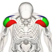
| Triceps brachii muscle | Insertion | The anterior surface of the coronoid process of the ulna |
| Anconeus muscle | Insertion | Olecranon process (lateral aspect) |
| Brachialis muscle | Insertion | The medial surface on the middle portion of the coronoid process (also shares its origin with the medial epicondyle of the humerus) |
| Pronator teres muscle | Origin | Olecranon process and posterior surface of the ulna (also shares origin with medial epicondyle of the humerus) |
| Flexor carpi ulnaris muscle | Origin | The anteromedial surface of the ulna (also shares its origin with the interosseous membrane) |
| Flexor digitorum superficialis muscle | Origin | Coronoid process |
| Flexor digitorum profundus muscle | Origin | The posterior surface of the distal ulna (also shares its origin with the interosseous membrane) |
| Pronator quadratus muscle | Origin | The distal portion of the anterior ulnar shaft |
| Extensor carpi ulnaris muscle | Origin | The posterior border of the ulna (also shares its origin with the lateral epicondyle of the humerus) |
| Supinator muscle | Origin | Proximal ulna (also shares origin with lateral epicondyle of the humerus) |
| Abductor pollicis longus muscle | Origin | The dorsal shaft of the ulna (also shares its origin with the dorsal shaft of the radius and the interosseous membrane) |
| Extensor pollicis longus muscle | Origin | The posterior surface of the distal ulna (also shares its origin with the interosseous membrane) |
| Extensor indicis muscle | Origin | The posterior surface of the distal ulna (also shares origin with the interosseous membrane) |
Blood Supply
The ulnar artery supplies the primary blood supply, as does its branch the common interosseous artery, which splits into the volar and posterior interosseous arteries, both of which continue to supply the ulna.
Nerve Supply Around the Bone
The ulna is innervated on the volar side by the anterior interosseous nerve, which branches from the median nerve, and posteriorly by the dorsal interosseous nerve, which branches from the radial nerve.
Primary Functions
Among the many essential functions performed by your ulna are:
- Facilitating movement, flexion, and rotation of your wrist and forearm.
- Retaining the position of more than 12 muscles.
- Supporting the remainder of your hand, wrist, and arm.
- Facilitating wrist and elbow movement. humerus and elbow joint formation
- Preserving and sculpting the forearm’s structure (together with the radius)
- Attaching to the hand’s and lower arm’s essential muscles and ligaments
- Rotating and moving the wrist by using the carpal and radius bones
For every task involving the hand, including eating, driving, typing, extending or flexing the arm, picking up, throwing, or holding something, the ulna and all of its connected muscles must operate properly.
Development of Bone
There are three distinct sites from which the ulna ossifies; the main center for the shaft appears during the eighth week of fetal development.
The distal and proximal ends’ secondary centers emerge between the ages of 5 and 7, and between 8 and 10 years, respectively.
Approximately between the ages of 18 and 20, all the centers come together.
Anatomical Conditions
Fractures of the ulna
Bone fractures commonly occur in the forearm. We will now examine the most prevalent types of ulna fractures.
An isolated ulnar fracture (without involving the radius)
is typically the result of an item striking the ulna. The most likely place for a fracture is the shaft. In this case, the proximal ulna will be pulled posteriorly by normal muscle tone.
Fractures to the olecranon process are less common. The patient’s landing on a flexed elbow is the cause of this. A portion of the fragment can be moved proximally by the triceps brachii.
The interosseous membrane connects the radius with the ulna. This membrane allows the force of an injury to one bone to transfer to the other.
The following are the most typical causes of ulna fractures:
- Injuries sustained in sports.
- Fall.
- Car accidents.
Among the signs of a fracture are:
- Pain.
- Swelling.
- Tenderness.
- A bone fracture
- Any Discolouration or bruises.
- A lump or malformation.
Particular kinds of ulna fractures consist of:
Monteggia fracture
A fracture involving the dislocation of the radius head and the proximal part of the ulna
radial head dislocation combined with a fracture of the proximal third of the ulnar shaft.
usually brought on by an external force behind the ulna. The head of the radius dislocates anteriorly at the elbow, and the proximal shaft of the ulna is shattered.
Hume fracture
An olecranon fracture accompanied by an anterior radial head dislocation
anterior radial head dislocation concomitant with olecranon fracture.
Galezzi’s fracture
Distal radius fracture combined with dislocation of the ulnar head at the distal radio-ulna joint.
When ulnar fractures occur in the distal two-thirds, solely affect the shaft, have no shortening, less than 10° angulation, and less than 50% displacement, conservative therapy may be an option.
ulna head dislocation at the distal radio-ulnar joint due to a distal radius fracture.
Osteoporosis
Osteoporosis weakens bones, increasing the likelihood of abrupt and unexpected fractures.
Many people don’t realize they have osteoporosis until it breaks a bone because it typically doesn’t present any noticeable symptoms.
People over 65 and women or those identified as female at birth (AFAB) are more likely to have osteoporosis.
Talk to your doctor about getting a bone density test to help avoid fractures and identify osteoporosis early.
Keeping the ulna in good health
You can take care of your bone health (as well as overall health) by eating correctly, exercising, and getting regular checkups from your doctor.
If you are over 50 or if osteoporosis runs in your family, talk to your doctor about having a bone density scan.
To lower your chance of getting hurt, heed this general safety advice:
- Wear your seatbelt at all times.
- When taking part in any sport or activity, wear the proper protective gear.
- Make sure nothing in your home or place of work could trip you or anybody else.
- When reaching for anything at home, always use the appropriate tools or equipment. Never stand on worktops, tables, or chairs.
- Adhere to a diet and exercise regimen that will support the health of your bones.
- If you have trouble walking or are prone to falling, use a cane or walker.
FAQs
What is the ulna bone?
The antebrachium is made up of the radius and the ulna, two of the long bones of the forearm. In its anatomical position, the bone on the medial side of the forearm extends from the elbow to the wrist. The ulna is characterized as being longer and larger than the radius. Make sure nothing could injure you or anybody else in your house or place of employment.
It’s crucial to always reach for objects around the house with the appropriate tools or equipment. Avoid standing on chairs, tables, or worktops.
What is the head of the ulnar bone?
The ulna’s lower extremity is tiny and has two eminences: the styloid process, a non-articular eminence that is more protruding, and the lateral, bigger, rounded eminence known as the head of the ulna.
Where is the ulna bone located?
One of the two bones that gives the forearm its structure is the ulna. On the other side of the forearm from the thumb is where the ulna is situated. It forms the elbow joint by joining the humerus at its bigger end with the hand’s carpal bones at its smaller end.
What is the structure and function of the ulna?
It forms the elbow joint with the humerus and articulates with the radius both distally and proximally. When the arm is in the anatomical position, it is situated in the medial forearm. Of the two forearm bones, it is the bigger one. The ulna aids in the forearm and hand’s pronation and supination.
Why is the ulnar called funny bone?
The funny feeling you experience after hitting the “funny bone” is what gave it its nickname. However, your amusing bone isn’t a bone at all. A nerve known as the ulnar nerve runs along the inside of your elbow. Your brain receives information about sensations in your fourth and fifth fingers from the ulnar nerve.
What is the ulna styloid bone?
The ulnar styloid process is a bony projection located close to your hand at the end of the ulna. It is a key component of the strength and flexibility of your wrist and forearm, fitting into the cartilage of your wrist joint. An ulnar styloid fracture is the term for any type of break in this region.
Reference
- Ulna. (n.d.). Physiopedia. https://www.physio-pedia.com/Ulna
- S. (2022, September 16). Ulna – Definition, Location, Anatomy, Functions, Labeled Diagram. TheSkeletalSystem.net. https://www.theskeletalsystem.net/arm-bones/ulna.html
- The Ulna – Proximal – Shaft – Distal – TeachMeAnatomy. (2020, January 20). TeachMeAnatomy. https://teachmeanatomy.info/upper-limb/bones/ulna/
- Professional, C. C. M. (n.d.). Ulna. Cleveland Clinic. https://my.clevelandclinic.org/health/body/24520-ulna
- Ulna. (2023, December 19). Wikipedia. https://en.wikipedia.org/wiki/Ulna


One Comment