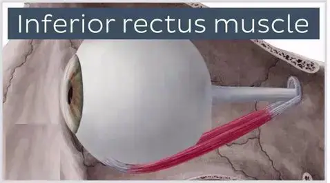
Inferior Rectus Muscle
Introduction :
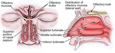
The inferior rectus is one of the seven extraocular muscles and is primarily responsible for letting the eye down (down gaze). The inferior rectus is one of the rectus muscles, which also has the superior rectus, the medial rectus, and the lateral rectus.
There are two oblique muscles also, the superior oblique and the inferior oblique. And the seventh extraocular muscle is the levator palpebrae superioris. The inferior rectus muscle is a muscle near the orbit of the eye. It is one of the four rectal muscles in the bunch of extra-ocular muscles. It starts from the common tendinous ring and inserts into the anteroinferior surface of the eye. It leads the eye down (downwards).
Structure and Function of inferior rectus muscle:
With the head looking straight and the eyes looking straight ahead, the eyes have to be in primary gaze. From this position, a movement of an extraocular muscle produces a secondary or tertiary action. Even the globe can be moved about 50 degrees from the primary position, usually while normal eye movement, only 15 degrees of extraocular muscle movement occurs before the head motion begins. The annulus of Zinn is the usual site of origin of the rectus muscles and fans to the superior orbital fissure. It consists of superior and inferior tendons.
The superior tendon is involved with the entire superior rectus muscle as well as parts of the medial rectus and lateral rectus muscles. The inferior tendon is involved with the inferior rectus muscle and parts of the medial rectus and lateral rectus muscles. The inferior rectus has the primary movement of depressing the eye, causing the cornea and pupil to move interiorly.
The inferior rectus starts from the Annulus of Zinn and courses anteriorly and laterally along the orbital floor, having an angle of 23 degrees to the visual axis. This angle causes the secondary and tertiary movement of the inferior rectus muscle to be adduction and extorsion (excycloduction). Each of the extraocular muscles has a functional insertion spot, which is at the nearest point where the muscle first contacts the globe. This point creates a tangential line from the globe to the muscle-originating point and is known as the arc of contact. The inferior rectus inserts in the vertical meridian, approximately 6.5 mm from the limbus.
Origin and Insertion of inferior rectus muscle :
The inferior rectus muscle is a narrow and strap-shaped muscle of the orbit that extends over the floor of the orbit. Alike most of the extraocular muscles, the inferior rectus muscle starts from the common tendinous ring, which is also called the annulus of Zinn, that is found in the posterior pole of the orbit covering the margins of the optic canal.
As the muscle goes anterolaterally across the floor of the orbit, its middle part is thickened and then slowly thinned into a tendon. The tendon ends by inserting obliquely on the anteromedial surface of the eyeball, which is below the limbus of the cornea.
Embryology :
The mesenchyme of the head, adding the orbit and its structures, rises in mesoderm and neural crest cells primarily. The extraocular muscles rise from the mesoderm, but the satellite and connective tissue of the muscle comes from neural crest cells. Most of the remaining connective tissue of the orbit also comes from neural crest cells.
Blood Supply and Lymphatics :
The inferior rectus conceives blood primarily from the inferior muscular branch of the ophthalmic artery, with a secondary contribution from the infraorbital artery. The primary blood supply for all of the extraocular muscles is the muscular distribution of the ophthalmic artery, the lacrimal artery, and the infraorbital artery. The two muscular parts of the ophthalmic artery are the superior branch and the inferior muscular branch.
Venous drainage is the same as the arterial system and empties into the superior and inferior orbital veins. Generally, there are a total of four vortex veins, and these are found on the lateral side and medial side of the superior and inferior rectus muscles. These vortex veins drain at the orbital venous system.
Nerves :
The inferior rectus is innervated by the lower branch of cranial nerve III (oculomotor nerve). Cranial nerve III is divided into upper and lower branches, with the upper branch innervating the superior rectus, levator palpebrae superioris, and the lower branch into the medial rectus, inferior rectus, and inferior oblique. The lateral rectus is innervated by cranial nerve VI (abducent), and the superior oblique is supplied by cranial nerve IV (trochlear).
Additionally, the parasympathetic innervation of the sphincter pupillae and ciliary muscle courses with the branch of the lower division of cranial nerve III that supplies the inferior oblique, and this pass near the inferior rectus. This has important surgical considerations that will be explained next.
Muscles
The inferior rectus and the superior rectus are the muscles that are vertically situated. The medial and lateral rectus muscles are the horizontally situated rectus muscles. Each of the rectus muscles arises posteriorly at the Annulus of Zinn and courses anteriorly.
Each of the rectus muscles inserts on the globe at different distances from the limbus, and the curved line drawn between the insertion points makes a spiral that is known as the Spiral of Tillaux. Originating at the medial aspect of the globe, the medial rectus inserts at 5.5 mm from the limbus, the inferior rectus ends at 6.5 mm from the limbus, the lateral rectus ends at 6.9 mm from the limbus, and the superior rectus at 7.7 mm from the limbus.
The inferior rectus is 9.8 mm wide at its ending point on the globe. The tendon is 7 mm, calculated from the origin. The whole length of the muscle is 40 mm.
Extraocular muscles have a large ratio of nerve fibers to skeletal muscle fibers. The ratio is 1:3 to 1:5, compared to other skeletal muscles, which are 1:50 to 1:125. Extraocular muscles are a specialized type of skeletal muscle with a variety of fiber types, adding both slow tonic types which resist fatigue, and also saccadic (rapid) type muscle fibers.
Development :
The inferior rectus develops from the embryonic mesoderm in the orbit of the skull. This is similar to the remaining extraocular muscles.
Relations :
The insertion of the inferior rectus muscle is 6 mm from the insertion of the medial rectus muscle, and 8 mm from the insertion of the lateral rectus muscle. A parasympathetic branch that supplies the ciliary muscles of the pupil goes close to the inferior rectus muscle.
Strabismus :
If the inferior rectus muscle is injured, weak, or paralyzed, this can cause strabismus. This can result in elevation of the eye, as the superior rectus muscle remains stronger. For minor muscles, prism glasses can be used to gradually realign the eye. Alternatively, for serious cases, it may be operatively corrected by slightly weakening the superior rectus muscle (opposite) – by this, there is a reduction in the elevation of the eye, and it corrects the strabismus. This procedure may result in overcorrection of the strabismus, but is otherwise generally successful.
Physiologic Variants :
The size of the inferior rectus muscle, as well as its insertion point on the globe from the limbus and other anatomical measurements, may vary largely from one individual to the next. The numbers described in this article may reflect average distances.
Congenital differences in extraocular muscles can cause ocular misplacement. See the Clinical Significance section for further details regarding strabismus.
Variation :
Rarely, the inferior rectus muscle is congenitally absent. This may lead to inferior rectus palsy, where the eye cannot be depressed.
Surgical Considerations :
Because the parasympathetic fibers of the sphincter pupillae and ciliary muscle travel with the innervation to the inferior oblique, and the inferior oblique crosses the inferior rectus laterally, there are potential complications from surgery in this area. If the parasympathetic fibers are injured during surgery in this area, pupillary abnormalities may result.
The inferior rectus also interacts with the lower eyelid via a facial connection. Weakening or recession of the inferior rectus may widen the palpebral fissure, and this can lead to the lower lid drooping. Conversely, strengthening or resection of the inferior rectus may lead to the fissure narrowing and elevating the lower lid.
The nerves of the rectus muscles and superior oblique muscles insert into the muscles at one-third from the origin to the insertion. This leads to injury to these nerves during anterior segment surgery difficult, but not impossible. Additionally, instruments that are advanced 26 mm posterior to the rectus muscle insertions can cause damage to the nerve.
The vessels of blood may be compromised during surgery of the inferior rectus muscle. The vessels which supply blood to the extraocular muscles also closely supply the temporal half of the anterior segment of the eye. Most of the nasal half of the anterior segment circulation also originates from blood vessels that supply the extraocular muscles. Therefore, care should be taken during surgery of the medial rectus or other extraocular muscles to avoid disrupting this blood supply.
There are other complications that result from inferior rectus surgery, which also results in other rectus muscle surgery. Unsatisfactory alignment is the most common complication and may need additional surgery to correct this. Refractive changes may happen when two rectus muscles of one eye are operated on, and this may resolve over months. Additional possible surgical complications include diplopia, perforation of the sclera, and postoperative infections.
Clinical Significance :
The function of the inferior rectus muscle can be assessed along with the other extraocular muscles during the clinical assessment. The action of the extraocular muscles can be assessed by having the patient look in nine directions, starting with a primary, followed by the secondary positions (up, down, left and right) and the tertiary positions (up and right, up and left, down and right, down and left). The clinician can test these positions by having the patient follow the clinician’s finger and trace a broad letter “H” in the air.
Further tests of ocular alignment can be tested further by different methods, including cover tests, corneal light reflex, dissimilar image tests, and dissimilar target tests. Since many patients with extraocular muscle abnormalities are young children, the clinician may have to employ various clever means such as using toys or other objects to elicit the cooperation of the child.
Strabismus, or ocular misalignment, can happen because of abnormalities in binocular vision or abnormalities of neuromuscular control. Weakness, damage, or paralysis that involves the inferior rectus muscle can be involved in strabismus.
Because the inferior rectus is near the orbital floor, orbital floor fractures involve this muscle. Inferior rectus muscle paresis can be a result of trauma to the inferior rectus muscle or nerve. This can happen either at the time of the initial injury or during surgical repair of the orbital floor. If inferior rectus muscle paresis is present without entrapment, the patient may show hypertropia in the primary position. If paresis is present with entrapment, the patient has a slight deviation or even slight hypotropia which decreases with downgaze. The treatment of inferior rectus paresis is observation for six months. If there is no improvement in this, muscle surgery may be recommended.
Other issues :
The inferior rectus has a muscle capsule that surrounds the muscle capsule of the inferior oblique. This fibrous connection is known as Lockwood’s ligament and attaches to the lower eyelid retractors.
FAQ :
1. Where is the inferior rectus muscle located?
orbit
The inferior rectus muscle goes along the floor of the orbit superior to the infraorbital canal, which gives space to the infraorbital artery. As it inserts onto the anteromedial surface of the sclera, the inferior rectus muscle is encircled by the inferior oblique muscle.
2. What do the superior and inferior rectus muscles do?
The superior rectus and inferior oblique muscles primarily move the eye superiorly. The inferior rectus and superior oblique muscles primarily move the eye inferiorly. The lateral rectus moves the eyes horizontally and laterally (abduction). The medial rectus muscle moves the eyes horizontally and medially (adduction).
3. What causes inferior rectus palsy?
Isolated inferior rectus (IR) muscle palsy may happen after orbital trauma. Many mechanisms have been proposed, including muscle contusion, longitudinal splitting of the muscle, transaction or destruction of the muscle, posterior muscle slippage within its sheath, and nerve injury (damage to the oculomotor nerve).
4. What is inferior rectus muscle entrapment?
Entrapment. Inferior rectus entrapment. Orbital fractures typically occur through blunt periocular trauma and are one of the most common types of facial fractures. And a very important complication of orbital fractures is entrapment of extraocular muscles and orbital fat in a trapdoor fracture
5. What causes eye muscle imbalance?
The reasons for eye misalignment are various and sometimes unknown. Potential reasons include high farsightedness, thyroid eye disease, cataract, eye injuries, myasthenia gravis, cranial nerve palsies, and in some patients it can be caused by brain or birth problems.