
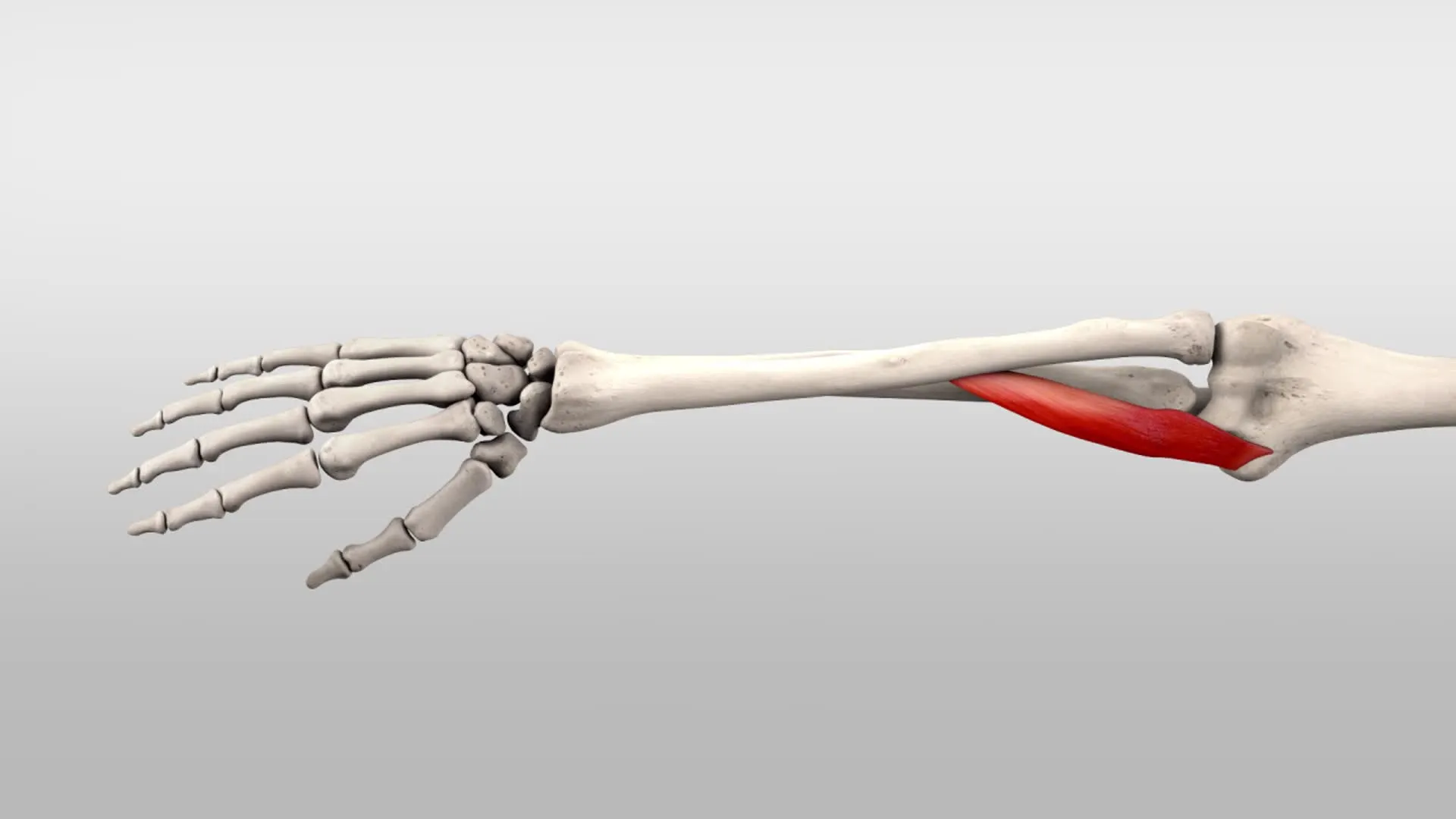
Pronator Teres Muscle
The pronator teres muscle is a forearm muscle that originates from two separate heads. The first head arises from the medial epicondyle of the humerus, while the second head originates from the coronoid process of the ulna. The two heads join together to form a single muscle belly, which inserts onto the lateral aspect of the radius bone.
What is Pronator Teres?
The anterior forearm is home to the fusiform muscle known as the pronator teres. Along with the flexor carpi radialis, palmaris longus, flexor digitorum superficialis, and flexor carpi ulnaris muscles, it is one of the forearm’s superficial flexors. This group’s most lateral muscle is the pronator teres. It has two heads that are named after the bones from which they come;
Head of the humerus that comes from the humerus’ distal aspect
Head of the ulna that comes from the ulna’s coronoid process
Origin of Pronator teres muscle
Origin of Humeral Head: immediately above the common flexor tendon, deep antebrachial fascia, and medial epicondyle of the humerus.
Origin of Ulnar Head: the medial side of the ulna’s coronoid process.
Insertion
middle of the radius’s lateral surface.
Relations
Pronator teres are the most lateral of the forearm’s superficial flexors because they are directly lateral to the flexor carpi radialis muscle. The brachioradialis muscle covers the muscle’s distal anterior surface, while the muscle lies deep to the flexor digitorum superficialis in its proximal portion.
The medial margin of the cubital fossa (or elbow pit) is formed by the Pronator teres muscle. There are several pronator’s teres-related neurovascular structures in this fossa. Near the top of the pronator teres, the brachial artery branches off into the ulnar and radial arteries. The radial artery follows its upper edge as the ulnar artery passes posteriorly to the muscle after branching off. Between the two pronator teres heads are the median nerve; The nerve and the ulnar artery are separated by the ulnar head of the muscle.
Innervation
The median nerve, which is a branch of the brachial plexus (C5-T1) and has root values C6 and C7, supplies the Pronator teres muscle with its innervation.
Blood supply
Three arteries are responsible for the pronator teres muscle’s vascularization:
Branches of an ulnar artery: typical interosseus artery, anterior ulnar recurrent artery
Branch of a radial artery: radial recurrent artery
Branches of a brachial artery: inferior ulnar collateral arteries
Function of the Pronator teres muscle
The primary function of the pronator teres muscle is to pronate the forearm, which means it rotates the forearm inwards so that the palm faces downwards. This movement is important for many activities such as turning a doorknob or using a screwdriver. The pronator teres muscle also assists in flexion of the elbow joint.
Pronation of the forearm, an exclusive upper limb movement, is the primary action of the pronator teres, as its name suggests. The muscle pulls the radius medially, causing the ulnar proximal radioulnar joint’s head to rotate around the ulnar proximal part.
The palm is also rotated by this action, causing it to pronate, or face the ground. The pronator teres also aid in forearm flexion because they cross the elbow joint.
Clinical significance
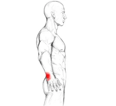
Injuries to the pronator teres muscle can occur due to overuse, trauma or repetitive strain, which can lead to pain, weakness, and limited range of motion in the forearm. Treatment for pronator teres muscle injuries typically involves rest, ice, compression, and physical therapy. In severe cases, surgery may be required.
One of the causes of wrist pain is pronator teres syndrome.
Patients with the pronator teres condition have deadness in middle nerve dissemination with tedious pronation/supination of the lower arm, not flexion and augmentation of the elbow
The early weariness of the lower arm muscles is seen with redundant unpleasant movement, particularly pronation
EMG might show just somewhat decreased conduction speeds
Regardless of their anatomic nearness, patients with pronator teres condition don’t have a higher frequency of AIN disorder
Further areas of compression:
- Ligament of Struthers
- Lacertus fibrosis
- Proximal arch of the FDS
- Rare reasons like observing tendon transfers for radial palsy
Contrast to CTS:
- Positive Tinel’s sign in the forearm instead than at the wrist
- Negative Phalen’s maneuver
- Dysesthesia of palmar triangle
- Pain on resistance to pronation
- Pain in the forearm on resistance to isolated flexion of the PIP joint of long and ring fingers
The pronator teres tendon can be rerouted to the extensor carpi radialis brevis tendon to restore wrist extension in patients with C5 tetraplegia or radial nerve palsy, a procedure known as tendon transfer.
Pronator teres syndrome
A neuropathy known as pronator teres syndrome is brought on by the compression of the median nerve at the forearm’s proximal aspect. The pronator teres muscle typically compresses the median nerve between its two heads. Acute nerve compressions as a result of muscle inflammation (myositis) or elbow injury can lead to this condition. Long-term compression, also known as honeymoon paralysis because it usually occurs when one partner sleeps on the other’s arm, is another possibility.
Numbness and/or pain in the median nerve’s innervation region, as well as malfunctioning of the index finger’s flexor pollicis longus, flexor digitorum profundus, and pronator quadratus, are the primary clinical signs. A neurological exam and imaging techniques (such as an MRI scan) are used to make the diagnosis.
Assessment
Palpation
At the medial epicondyle of the humerus, the common flexor tendon gives rise to the pronator. The cubital fossa’s medial border is formed by its oblique path from medial to lateral. To palpate it, pronate or supinate the forearm and feel the antecubital fossa most slightly medially.
Strength
The patient is lying down or supine. To prevent shoulder abduction, the examiner should hold the elbow against the patient’s side or stabilize it.
Test: Forearm pronation with the elbow partially flexed. Pressure: In the direction of supinating the forearm, at the lower forearm, above the wrist (to avoid twisting the wrist).
Pronator teres stretching
Common wrist flexors stretch
Stretch your forearm muscles by doing this. Rotate your wrist so that your fingers point toward the ground and place your hand against a wall. Hold for 3-5 exhalations.

Pronator teres strengthening
Dowel wrist stars
This is an exercise for strengthening the forearm and wrist in multiple directions. While situated, clutch one finish of a dowel/broomstick while supporting the lower arm with your thigh. From here, use the broomstick’s tip to make a star by moving the wrist. When drawing the star, make the largest possible movements to achieve maximum intensity.
Dowel wrist rotation
The forearm muscles are the focus of this wrist-strengthening exercise. While sitting, support your forearm on your thigh with one end of a dowel or broomstick. To rotate the stick-up and over, turn your wrist. This exercise will be easier if you grab the stick closer to the center while grabbing the stick closer to the end will make it harder.
Banded wrist pronation
Banded wrist pronation is an exercise for strengthening the wrist’s pronators. Beginning with the pinky side and working your way toward the thumb, place a band in your palm and loop it once around your hand. Make sure the loop ends by wrapping around the back of your hand and grip the band with your thumb. With the hand that is not the target, hold the other end of the band. Rotate the band until your palm is facing down, starting with your palm facing upward. To finish a repetition, reverse the rotation.

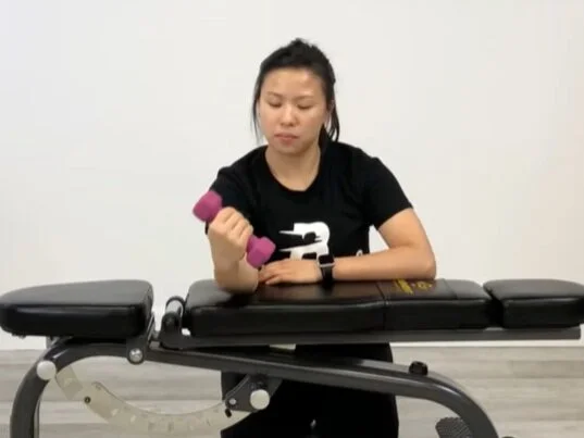
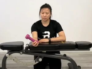
Wrist flip
Wrist flip This exercise strengthens the wrist. Hold a dumbbell while supporting your forearm with a bench, table, or chair. To lift the weight against gravity, extend your wrist, starting with your wrist facing down. After that, flex the wrist and rotate the wrist so that the palm is facing upward to raise the weight against gravity once more.
Half-wrist rotation holds
This is a reinforcing exercise for the wrist and lower arm. A bench, table, or chair can help support your forearm. While the hand is hanging off the edge of the bench, grasp one end of a small weight. After five seconds of holding the weight at a 45-degree angle, rotate the wrist to hold the weight at a 135-degree angle. Alternate sides after holding for five seconds and repeat as necessary.
Isometric wrist pronation
Isometric wrist pronation This exercise places rotational stress on the wrist and forearm muscles with a kettlebell. To help isolate the muscles in your forearms, pin a rolled-up towel or yoga block between your elbow and body. Flex the elbow to 90 degrees of flexion while holding the weight by the handle. To bring the kettlebell into a bottoms-up position and allow it to slightly tilt outward of the forearm, supinate the wrist. Maintain this stance for 5-7 seconds. Avoid full-range supination because it will rely on your passive tissues rather than your wrist and forearm pronators to hold the weight.
Dowel ABC’s
This is a wrist and forearm strengthening exercise called Dowel ABCs. Hold one finish of a dowel or broomstick out before you so the opposite end contacts the ground. From here, move the wrist to use the broomstick’s tip to draw the alphabet. While writing the ABCs, try to move as much as you can to get the most intensity.
FAQ
In the pronator teres muscle, which nerve has been damaged?
When the median nerve in the upper forearm is compressed, pronator syndrome, also known as pronator teres syndrome (PST), occurs. The middle nerve is one of the three nerves that permit our furthest point to detect and development starts in the upper arms and its branches stretch out into the fingers.
How long do pronator teres take to recover?
Conservative treatment works well for the vast majority of pronator syndrome patients. Under the watchful eye of a hand specialist, three to six months of rest from the problematic activity, splinting, and the use of nonsteroidal anti-inflammatory drugs (NSAIDs) to reduce inflammation may be all that is required for symptoms to improve.
How should the pronator teres muscle be massaged?
The press-and-move massage suggests pressing on the muscle and moving it afterward. Thus, pronate your lower arm palms down position, put a few fingers on the muscle, and afterward leisurely supinate and pronate your lower arm around 10 to multiple times.
What sets pronator teres syndrome apart from carpal tunnel syndrome?
Compression of the median nerve as it travels through the forearm’s anatomical structures can lead to pronator syndrome, whereas carpal tunnel syndrome refers to a specific topographic area where compression occurs, the carpal tunnel.
What exactly is pronator teres inflammation?
Tingling or numbness in the palm, thumb, and three fingers—but not in the little pinky finger—are signs of pronator teres syndrome, which can be very similar to carpal tunnel syndrome. When you feel or press on the arm’s pronator teres muscle, you’ll feel pain and tenderness in your forearm.


3 Comments