
Internal abdominal oblique muscle
What is the Internal abdominal oblique muscle?
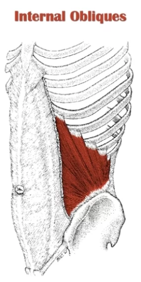
The internal abdominal oblique muscle, also known as the internal oblique muscle, is one of the muscles that make up the anterolateral abdominal wall. It is located deep to the external abdominal oblique muscle and superficial to the transverse abdominal muscle. The internal oblique muscle is paired and present on both sides of the abdomen.
The three distinct layers of the lateral abdominal wall are the external abdominal oblique, internal abdominal oblique, and transversus abdominis, which run from the superficial to the deep.
Originating from the lumbar fascia, the iliac crest, and the inguinal ligament, the internal oblique muscle inserts onto the lower three ribs, linea alba (a fibrous structure that runs vertically down the midline of the abdomen), and the pubic crest. It runs at an angle that is roughly perpendicular to the external oblique muscle.
The primary function of the internal abdominal oblique muscle is to assist in trunk rotation, lateral flexion (side-bending), and compressing the abdominal contents. When both sides of the internal oblique muscle contract simultaneously, they flex the trunk forward, compressing the abdominal viscera (organs) and increasing intra-abdominal pressure. When the muscles contract on one side, they laterally flex the trunk to the same side and rotate it to the opposite side.
The internal oblique muscle also plays a role in stabilizing the trunk, especially during movements like lifting, coughing, sneezing, and defecation. It works in conjunction with the other abdominal muscles, including the external oblique, rectus abdominis, and transverse abdominis, to provide overall core stability and support.
The oblique orientation of its fibers is perpendicular to those of the external abdominal oblique, as its name suggests. Together with the further abdominal muscles, the internal abdominal oblique is essential for motions of the trunk, keeping normal abdominal tension, and raising intra-abdominal pressure.
Origin
Thoracolumbar fascia, the lateral two-thirds of the inguinal ligament, and the anterior two-thirds of the iliac crest’s intermediate line.
Insertion
Four lower ribs, an abdominal linea alba aponeurosis, and a pubic crest.
Relations
The internal abdominal oblique muscle is one of the lateral abdominal wall’s three layers. It is located superficial to the transverse abdominis and deep in the external abdominal oblique.
The large aponeurosis of the anterior abdominal wall, the rectus sheath, and the lateral fibers of the internal abdominal oblique muscle are continuous. The rectus abdominis and pyramidalis muscles, as well as numerous neurovascular structures of the anterior abdominal wall, are largely enclosed within the rectus sheath.
The rectus abdominis muscle was surrounded by the upper three-quarters of the internal abdominal oblique muscle’s aponeurosis, which split into deep and superficial layers below the costal margin. The rectus sheath’s posterior layer is made up of the deep layer and the transversus abdominis aponeurosis. In contrast, the anterior layer of the rectus sheath is formed when the superficial layer and the aponeurosis of the external abdominal oblique combine.
The aponeuroses of the external abdominal oblique, internal abdominal oblique, and transversus abdominis converge and are located anterior to the rectus abdominis muscle around the level of the lower quarter of the aponeurosis of the internal oblique muscle. The arcuate line, which is 2.5 centimeters below the umbilicus, marks the point at which they meet.
Innervation
The lower six thoracic spinal nerves are the terminal branches of the lower five intercostal nerves and the subcostal nerve, which supply the internal abdominal oblique muscle (T7-T12). The iliohypogastric and ilioinguinal nerves (L1) also contribute to this muscle’s nervous supply.
Blood supply
The following arteries supply the internal abdominal oblique muscle with arterial blood:
- Lower posterior intercostal and subcostal arteries
- Superior and inferior epigastric arteries
- Superficial and deep circumflex iliac arteries
- Posterior lumbar arteries
Function
The internal abdominal oblique muscle performs various functions depending on which areas of the muscle contract. The internal abdominal oblique flexes the trunk when both muscles contract. It simultaneously increases intra-abdominal pressure by compressing the intra-abdominal viscera. For functions like forced expiration, urination, and defecation, among others, this action is used. The anterior part of the pelvis is lifted and the degree of pelvic tilt is altered by the bilateral contraction of the internal abdominal oblique if the rib cage is fixed. Upon one-sided contraction, the internal abdominal oblique causes ipsilateral flexion of the trunk and an ipsilateral turning of the trunk.
The internal abdominal oblique muscle is essential for maintaining normal abdominal wall tension, along with other abdominal wall muscles. As a result, these muscles’ tonic contraction serves both a supporting and protecting function. Abdominal hernias are also more likely to occur when the internal abdominal oblique or other abdominal muscles are weak.
Clinical relevance
Injury or strain to the internal abdominal oblique muscle can occur during activities involving sudden twisting or bending of the trunk, excessive lifting, or repetitive movements. Symptoms may include pain, tenderness, and weakness in the affected area. Rest, ice, compression, and physical therapy exercises are typically prescribed for the treatment and rehabilitation of such injuries.
Abdominal hernias are more likely to occur if the internal abdominal oblique and other abdominal muscles are weak.
Internal oblique muscle exercise
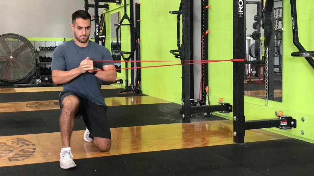
Single leg pallof press ( Contralateral )
This is a rotation-preventing core stability exercise. Stand just with the leg that is nearest to the band and anchor a band close to you so it is about midsection level. Begin by holding the band in both hands and slowly extending your elbows out in front of you. With no side bending or body rotation, hold the outstretched position for up to five seconds.
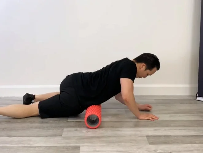
Oblique foam rolling
This is a technique for oblique soft tissue mobilization. Place a foam roller between your pelvis and your 12th rib, also known as your bottom rib, and lie down on it. By supporting the body with your elbow and top leg as needed, you can lessen the amount of stress on the obliques. On a tender spot, hold for up to 30 seconds, or 3-5 breaths. There may be three to four tender areas; repeat as required. Foam rolling is typically restricted to two to three times per week to maximize recovery.
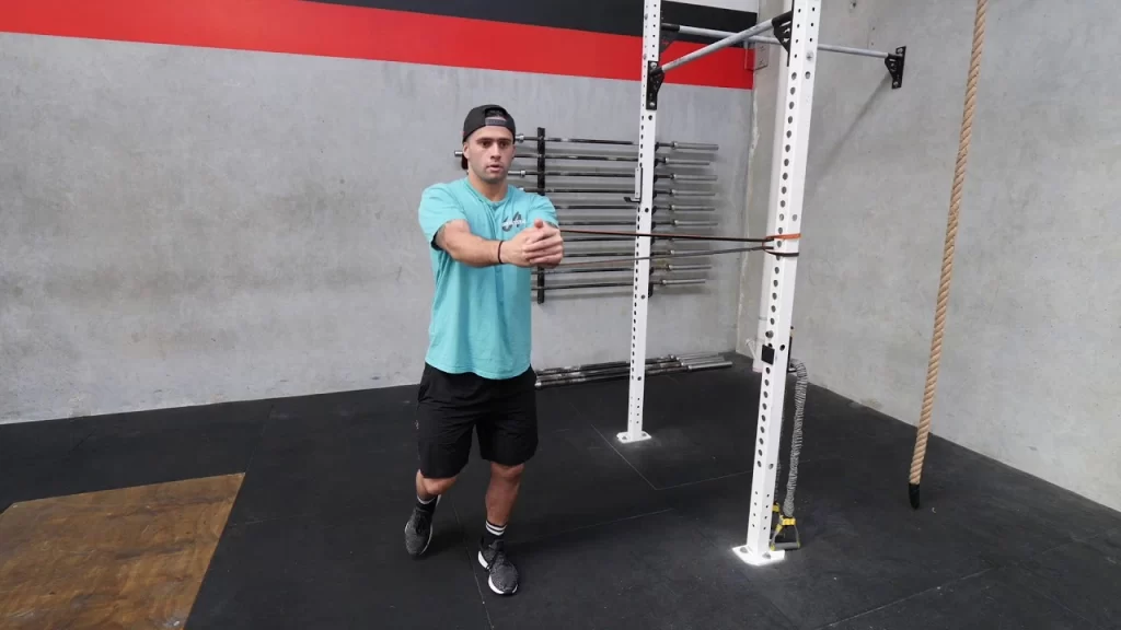
Half kneel pallof press
The purpose of this exercise is to strengthen the spine’s anti-rotation stability. Using a cable pulley system or a band, you can perform this exercise. Hold a band in the middle of your chest with some tension by anchoring it to the side of a sturdy object. Kneel on one leg with the leg in front of you that is closest to the band. Press your hands forward straightforwardly before you at generally shoulder level and hold for 5-10 seconds or for 3-5 full breaths. Maintain an upright, neutral spine by stacking your shoulders directly above your hips.
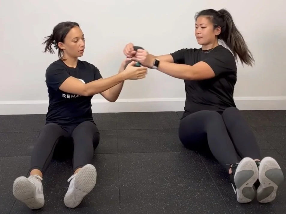
Russian twist pass
Using a kettlebell is a partner core exercise. Sit back to back with your exercise partner to get started. Keep your legs together in front of you with your knees bent. To begin, one person holds a kettlebell and pulls the weight across their body by rotating their torso. From this point on, give the kettlebell to your partner and have them perform the same movement. Pivot the body back to get the iron weight. In both directions, complete.
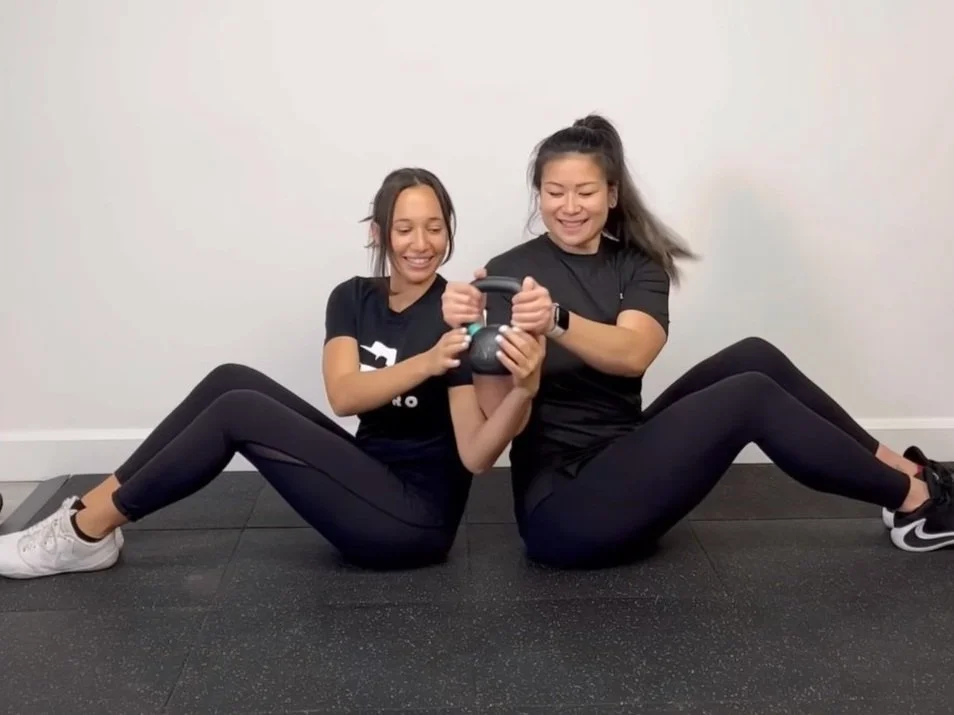
Partner Russian twist
Using a kettlebell is a partner core exercise. Begin by sitting side by side with your partner in exercise. Keep your legs together in front of you with your knees bent. To begin, one person holds a kettlebell and pulls the weight across their body by rotating their torso. From this point on, give the kettlebell to your partner and have them perform the same movement. Pivot the body back to get the iron weight. In both directions, complete.
FAQ
What muscles are in the internal oblique?
The transversus abdominis, external abdominal oblique, rectus abdominis, and pyramidalis muscles are all members of the anterolateral abdominal muscles. The internal abdominal oblique is one of these muscles.
What are external and internal oblique?
While the internal oblique rotates in the same direction, the external oblique rotates in the opposite direction. They perform together. The spine is rotated LEFT by the right external oblique and left internal oblique.
What is the internal oblique?
The internal abdominal oblique is a muscle located on the lateral side of the abdomen. It is thin and wide. Along with the external oblique on one side and the transverse abdominis on the other, it is one of the layers of the lateral abdominal wall.
What are the internal oblique structures?
One of the four abdominal muscles is the internal obliques. They end at the costal margin, aponeurosis of the rectus sheath, and pubic crest and begin at the lumbar fascia, inguinal ligament, and iliac crest. As a result, under the rib cage, any movement of the internal obliques causes a contraction.
What direction is internal oblique?
The internal oblique spreads out, causing the highest and lowest fibers to travel in distinctly opposite directions. The internal oblique’s fibers run steeply upward. Here they run less steeply, here they cross over, and here towards the inguinal area they run descending.

3 Comments