
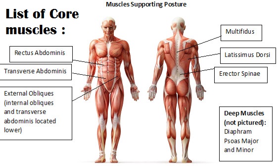
List of Core Muscles
Major list of Core muscles are the pelvic floor muscles, transverses abdominis, multifidus, internal and external obliques, rectus abdominis, erector spinae (sacrospinalis) mainly the longissimus thoracis and Diaphragm. Minor core muscles list are the latissimus dorsi, gluteus maximus and trapezius.
The core muscles have two main functions:
- 1) to spare the spine from excessive load.
- 2) to transfer force from the lower body to the upper body and vice versa. Having a strong, stable core helps us to prevent injuries and allows us to perform at our best.
List of Core muscles:
Major List of CORE Muscles
| Muscles Name | Origin | Insertion | Action |
| Transversus abdominis | Internal surfaces of costal cartilages of ribs 7-12, thoracolumbar fascia, anterior two thirds of iliac crest, iliopectineal arch | Linea alba, aponeurosis of internal abdominal oblique muscle; pubic crest, pectinal line of pubis | Bilateral contraction – Compresses abdominal viscera, expiration Unilateral contraction – Trunk rotation (ipsilateral) |
| external obliques | External surfaces of ribs 5-12 | Linea alba, pubic tubercle, anterior half of iliac crest | Bilateral contraction – Trunk flexion, compresses abdominal viscera, expiration Unilateral contraction – Trunk lateral flexion (ipsilateral), trunk rotation (contralateral) |
| Internal Abdominal Oblique | Lateral two-thirds of the inguinal ligament, anterior two-thirds of the intermediate line of the iliac crest, thoracolumbar fascia | Lower four ribs, abdominal aponeurosis of linea alba, crest of pubis | Internal abdominal oblique flexes the trunk when bilaterally contracted and lateral flexion upon unilateral contraction, compression causes increase in the intraabdominal pressure |
| diaphragm | Sternal part: Posterior aspect of xiphoid process Costal part: Internal surfaces of lower costal cartilages and ribs 7-12 Lumbar part: Medial and lateral arcuate ligaments (lumbocostal arches), bodies of vertebrae L1-L3 (+intervertebral discs), anterior longitudinal ligament | It inserts onto the central tendon of the diaphragm. In other words, it inserts on itself. | Depresses costal cartilages, primary muscle of breathing (inspiration) |
Minor core muscles list:
| Muscles Name | Origin | Insertion | Action |
| latissimus dorsi | Vertebral part: Spinous processes of vertebrae T7-T12, Thoracolumbar fascia Iliac part: Posterior third of crest of ilium Costal part: Ribs 9-12 Scapular part: Inferior angle of scapula | Intertubercular sulcus of the humerus, between pectoralis major and teres major muscles Mnemonic: Lady between two majors (lady refers to latissimus dorsi) | Shoulder joint: Arm internal rotation, Arm adduction, Arm extension; Assists in respiration |
| gluteus maximus | Lateroposterior surface of sacrum and coccyx, gluteal surface of ilium (behind posterior gluteal line), thoracolumbar fascia, Sacrotuberous ligament | Iliotibial tract, gluteal tuberosity of femur | Hip joint: Thigh extension, thigh external rotation, thigh abduction (superior part), thigh adduction (inferior part) |
| trapezius | Descending part (superior fibers): medial third of the superior nuchal line, external occipital protuberance Transverse part (middle fibers): nuchal ligament attached to the spinous processes of C1-C6 vertebrae, spinous processes and supraspinous ligaments of vertebrae C7-T3 Ascending part (inferior fibers): spinous processes and supraspinous ligaments of vertebrae T4-T12 | Descending part (superior fibers): lateral third of clavicle Transverse part (middle fibers): medial acromial margin, superior crest of spine of scapula Ascending part (inferior fibers): lateral apex of the medial end of scapular spine | The function of the trapezius is to stabilize and move the scapula. The upper fibers can elevate and upwardly rotate the scapula and extend the neck. The middle fibers adduct (retract) the scapula. The lower fibers depress and aid the upper fibers in upwardly rotating the scapula. |
Major muscles:
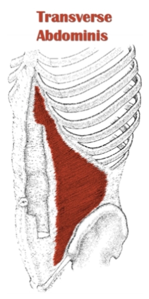
Transversus abdominis:
Origin
Internal surfaces of costal cartilages of ribs 7-12, thoracolumbar fascia, anterior two thirds of iliac crest, iliopectineal arch.
Insertion
Linea alba, aponeurosis of internal abdominal oblique muscle; pubic crest, pectinal line of pubis.
Action
Bilateral contraction – Compresses abdominal viscera, expiration
Unilateral contraction – Trunk rotation (ipsilateral).
Innervation:
Intercostal nerves (T7-T11), subcostal nerve (T12), iliohypogastric nerve (L1), ilioinguinal nerve (L1)
Blood supply:
Lower posterior intercostal and subcostal arteries, superior and inferior epigastric arteries, superficial and deep circumflex arteries,posterior lumbar arteries.
External obliques:
Origin:
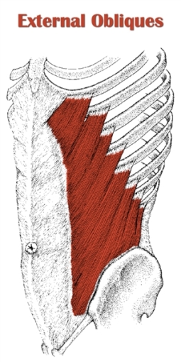
External surfaces of ribs 5-12.
Insertion:
Linea alba, pubic tubercle, anterior half of iliac crest
Innervation:
Motor: Intercostal nerves (T7- T11), Subcostal nerve (T12)
Sensory: Iliohypogastric nerve (L1)
Blood supply:
Lower posterior intercostal arteries, subcostal artery, deep circumflex iliac artery.
Action:
Bilateral contraction – Trunk flexion, compresses abdominal viscera, expiration
Unilateral contraction – Trunk lateral flexion (ipsilateral), trunk rotation (contralateral)
Internal Abdominal Oblique:
Origin:
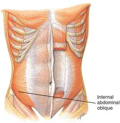
Lateral two-thirds of the inguinal ligament, anterior two-thirds of the intermediate line of the iliac crest, thoracolumbar fascia.
Insertion:
Lower four ribs, abdominal aponeurosis of linea alba, crest of pubis
Blood supply:
Lower posterior intercostal and subcostal arteries,
superior and inferior epigastric arteries,
superficial and deep circumflex arteries,
posterior lumbar arteries
Action:
Internal abdominal oblique flexes the trunk when bilaterally contracted and lateral flexion upon unilateral contraction, compression causes increase in the intraabdominal pressure.
Diaphragm:
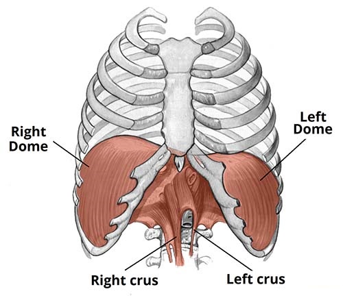
Origin :
Sternal part: Posterior aspect of xiphoid process
Costal part: Internal surfaces of lower costal cartilages and ribs 7-12
Lumbar part: Medial and lateral arcuate ligaments (lumbocostal arches), bodies of vertebrae L1-L3 (+intervertebral discs), anterior longitudinal ligament
Insertion:
It inserts onto the central tendon of the diaphragm. In other words, it inserts on itself.
Blood supply:
The blood supply to the diaphragm is from the superior phrenic, musculophrenic, inferior phrenic, pericardiacophrenic, and lower internal intercostal arteries. The superior phrenic arteries arise from the thoracic aorta.
Action:
Depresses costal cartilages, primary muscle of breathing (inspiration)
Minor core muscles list:
Latissimus dorsi:
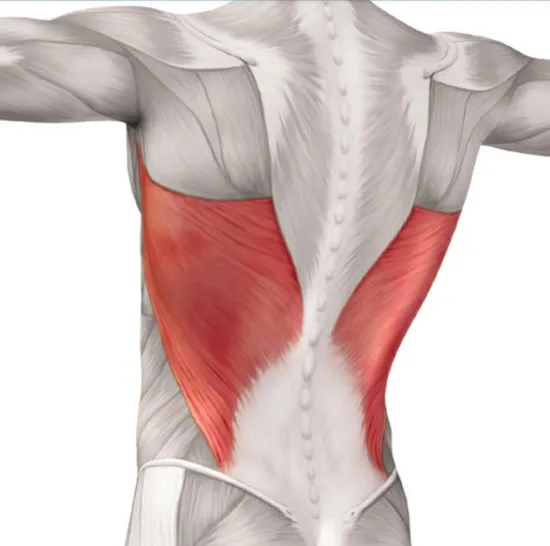
Origin:
Vertebral part: Spinous processes of vertebrae T7-T12, Thoracolumbar fascia
Iliac part: Posterior third of crest of ilium
Costal part: Ribs 9-12
Scapular part: Inferior angle of scapula
Insertion:
Intertubercular sulcus of the humerus, between the pectoralis major and teres major muscles
Mnemonic: Lady between two majors (lady refers to latissimus dorsi)
Blood supply:
Thoracodorsal artery, perforating arteries of the 9th-11th posterior intercostal arteries, and 1st-3rd lumbar arteries.
Action:
Shoulder joint: Arm internal rotation, Arm adduction, Arm extension; Assists in respiration.
Gluteus maximus:
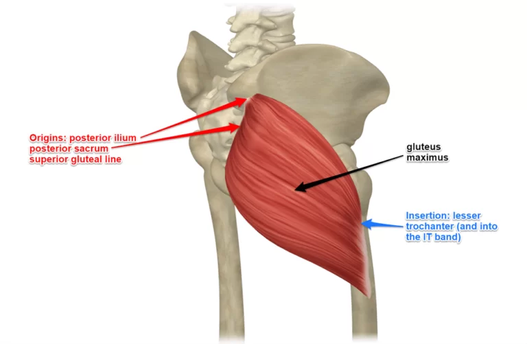
Origin:
Lateroposterior surface of the sacrum and coccyx, the gluteal surface of the ilium (behind the posterior gluteal line), thoracolumbar fascia, and Sacrotuberous ligament.
Insertion:
Iliotibial tract, gluteal tuberosity of femur
Blood supply:
Inferior gluteal and superior gluteal arteries.
Action:
Hip joint: Thigh extension, thigh external rotation, thigh abduction (superior part), thigh adduction (inferior part)
Trapezius:
Origin:
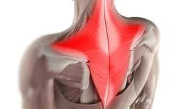
Descending part (superior fibers): medial third of the superior nuchal line, external occipital protuberance
Transverse part (middle fibers): nuchal ligament attached to the spinous processes of C1-C6 vertebrae, spinous processes and supraspinous ligaments of vertebrae C7-T3
Ascending part (inferior fibers): spinous processes and supraspinous ligaments of vertebrae T4-T12
Insertion:
Descending part (superior fibers): a lateral third of the clavicle
Transverse part (middle fibers): medial acromial margin, a superior crest of the spine of the scapula
Ascending part (inferior fibers): lateral apex of the medial end of the scapular spine.
Blood supply:
Occipital artery (descending part), superficial or transverse cervical artery (transverse part), dorsal scapular artery (ascending part)
Action:
The function of the trapezius is to stabilize and move the scapula. The upper fibers can elevate and upwardly rotate the scapula and extend the neck. The middle fibers adduct (retract) the scapula. The lower fibers depress and aid the upper fibers in upwardly rotating the scapula.
What is the function of your core muscles?
These muscles act to stabilize your spine providing a firm support for all the activities we do. They transfer force through your body and prevent you from having undesired back, hip, knee, and even neck pain.
What is muscle imbalance?
Your core muscles have to work together in symmetry in order to avoid abnormal movement. Muscle imbalance means that there is not a good balance between muscles that are too tight or loose versus muscles that are weak or strong.
Let me give you an example: in the pelvic area is very common to see hip flexors and lumbar paraspinals being very tight and gluteal/core muscles being very weak. This combination could result in an anterior pelvic tilt which can cause lower back pain as well as knee and hip problems.
Usually, Physiotherapists perform functional movement assessments to identify these imbalances and correct them with stability and strengthening exercises, myofascial release, active release, stretching exercises, soft tissue, and joint mobilizations.
Core Muscles Strengthening Exercise :
Core Muscles Strengthening exercise helps to improve the Balance and stability of the Back and the overall function of the Body, also Helps in Low Back pain management.
Pelvic Tilt with External Rotation :
Lie on the back with both knees bent and feet on the mat. Place band around both knees. Tighten your stomach muscles and tilt your pelvis in order to flatten back against the table. Bring both knees out to the side about 45 deg and hold for 3 seconds. Slowly return to the starting position.
Perform 2 sets of 10 repetitions
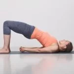
Bridging Exercise:
While lying on your back, tighten your lower abdominals, squeeze your buttocks, and then raise your buttocks off the floor/bed to create a “Bridge” with your body. Hold for 3 seconds. Slowly return to the starting position.
Perform 2 sets of 10 repetitions
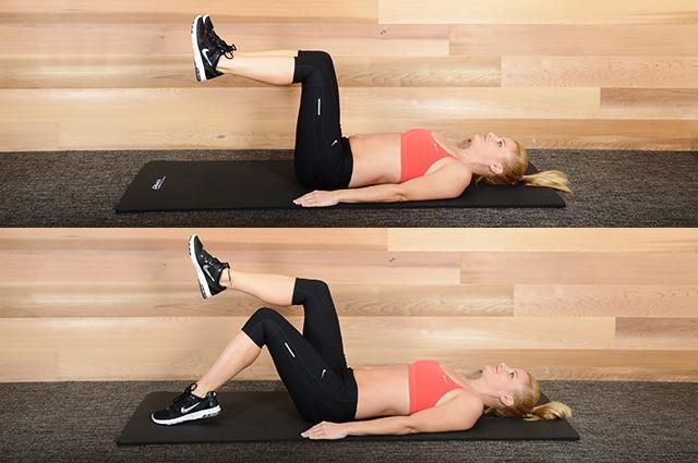
Pelvic Tilt with Toe Taps:
Supine Lie on the back and lift both feet into the air, keeping a 90-degree bend at both the hips and the knees. Tighten stomach muscles to flatten back against the table.
Slowly lower one foot to the mat, keeping the knee bent. Lift the leg back to the starting position. Alternate between left and right legs. Do not allow your back to arch.
Perform 2 sets of 10 repetitions.
Side Plank:

Start lying on one side, holding yourself up on your elbow. Place one foot on top of the other, and lift the torso up, keeping shoulders, hips, and ankles in a line.
Start by holding for 20 seconds and repeat 3 times on each side
FAQs
What muscles are the core?
The muscles that join the spine or pelvis deep within your back and abdomen are known as your core muscles. The transversus abdominis, pelvic floor muscles, and oblique muscles are a few of these muscles. The multifidus is another muscle that is used to move the trunk.
What are the 5 core muscles?
The transverse abdominis, multifidus, internal and external obliques, erector spinae, diaphragm, pelvic floor muscles, and—of course—your abs, the rectus abdominis, are the main muscles of your core. To the surprise of many, your lesser core muscles include your glutes, lats, and traps.
What are the 4 types of core muscles?
Up to 35 distinct muscle groups that connect the hip and spine to the pelvis make up the Core. I’ve broken down the core muscles into four areas to make them easier to understand: the hip muscles, lateral trunk muscles, abdominal muscles, and back extensors.
How can I improve my core?
Try Pilates or yoga.
Engaging in these exercises strengthens your core muscles and improves flexibility and balance. Develop a comprehensive fitness program that includes strength training, aerobic and plyometric workouts, and core exercises.
Why is my core so weak?
Surgery, injuries, or inactivity can all lead to muscle weakening or reduced control over certain muscles. Common indications of weakening or losing control over your core muscles can cause movement dysfunction, which will ultimately lower your quality of life.
Is core and abs the same?
No, core and abs are not the same. The core includes the muscles in the midsection of the body, such as the abdominals, but it also encompasses other muscle groups like the lower back, obliques, and more. The abs, or abdominals, specifically refer to the muscles in the front of the core.
Where is the core in the body?
The set of muscles in the trunk and hips that round the spine, abdominal viscera, and hip is known as the core. For the spine, pelvis, and kinetic chain to all balance properly under stress, core muscles are necessary.
Where is my core located?
The primary function of the human body’s core, which is situated between the diaphragm and the pelvic floor, is to support and cover the spine. Although they make up a portion of your core, your abdominal muscles are only one aspect of it.

93 Comments