Where is SI Joint located?
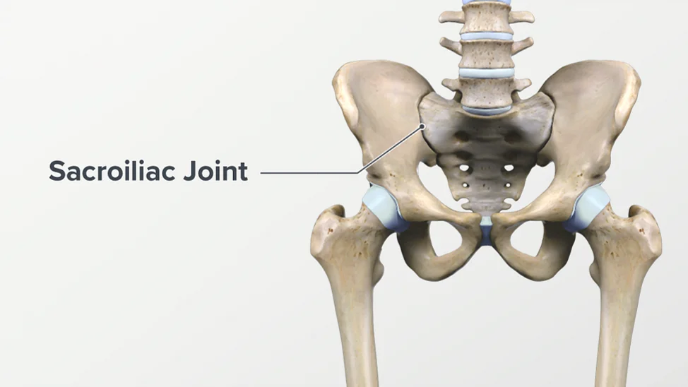
The SI joint is located in the pelvis. It is formed by the connection between the sacrum, which is the triangular bone at the base of the spine, and the ilium, which is one of the large bones of the pelvis. The SI joint is located on each side of the sacrum, where it connects with the ilium.
It is found at the back of the pelvis, just below the waistline, and can be felt as two small dimples or bony prominences on either side of the lower back. The SI joint plays a role in supporting the weight of the upper body and transferring forces between the spine and the lower limbs.
Anatomy of SI joint
The sacroiliac joint (SI joint) is a diarthrodial joint that connects the sacrum, which is the triangular bone at the base of the spine, to the ilium, one of the large bones of the pelvis. Here’s a breakdown of the anatomy of the SI joint:
- Sacrum: It is a triangular-shaped bone where the fusion of five vertebrae is joined together. It is located at the base of the spine, between the two hip bones (ilium). The upper part of the sacrum articulates with the last lumbar vertebra, and the lower part connects with the coccyx.
- Ilium: The ilium is one of the three bones that make up the hip bone, along with the ischium and pubis. It forms the upper and outer part of the pelvis and provides support for the abdominal organs.
- Joint Structure: The SI joint is a synovial joint, meaning it has a joint capsule and synovial fluid to facilitate movement. The joint is covered with articular cartilage, which helps reduce friction and allows for smooth movement between the sacrum and the ilium.
- Ligaments: The SI joint is stabilized by a network of ligaments that surround and support it. The most important ligaments include:
- Anterior sacroiliac ligament: It connects the front of the sacrum to the ilium.
- Posterior sacroiliac ligament: It connects the back of the sacrum to the ilium.
- Interosseous sacroiliac ligament: It connects the rough surfaces between the sacrum and ilium.
- Sacrotuberous ligament: It connects the sacrum to the ischial tuberosity (a bony prominence in the pelvis).
- Sacrospinous ligament: It connects the sacrum to the spine of the ischium.
- Movement: The SI joint has limited mobility compared to other joints in the body. It allows for small movements, such as rotation, tilting, and a small amount of gliding. These movements play a role in providing stability to the pelvis during activities such as walking, running, and lifting.
It’s worth noting that the SI joint can be a source of pain and dysfunction in some individuals, leading to a condition known as sacroiliac joint dysfunction. Proper diagnosis and treatment by a healthcare professional are important in managing SI joint-related issues.
Sacrum
The sacrum is a large triangular bone located at the base of the spine, between the fifth lumbar vertebra (L5) and the coccyx (tailbone). It forms the posterior part of the pelvis and serves as a connection point for the spinal column and the pelvic girdle.
Here are some key features and characteristics of the sacrum:
- Shape: The sacrum has a triangular shape and consists of five fused vertebrae known as sacral vertebrae (S1-S5). The shape and structure of the sacrum are specialized for weight-bearing and transferring forces between the spine and the lower limbs.
- Articulations: The sacrum articulates with several bones, forming different joints:
- Sacroiliac Joints: The sacrum connects with the ilium bones on each side, forming the sacroiliac joints. These joints are responsible for transferring weight and forces between the spine and the lower limbs.
- Lumbosacral Joint: The sacrum connects with the fifth lumbar vertebra (L5) above it, forming the lumbosacral joint. This joint allows for limited movement and plays a role in transferring loads between the lumbar spine and the sacrum.
- Coccyx: The lower end of the sacrum articulates with the coccyx, a small bone composed of fused coccygeal vertebrae.
- Prominent Landmarks: The sacrum has several prominent landmarks that can be palpated:
- Sacral Base: It is the broad upper part of the sacrum that articulates with the fifth lumbar vertebra (L5).
- Sacral Apex: It is the narrow, pointed lower end of the sacrum that connects with the coccyx.
- Sacral Promontory: It is the anterior projection of the sacrum into the pelvic cavity, forming part of the pelvic brim.
- Sacral Foramina: There are four pairs of sacral foramina on the sides of the sacrum. These foramina allow for the passage of nerves and blood vessels.
- Function: The sacrum plays a crucial role in providing stability and support to the spine and the pelvis. It transmits the weight of the upper body to the pelvis and lower limbs and serves as an attachment point for various muscles, ligaments, and tendons.
Understanding the anatomy of the sacrum is important in diagnosing and treating conditions related to the lower back, pelvis, and sacroiliac joint.
Ilium
The ilium is one of the three bones that make up the hip bone, along with the ischium and pubis. It is the largest and uppermost portion of the hip bone, forming the superior and lateral aspects of the pelvis. Here are some key features and characteristics of the ilium:
- Location: The ilium is located on each side of the pelvis, forming the upper part of the hip bone. It extends from the sacrum, where it articulates with the sacroiliac joint, to the acetabulum, which is the socket that receives the head of the femur (thigh bone) to form the hip joint.
- Shape: The ilium has a fan-shaped structure, with a broad upper portion called the iliac crest and a narrower lower portion called the body or wing of the ilium. The iliac crest is the curved ridge that can be felt on the lateral side of the pelvis.
- Landmarks: The ilium has several landmarks that are important in anatomical reference and clinical examinations:
- Iliac Crest: It is the upper curved ridge of the ilium and serves as an attachment site for various muscles, including the abdominal muscles and the muscles of the lower back.
- Anterior Superior Iliac Spine (ASIS): It is a bony prominence located at the front end of the iliac crest. The ASIS can be palpated and is commonly used as a reference point for measuring and locating structures on the body.
- Posterior Superior Iliac Spine (PSIS): It is a bony prominence located at the back end of the iliac crest. The PSIS is another reference point often used in anatomical measurements and assessments.
- Articulations: The ilium forms several important joints:
- Sacroiliac Joint: The ilium articulates with the sacrum at the sacroiliac joint, which is a synovial joint that allows limited movement and provides stability to the pelvis.
- Acetabulum: The ilium contributes to the formation of the acetabulum, which is the socket that receives the head of the femur to form the hip joint.
- Function: The ilium plays a crucial role in providing support and stability to the pelvis and transmitting forces between the trunk and the lower limbs. It serves as an attachment site for numerous muscles involved in hip movement, posture, and locomotion.
Understanding the anatomy of the ilium is essential in areas such as orthopedics, anatomy, physical therapy, and sports medicine, as it helps in diagnosing and treating conditions related to the hip, pelvis, and sacroiliac joint.