Pectineus muscle
What is the Pectineus muscle?
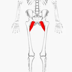
The pectineus is a flat muscle in the anterior thigh’s superomedial part. Each of the thigh muscles’ facial compartments is unique in that it is innervated by a distinct nerve. The pectineus is one of the few muscles that can be divided into two compartments at the same time because it has dual innervation; anterior and medial. The others are adductor magnus and adductor longus.
The five muscles of the medial thigh are typically used to define the pectineus for instructional purposes. The gracilis, adductor longus, adductor brevis, and adductor magnus are the names of these muscles. This is the thigh adductors functional group.
Pectineus functions include thigh flexion, external (lateral) rotation, and internal (medial) rotation, in addition to adducting the thigh. The pectineus is a postural muscle as well as a prime mover because it balances the trunk on the lower extremity while walking and stabilizes the pelvis.
Origin
pelvic bone and the pectineal line (pecten pubis)
Insertion
An oblique line that connects the linea aspera on the femur’s posterior surface with the base of the lesser trochanter.
Relations
The muscle is medially adjacent to the adductor longus and in the same plane as it. Laterally, it is connected to the medial circumflex femoral artery and vein and the psoas major muscle.
Together with the adductor longus, the anterior surface of the pectineus forms the medial portion of the floor of the femoral triangle. The iliacus and psoas major complete the lateral portion of the floor. The deep layer of fascia lata that separates the pectineus from the femoral artery, femoral vein, and great saphenous vein that runs through the femoral triangle covers this surface of the pectineus.
The adductor magnus, adductor brevis, and obturator externus muscles, in addition to the obturator nerve’s anterior branch, are located posterior to the pectineus. Note that the fascia lata peripherally limits the anterior and posterior thigh compartments. These two are internally isolated by medial and lateral intermuscular septa that connect to the femur.
Innervation
The femoral nerve (L2, L3) innervates the pectineus most of the time. Nonetheless, in certain individuals, the pectineus may get innervation from two separate lumbar plexus nerves.
The femoral nerve (L2, L3), which is characteristic of muscles of the anterior thigh, innervates the anterior portion of the muscle that sits in these instances. The accessory obturator nerve, a branch of the obturator nerve, supplies the muscle’s posterior, smaller portion (L3, L4).
Blood supply
The superficial portion of the muscle is provided by the medial circumflex femoral artery, a part of the femoral artery. The anterior branch of the obturator artery, which is itself a branch of the internal iliac artery, vascularizes the deep muscle portion.
Function
The pectineus flexes and adducts the thigh at the hip joint when it contracts considering the course of its strands. The hip joint flexes first when the muscle contracts when the lower limb is in the right anatomical position. This flexion can be extended when the thigh is positioned at a 45-degree angle to the hip joint.
The contracted muscle fibers are now dragging the thigh toward the midline, resulting in thigh adduction, as the angulation of the fibers has increased by that point. Crossing your legs at the knee or ankle is an example of a sequential movement that involves both actions of the pectineus muscle. The carry-through phase of the gait cycle is made possible by hip flexion alone, in conjunction with the psoas major, iliacus, rectus femoris, and sartorius muscles.
The pectineus muscle is also said to play a role in the thigh’s internal and external rotation, according to some sources. Even though the insertion of the pectineus is lateral to the midline and extends beyond the femur’s mechanical axis of rotation, there is still a lot of debate about this.
Other than being the main player, this muscle is likewise a significant stabilizer of the pelvis. It is hypothesized that all of the muscles that attach the pelvis to the proximal femur serve as hip joint adaptable ligaments, preserving its integrity during body movements.
Clinical Significance:
Pectineus muscle pain
Pain in this muscle can occur due to a variety of reasons, including overuse, strain, or injury.
Symptoms of pectineus muscle pain can include:
- Pain in the groin area
- Difficulty walking or standing
- Pain during hip flexion or adduction
- Tightness or stiffness in the muscle
- Swelling or inflammation in the muscle
To treat pectineus muscle pain, it is important to rest the affected area and avoid any activities that aggravate the pain. The use of ice in the area can also help reduce swelling and inflammation. Gentle stretching and massage can also help to relieve pain and improve flexibility.
Pectineus muscle strain
Pectineus muscle strain refers to a tear or injury to the pectineus muscle located in the groin area. Symptoms of a strain may include pain, stiffness, and difficulty with movement. Treatment options may include rest, ice, and physical therapy. In severe cases, surgery may be necessary. Early intervention and proper treatment can help to prevent further injury and promote healing.
Pectineus muscle stretching
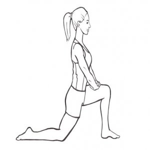
Kneeling Lunge and Overhead Stretch
With your right hand on your right knee, bring your left arm slightly to the right and up over your head. As you lunge slightly forward with your right leg, notice how your pectineus and upper body (shoulder and lat muscles) stretch.
Pectineus muscle strengthening exercise
Standing hip flexion exercise
Stand with your back to a bench or other elevated surface, like a steady chair. Your distance from it should be roughly the length of your leg. With your hands on your hips, put one foot on the seat or raised surface. Twist your raised leg gradually, making you lunge forward while keeping your spine straight. Hold the stretch for at least two seconds by moving your hips forward. As your strength improves, you may be able to hold it for longer. Do the same with the other leg ten times on one side.
Lateral Lunges
To begin, stand with your feet about shoulder-width separated. To assist with balance, place your hands on your hips or out to the side. Step your right foot out to your right side and into a side squat. Go as far down as you are open to doing, however not beyond the place where the thigh is lined up with the ground. To get back up, gently push with your right foot and return the right foot to its starting position. During this exercise, contract your abdominal muscles to keep your core stable. On each side, perform this exercise four times, increasing the number of sets and repetitions as your strength improves.

FAQ
How does a strain of the pectineus feel?
Pain, bruising, swelling, tenderness, and stiffness are the most typical signs of a pectineus muscle injury. The primary hip flexor muscles, the hip adductor muscles, or a combination of the two could be to blame for pain in the front of your hip.
What causes tight pectineus?
The most common injury to the pectineus muscle is a groin strain, which is caused by power walkers and some runners overworking or overextending their stride. A primary sign of injury to the pectineus is localized pain in the groin on one side or the other.
How can you tell if your pectineus has been pulled?
Pain is the most typical symptom of an injured pectineus muscle. Other symptoms include swelling, tenderness, stiffness, and bruising.
What exactly is referred pectineus pain?
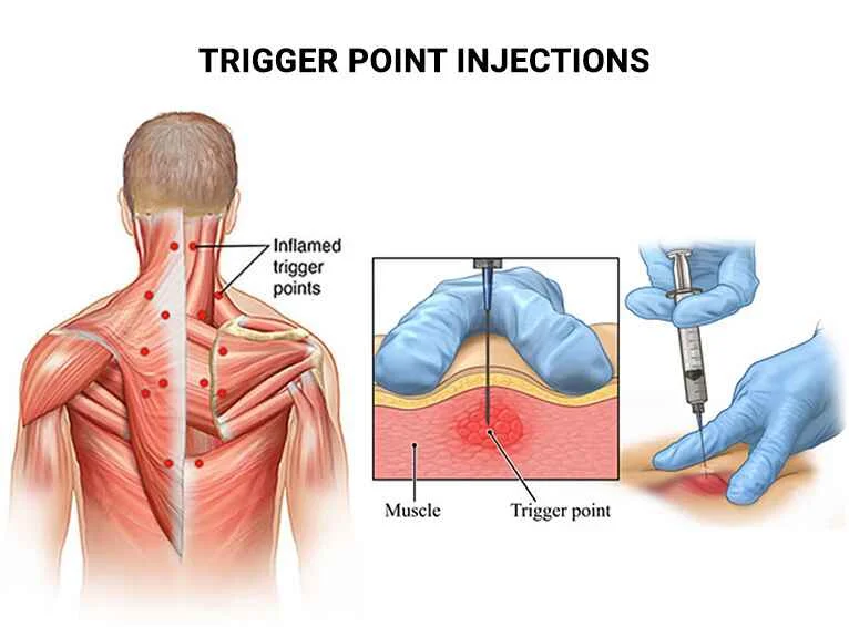
The femoral nerve, and occasionally the obturator nerve, innervate the pectineus. referred leg pain is caused by the trigger point, which is in the upper thigh near the pubic bone; medial thigh ache, and also anterior thigh ache.
How can one tell the difference between a hernia and a pectineus strain?
Both muscle strain and a hernia are accompanied by dull aches and pains in the groin. However, if you have a small lump or bulge on one side of your groin, this is a strong indication that you may have a hernia.

