
Rectus abdominis muscle
What is the Rectus abdominis muscle?
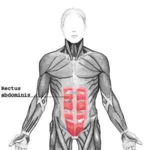
The rectus abdominis muscle, commonly known as the “abs” or “six-pack,” is a paired muscle located in the anterior (front) part of the abdomen. It is a long, strap-like muscle that extends vertically from the pubic bone to the lower ribs. The rectus abdominis is enclosed by a layer of connective tissue called the rectus sheath.
Together with the pyramidalis muscle, the rectus abdominis muscle is one of the anterior abdominal muscles. However, these two muscles, along with the three lateral abdominal muscles, make up the anterolateral abdominal wall, according to functional anatomy; transversus abdominis, internal oblique, and external oblique.
Origin and Insertion
Two tendons connect the rectus abdominis muscle in the inferior region; From the pubic tubercle to the pectineal line, the larger tendon attaches to the pubic crest, while the smaller, medial tendon attaches to the pubic symphysis.
The fibers of the rectus abdominis then extend vertically superiorly and insert into the xiphoid process of the sternum and the costal cartilages of the fifth, sixth, and seventh ribs. Most lateral fibers attach to the anterior end of the fifth rib, but in some cases, this slip extends to the third and fourth ribs or is absent entirely.
Structure
On the anterior surface of the abdominal wall, the rectus abdominis muscle is a pair of muscles that run vertically on either side of the linea alba. The muscle’s two halves are separated vertically by the linea alba, a band of connective tissue.
The rectus abdominis muscle is divided into distinct segments by tendinous intersections, giving it the appearance of several muscle bellies, which is why it is often referred to as the “six-pack.”
The tendinous intersection that separates the muscle’s lateral edge from the external and internal oblique muscles on the lateral surface of the anterior abdominal wall is called the linea semilunaris. It typically connects the pubic tubercle to the tip of the ninth costal cartilage.
Rectus abdominis muscle has 3 tendinous intersections. There are three of these horizontal intersections: one at the umbilicus, one at the xiphoid process, and one in the middle of the other two. In individuals with low body fat, these fibrous bands divide the muscle into segments, creating a gridiron six-pack shape. It is thought that the intersections represent myosepta, which separate the myotomes that make muscles.
Relations
The aponeurosis of the transversus abdominis and the external and internal oblique abdominal muscles combine to form the rectus sheath, which contains the rectus abdominis muscle itself.
Innervation
The thoracoabdominal nerves innervate the rectus abdominis muscle by piercing the rectus sheath’s anterior surface.
They penetrate the rectus abdominis muscle sheath and travel between the layers of the transversus abdominis and internal oblique muscles.
The nerves are the anterior divisions of the seventh through eleventh lower intercostal nerves. These nerves continue to supply the abdominal wall after the intercostal spaces they provide medially end.
Blood supply
Several vessels supply the rectus abdominis muscle with blood, but the inferior and superior epigastric arteries provide the majority of the supply.
Small terminal branches of the subcostal, deep circumflex, and lower three posterior intercostal arteries also contribute.
Functions
The main function of the rectus abdominis muscle is to flex the lumbar spine, bringing the ribcage closer to the pelvis. This movement is commonly referred to as “crunching” or “sit-ups.” Additionally, the muscle assists in stabilizing the trunk and maintaining posture. It plays a crucial role in activities that involve bending forward, such as lifting heavy objects or performing exercises like planks and squats.
This muscle also compresses the abdominal viscera and raises the intra-abdominal pressure when it works with other abdominal muscles. This is essential for deep breathing, labor, defecation, and micturition.
The anti-lordotic tilt of the pelvis is also stabilized and controlled by the rectus abdominis.
Clinical points
The core’s stability is partly influenced by the abdominal’s recti muscles. The rectus abdominis, transversus abdominis, erector spinae, and obliques are core muscles that protect the lower back from injury like a natural weight belt. People who have weak core muscles are more likely to have spinal problems.
A midline separation at the linea alba is referred to as diastasis recti abdominis. A diastasis is a palpable midline gap of more than 2.5 centimeters or any visible bulging when exerted. It can occur anywhere between the xiphoid process and pubic bone, but it usually occurs around the umbilicus.
This belly spread can also occur in newborns, and it should go away on its own. African American and premature infants are the most frequently affected groups.
Assessment
Palpation
To ensure the muscle relaxes, place the patient supine with a pillow under the knees. On both sides, palpate along the muscle from the xiphoid process and adjacent ribs down to the pubic bone. Relax after asking the patient to raise the trunk or at least try to raise it.
Muscle power
We must focus more on the abdominal muscles’ strength than their flexibility because weak abdominal muscles will cause severe issues in the future.
In most cases, MMT for rectus abdominis comprises having the patient lift their head, trunk, and shoulders against gravity in a supine position until the inferior angles of the scapula are off the table. The therapist will then start grading the patient based on how well the patient does the movement and how well they can do it.
The ability to bear the resistance (the resistance here is generated by the patient’s arms behind the head, crossed over the chest, or overstretched above the body plane) to only minimal contraction felt by the examiner’s hand will determine the grade, which will range from 5 to 1.
Exercise of Rectus Abdominis muscle
To strengthen and tone the rectus abdominis muscle, exercises such as sit-ups, crunches, leg raises, and planks are commonly performed. It’s important to note that proper form and technique should be observed to avoid strain or injury to the lower back.
Mainly of 2 types:
- Rectus Abdominis Stretching exercise
- Rectus Abdominis Strengthening exercise
Rectus abdominis muscle stretching
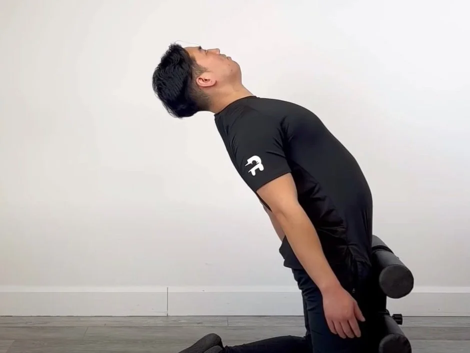
Thigh block back extension
This is a mobility exercise that requires you to contract your abdominal wall eccentrically during full spine extension. Start by using a stable object like a wall to block the front of your thighs while kneeling. To protect your knees, kneel on a padded surface. Begin to slowly resist gravity by extending your spine backward. Return to your starting position in a tempo of two seconds (concentric phase) after reaching your tolerable end range in 5 to 7 seconds. Repeat as necessary.
Thigh block full extension
This is a mobility exercise that requires you to contract your abdominal wall eccentrically during full spine extension. Start by using a stable object like a wall to block the front of your thighs while kneeling. To protect your knees, kneel on a padded surface. Slowly resist gravity by raising your arms straight up high and extending your spine backward. Return to your starting position in a tempo of two seconds (concentric phase) after reaching your tolerable end range in 5 to 7 seconds. Repeat as necessary.
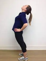
Standing lumbar extension
This is a low back or lumbar spine mobility exercise based on extension. Standing, place both of your hands on the back of your hips to begin. As you extend your low back, move your hips forward. The lumbar spine should have a relatively even curvature throughout. Return to the standing position after holding this position for up to ten seconds.
Rectus abdominis muscle strengthening
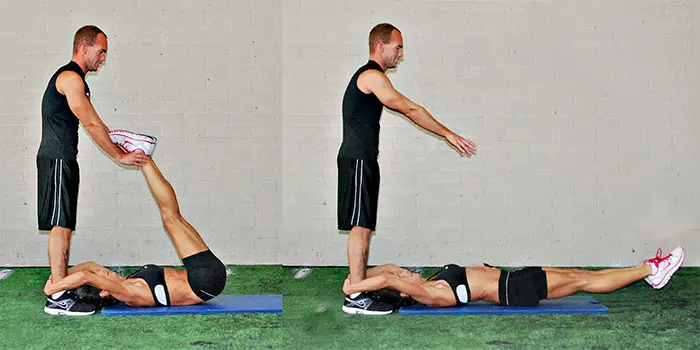
Partner leg raise
This partner exercise targets the hip flexors and core muscles. To get things started, have your partner stand while you are lying on your back. During the exercise, hold onto your partner’s ankles as a base of support. Have your partner push your feet toward the floor while you raise both legs with straight legs. Repeat with the legs raised back up just before your feet touch the ground.
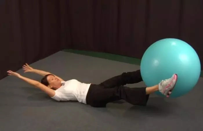
Swiss ball pass
A Swiss ball or stability ball is used in this exercise to strengthen the abdominal muscles. To begin, lie on your back and brace your core to maintain a neutral spine. Hold the ball in your hands with your hips extended and arms overhead. Flex your hips and lower your arms to transfer the ball from your hands to your feet. Extend your hips once more and raise your arms overhead to get back into a long position. Return the ball to your hands from your feet in this manner.
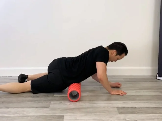
ABS foam rolling
For the abs muscle group, this is a soft tissue mobilization technique. Lay down on a foam roller that is positioned between your pelvis and your 12th rib, also known as your bottom rib. Reduce the amount of pressure placed on the abs as necessary by supporting the body with your elbows and legs. Hold for up to 30 seconds, or 3-5 breaths, on a tender spot. You might find three to four tender spots; repeat as necessary. To maximize recovery, foam rolling is typically limited to two to three times per week.
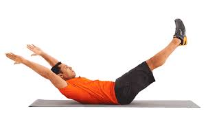
Banana superman roll
This is a core-strengthening exercise that works the lower back and abdominal wall simultaneously. Begin by lying on your back and lifting your legs and upper back off the ground. Log roll onto your stomach without leading with your hips or shoulders while keeping your shoulders raised. By using your low back muscles, keep your arms and legs off the ground. Roll back onto your back after holding for 3 to 5 seconds. To strengthen both sides of your obliques, roll in a different direction.
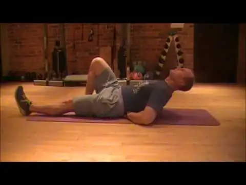
MCGILL crunch
The traditional crunch has been modified for this core-strengthening exercise to reduce lumbar compression. Maintain a crouching position on the floor while lying down with one leg straight and the other knee bent and the foot flat on the floor. Lift your upper back off the floor while performing a crunch with both hands below the small of your low back.
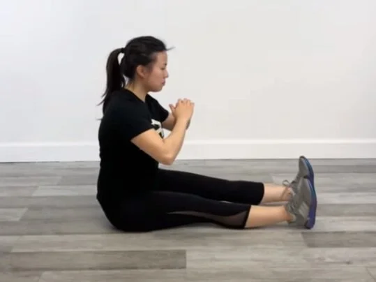
Butt walks
You will also mobilize the hip flexors and spinal rotators with this pelvis rotation exercise. Keep your legs out in front of you and take long sit position on the floor. Take a step forward with your buttock while lifting the pelvis. From one end of a yoga mat to the other, slowly move forward. Then walk in the opposite direction by moving your pelvis backward.
FAQ
What is a weakness of the rectus abdominis?
Diastasis recti is an abdominal wall weakness that results in an increased distance between the muscles of the rectus abdominis at the midline. The majority of medical professionals concur that the linea alba is weak, thinning, and widening, as is the abdominal muscle.
Why strengthen rectus abdominis?
During many movements, the rectus abdominis helps you bend forward, lift your pelvis, and stabilize your body. Additionally, it strengthens your abdominals, protects your spine, and assists you in exhaling strongly during exercises like the deadlift and squat.
What are the symptoms of rectus abdominis?
As a result, when the rectus muscle is injured, pain often radiates down the inner thigh and into the front of the groin. On the other hand, rectus pain can also be felt in the upper, lower, or even the testicles of the body. Heartburn, indigestion, and bloating are common symptoms.
How do you treat rectus abdominis?
Surgery may be considered if physiotherapy does not resolve or improve the rectus diastasis. Plication can be done during open or laparoscopic surgery to correct rectus diastasis, and both methods have high success rates.
What is the rectus abdominis strain?
The rectus abdominis muscle, also known as the “six-pack” muscle, is the abdominal muscle that is most frequently injured. It is situated in the front of the abdomen. Heavy lifting, twisting, and sudden movements all put a lot of strain on this muscle.


4 Comments