Sacral plexus
The sacral plexus is a network of nerve fibers that supply the skin & muscles of the pelvis & lower limb. the sacral plexus is present on the surface of the posterior pelvic wall, anterior to the piriformis muscle.
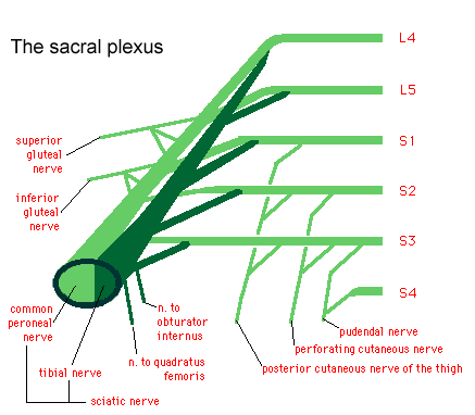
The plexus is formed by the anterior rami (divisions) of the sacral spinal nerves S1, S2, S3 & S4. It also receives benefactions from the lumbar spinal nerves L4 & L5.
The sacral plexus is present on the posterolateral wall of the pelvic cavity, situated anterior to the Piriformis. The sacral contributions pass out of the anterior sacral foramina & course laterally & inferiorly on the pelvic wall. A majority of the nerves originating from the sacral plexus pass through the greater sciatic foramen – inferior to the piriformis muscle & enter the gluteal region of the lower limb.
Root
The sacral plexus is established by the anterior rami of S1 to S4 as well as the lumbosacral trunk (anterior ramus of L4 & L5). The lumbosacral trunk courses vertically into the pelvic cavity from the abdomen & pass immediately anterior to the sacroiliac joint.
Branches of Sacral plexus
The sacral plexus provides motor & sensory supply through the following nerves:
- Sciatic Nerve (L4 – S3)
- Pudendal Nerve (ventral divisions from S2 – S4)
- Superior Gluteal Nerve (dorsal divisions from L4 – S1)
- Inferior Gluteal Nerve (dorsal divisions from L5 – S2)
- Nerve to Obturator Internus (ventral divisions L5 – S2)
- Nerve to Quadratus Femoris
- Posterior Femoral Cutaneous Nerve
- Perforating Cutaneous Nerve
- Nerve to Piriformis
- Obturator Nerve (L2 – L4)
Spinal Nerves
The spinal nerves S1 – S4 structure the base of the sacral plexus.
At each vertebral level, pair of spinal nerves leave the spinal cord via the intervertebral foramina of the vertebral column.
Each nerve then divides into anterior & posterior nerve fibers. The sacral plexus is initiated as the anterior fibers of the spinal nerves S1, S2, S3, and S4. They are joined by the 4th & 5th lumbar roots, which combine to form the lumbosacral trunk. This runs down into the pelvis to meet the sacral roots as they come out from the spinal cord.
- The Branches
The anterior rami of the S1-S4 spinal roots (& the lumbosacral trunk) divide into several cords. These cords then join together to make the 5 major peripheral nerves of the sacral plexus. - These nerves then run down toward the posterior pelvic wall. They have two main destinations:
- Leave the pelvis along the greater sciatic foramen – these nerves enter the gluteal region of the lower limb, innervating the structures there.
Remain in the pelvis – these nerves supply the pelvic muscles, organs & perineum.
Superior Gluteal Nerve
The superior gluteal nerve exit the pelvis along the greater sciatic foramen, entering the gluteal region superiorly to the piriformis muscle. It goes along with the superior gluteal artery & vein for much of its course.
- Roots: L4, L5, S1.
- Motor Functions: supplies the gluteus minimus, gluteus medius & tensor fascia lata.
- Sensory Functions: None.
- A helpful memory support for the main branches of the sacral plexus is ‘Some Irish Sailor Pesters Polly’. This stance is for the Superior Gluteal, Inferior Gluteal, Sciatic, Posterior cutaneous nerve of the thigh, & Pudendal.
Inferior Gluteal Nerve
The inferior gluteal nerve leaves the pelvis along the greater sciatic foramen, entering the gluteal region inferiorly to the piriformis muscle.
It is accompanied by the inferior gluteal artery & vein for much of its course.
- Roots: L5, S1, S2.
- Motor Functions: supplies gluteus maximus.
- Sensory Functions: None.
Sciatic nerve
- Roots: L4, L5, S1, S2, S3
- Motor Functions:
Tibial portion – Innervates the muscles in the posterior compartment of the thigh (apart from the short head of the biceps femoris), and the hamstring component of the adductor Magnus. supplies all the muscles in the posterior compartment of the leg & sole of the foot.
Common fibular portion – Short head of biceps femoris, all muscles in the anterior & lateral compartments of the leg & extensor digitorum Brevis.
- Sensory Functions:
- Tibial portion: supplies the skin of the posterolateral leg, lateral foot & the sole of the foot.
- Common fibular portion: supplies the skin of the lateral leg & the dorsum of the foot.
Posterior Femoral Cutaneous
The posterior cutaneous nerve of the thigh leaves the pelvis via the greater sciatic foramen, entering the gluteal region inferiorly to the piriformis muscle. It goes down deep to the gluteus maximus & runs down the back of the thigh to the knee.
- Roots: S1, S2, S3
- Motor Functions: None
- Sensory Functions: supplies the skin on the posterior surface of the thigh & leg.
Pudendal Nerve
This nerve exit the pelvis along with the greater sciatic foramen, then again it enters along with the lesser sciatic foramen. It moves anterosuperiorly along with the lateral wall of the ischiorectal fossa,& terminates by separating into several branches.
- Roots: S2, S3, S4
- Motor Functions: supplies the skeletal muscles in the perineum, the external urethral sphincter, the external anal sphincter, & the levator ani.
- Sensory Functions: Innervates the penis & the clitoris & most of the skin of the perineum.
Other Branches
In addition to the five major nerves of the sacral plexus, there are a number of smaller branches. These tend to be nerves that directly supply muscles (with the exception of the perforating cutaneous nerve, which supplies the skin over the inferior gluteal region & the pelvic splanchnic nerves, which innervate the abdominal viscera):
Nerve to piriformis
Nerve to obturator internus
Nerve to quadratus femoris
Clinical relevance
- Lesions of the sacral plexus can be caused by pelvic fractures, hip surgery, malignant infiltration, local radiotherapy, & the use of orthopedic traction tables. These lesions are usually unilateral & do not result in significant sexual dysfunction unless the sensory symptoms become disruptive.
- Cancers can occupy the sacral plexus causing pain radiating pain syndromes similar to sciatic nerve lesions. Numbness in the perianal region as well as the involvement of sympathetic fibers causing “hot & dry foot” can be found.
- The sacral plexus can be entrapped by the fetal head at the pelvic brim during the final trimester of pregnancy or labor. The clinical syndrome will resemble L5 radiculopathy & symptoms will usually resolve completely on their own 4 – 6 months post-birth.
Assessment
- Sensory testing has cautious sensitivity in the detection of lumbosacral radiculopathy
- previous knowledge of MRI results is a source of bias in sensory testing
- In clinical practice, diagnosis of lumbosacral radiculopathy should always be reached through the combined use of the sensory, motor, and deep tendon reflex tests – not through single test results
Physiotherapy Treatment
- Patients with severe neuropathic incontinence should first undergo severe conservative management including physiotherapy/pelvic floor retraining (biofeedback) before surgical treatment is considered.
- Sacral Nerve Stimulation- there is good emerging evidence for the treatment of patients with end-stage fecal incontinence.
FAQ
What happens when the sacral plexus is damaged?
A sacral plexus lesion may cause manifestations in the distributions of the gluteal, sciatic, tibial, & peroneal nerves. This manifests in the weakness of the hip extensors, hip abductors, knee flexors, & all foot & toe functions.
What does the sacral plexus give rise to?
The sacral plexus gives rise, craniocaudally, to the superior & inferior gluteal nerves, the posterior cutaneous nerve to the thigh, the sciatic nerve containing the common peroneal nerve (lateral part of sciatic) & the tibial nerve (medial part of sciatic), & the pudendal nerve.
What muscles does the sacral plexus innervate?
It travels superficial to the sciatic nerve & supplies the gluteus maximus muscle.
How many nerves are in the sacral plexus?
The sacral plexus is formed by the lowest lumbar spinal nerves, L4 & L5, as well as sacral nerves S1 through S4. Several combinations of these six spinal nerves combine together & then divide into the branches of the sacral plexus.
What does the sacral plexus control?
The sacral plexus is a nerve plexus that provides motor & sensory nerves for the posterior thigh, most of the lower leg, the entire foot,& part of the pelvis
What causes sacral plexus pain?
Lesions of the sacral plexus can be caused by pelvic fractures, hip surgery, malignant infiltration, local radiotherapy, & the use of orthopedic traction tables. These lesions are usually unilateral & do not result in significant sexual dysfunction unless the sensory symptoms become disruptive.
What is the thickest nerve of the sacral plexus?
sciatic nerve
the sciatic nerve, the largest & thickest nerve of the human body is the principal continuation of all the roots of the sacral plexus.
Where does the sacral plexus supply?
In human anatomy, the sacral plexus is a nerve plexus that provides motor & sensory nerves for the posterior thigh, most of the lower leg & foot, & part of the pelvis. It is part of the lumbosacral plexus & emerges from the lumbar vertebrae & sacral vertebrae (L4-L3.
What does the sacral plexus control?
A sacral plexus lesion might cause manifestations in the distributions of the gluteal, sciatic, tibial, & peroneal nerves. This manifests in the weakness of the hip extensors, hip abductors, knee flexors & all foot and toe functions.


6 Comments