
Lateral Pterygoid muscle
Introduction
The lateral pterygoid muscle is a craniomandibular muscle that plays an essential role in the inferior temporal region. It is active during mastication and mandibular motions – including protrusion (forward movement of the mandible), abduction (depression of the mandible), and mediotrusion (mandibular condyle movement towards the midline). It functions particularly during speaking, singing, and clenching.
The lateral pterygoid muscles have two heads or bellies. The inferior belly is three periods larger than the superior belly. Among all the masticatory muscles, it is just one with horizontally arranged fibers.
Functionally, the muscle is equivalent to the temporalis muscle. The fan-shaped temporalis muscles exhibit a continuous range of anteroposterior movements, while the lateral pterygoid muscle exhibits a range of mediolateral and superoinferior movements.
Origin of Lateral Pterygoid muscle
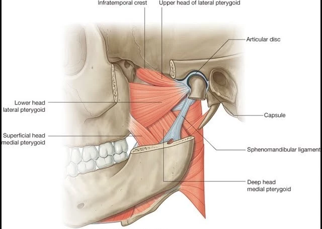
The lateral pterygoid is located deep in the temporalis and masseter muscles, traversing between the sphenoid bone and temporomandibular joint. Its muscles’ belly is divided by a small horizontal fissure into two heads; superior (upper) and inferior (lower).
The superior head is formed by the most superomedial fibers of the muscles. It consists of muscular slips that originate from the infratemporal crest of the grander wing of the sphenoid bone.
The inferior head is greatly wider than the superior one. It arises from the lateral surface of the lateral pterygoid plate of the sphenoid bone.
Insertion of Lateral Pterygoid muscle
Fibers from both heads congregate to the course posterolaterally, in a predominately horizontal plane. The superior fibers insert into the anteromedial portion of the articular capsule and also the articular disc of the temporomandibular joint. Whilst the inferior fibers insert into the pterygoid fovea on the neck of the condyloid process of the mandible bone. The superior attachment onto the temporomandibular joint (TMJ) permits the muscle head to act on the superior compartment of the joint and to produce the gliding motions of the disc and mandibular condyle. The inferior head works on the inferior compartment of the TMJ. Facilitating the hinge-like rotation that happens between the mandibular condyles and the inferior surface of the articular disc.
The lateral pterygoid muscle includes the medial wall of the infratemporal fossa and is contained within the masticator space. This space is determined by the superficial layer of the deep cervical fascia, which, at the level of the mandibular ramus, splits into superficial and deep laminae. The two laminae cover the outer surface of the masseter muscles and the deep surface of the medial pterygoid muscles, respectively. Besides the lateral pterygoid muscles, the masticator space also contains the temporalis tendon, masseter, medial pterygoid muscles, and the pterygoid venous plexus, along with several structures anatomically associated with the lateral pterygoid muscle.
The lateral pterygoid located deep to the medial pterygoid muscle and the tendon of the temporalis muscle. While it is superficial to the deep head of the medial pterygoid, sphenomandibular ligament, middle meningeal artery, and mandibular nerve (CN V3).
The superior margin of the muscles is crossed by the temporal and masseteric branches of the mandibular nerve, and the lingual and inferior alveolar nerves cross their inferior margin. Of note, the maxillary artery and buccal nerve (branch of the mandibular nerve) pass via the split between the superior and inferior heads of the lateral pterygoid muscle.
Nerve supply
The lateral pterygoid muscles obtain innervation from the mandibular branches of the trigeminal nerve. The primary trunk of the mandibular nerve separates into the anterior and posterior divisions. The anterior division passes off three motor branches. One of the motor branches is the nerve to the lateral pterygoid, paired nerves (one for each belly) innervating the lateral pterygoid muscle. According to some authors, these muscles may also obtain innervation from the anterior, middle, and posterior deep temporal nerves, the masseteric nerve, and sometimes from the buccal nerve. They innervate different muscle parts, resulting in selective activation, which provides the coordination of activities and reduces stress on the temporomandibular joint.
It is generally accepted that the lateral pterygoid muscles are innervated by the lateral pterygoid nerve, which originates from the mandibular nerve. Although both bellies receive their innervation from the mandibular nerve, additional nerves may innervate each of them. Kim et al. represented a specific nerve distribution design where the superior belly is innervated by the buccal nerve and the inferior belly by the mandibular nerve trunk.
Blood supply
Vascular supply to the lateral pterygoid muscles arrives from the pterygoid branches of the maxillary artery and the ascending palatine branch of the facial artery.
Function of Lateral Pterygoid muscle
Being a masticatory muscle, the lateral pterygoid assists in chewing and biting actions by controlling the movements of the mandible. The sphenoid attachment of the muscle is always fixed, indicating that the direction of the pull is oriented towards it.
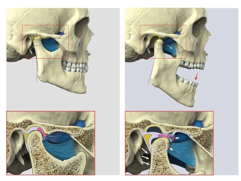
Bilateral contraction of the left and right lateral pterygoid muscles results in translation and also rotation within both temporomandibular joints. The inferior heads pull the mandibular condyle anteriorly, which occurs in the rotation of the condyle against the inferior surface of the articular disc. At the identical time, the superior heads pull the articular capsule and disc in the same direction (anteriorly) to cushion the movements of the condyle. The result is the anterior translation of the disc and condyle, happening concurrently with the rotation of the condyle. Which we notice as protrusion and depression of the mandible. Alternate contraction of the digastric and geniohyoid muscles concludes the movement of opening the jaw.
The retrodiscal fat pad within the TMJ stretches and alters anterior joint translation during protrusion and depression. Its inherent elasticity generates the force that initiates the closing of the mouth. In jaw closing, the same translation and rotation movements happen within the TMJ, they just happen in the opposite direction. Here, the inferior heads of the muscle eccentrically contract to smoothen the posterior translation of the articular disc and mandibular condyle, balancing the net pull of temporal and masseter muscles which draw the mandible posteriorly.
Side-to-side movements of the jaw occur when the inferior head contracts unilaterally, rotating the mandibular condyle anteromedially. The movement occurs in synergy with the contraction of the ipsilateral medial pterygoid muscle. This combined muscle motion swings the jaw to the opposite side and is seen, for example, when grinding food between the teeth on one side of the mouth.
Clinical Significance
The spasm of the lateral pterygoid muscles can be painful and it results in trismus (locked jaw), and the patient may require analgesics or muscle relaxants. Some authors acknowledge that during temporomandibular disorders, these muscles play an important role. This involvement may be due to the deficiency of coordination between the muscle’s superior and inferior bellies. This lack of coordination conducts to the disturbance in the horizontal positioning of the intra-articular disc proximate to the condyle. Clinical examination of patients with such disorders indicates pain in this muscle region during jaw movements and with palpation behind the tuberosity region. In the internal derangement of the temporomandibular joint, there is the importance that the superior belly of the lateral pterygoid muscle plays a role in causing anterior dislocation of the disk. The prolonged contraction of the muscle puts forward traction on the disk, resulting in anterior displacement of the disk.
Long-term completely edentulous patients wearing dentures with worn-out occlusal surfaces of the artificial teeth manage to position their mandible in the forward and lateral position. The dentist may see different anteroposterior/lateral teeth contact positions during subsequent appointments, which poses difficulty in fabricating a new complete denture, particularly during the recording of the maxillomandibular relations. There have been recommendations that this variation in the position of the mandible during relaxing jaw posture is a result of an attempt to attain a reasonable bite with worn-out dentures. Some authors acknowledge that the lateral pterygoid muscles lower and upper belly plays a substantial role in maintaining the mandible at this new anterior position. The variations in the activity levels between the two bellies on the individual side determine the lateral and anteroposterior positioning of the jaw.
The origin of both the bellies of the lateral pterygoid muscles is medial to their insertions. Thus, the wide opening of the mandible may temporarily occur in the mandible undergoing distortion in the transverse plane. Thus, during the appearance making of the lower angle, if the mouth is widely open, it may result in an impression that is not exactly in the transverse dimension due to mandibular flexion.
Some parafunctional motions of the jaw are distinguished by protrusion and side-to-side movements of the mandible bone. Such activities are associated with large jaw-closing muscle activity, necessitating sizeable horizontal force vectors to overwhelm the frictional resistance between the teeth. The inferior belly of the lateral pterygoid muscle and also the superior belly recreate a vital role in inducing these horizontal forces.
Treatment
Jaw mobility exercises
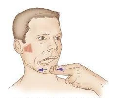
This can be performed to target the lateral pterygoid muscles. Open your jaw around one inch. Then gradually move your jaw from one side to the other without opening your jaw any further. If you have difficulty holding a one-inch gap you can clench a pen or pencil between your teeth while you perform the side-to-side movement. Do not force your jaw so distant to the side that you experience pain. Complete three sets of ten times side-to-side jaw movements.

Myofascial release
Put index finger inside the mouth, under the cheekbone. Point the finger up and towards the opposing eye. Apply pressure to the muscles until it relaxes. To check the positioning of the finger, actively move the jaw in the opposite direction and the muscle will contract under the finger. Hold until relaxed and do one-two terms per day.
Massage
The pterygoid muscles are deep in your jaw and aren’t effortless to contact. Wash your hands, then, with your palm facing out from your body, insert your index finger into your mouth between your upper teeth and at the cheek. You’ll reach a crevice where the jawbone encounters the roof of your mouth. Press your finger lightly against the space, moving from the joint area upward and toward the front of your mouth. Move your jaw gradually while you massage the muscles.
Mobility Exercises
You can test a variety of mobility exercises to relax your pterygoid muscles and the TMJ region in general. Several exercises are, in development, dynamic stretches. For example, conduct a simple opening and stretch exercise by opening your mouth slowly, as widely as you can without experiencing any pain. Target your lateral pterygoids additionally directly by opening your mouth about 1 inch wide, then moving your jaw from side to side as far as you can, provided you stay pain-free. Don’t open your jaw wider as you move laterally; keep the 1-inch gap. Try to complete 10 repetitions of both exercises.
Correcting Muscle Imbalances
Patients with TMJ problems may suffer from a muscle inequality between the lateral pterygoids and the suprahyoid along the lower jaw. To help correct the imbalance, perform a two-part exercise as often as potential throughout the day. Place your tongue as far back along the roof of your mouth as is attainable. Open and close your mouth, gradually and steadily, for several repetitions. If you can perform the exercise pain-free, advance to the second step by holding your chin with one hand, then creating a fist with your opposite hand and placing it below the jaw line. Replicate the exercise against the resistance from your hands.
Head Resistance Exercises
If you have access to a chair with a headrest you can utilize it as resistance for another two-part exercise. Place the back of your head against the headrest and close your mouth, but don’t clench your teeth tightly. Place your palm against your jaw so the loose skin between your thumb and index finger pushes on the front of your chin. Use your jaw muscles to push your head back into the headrest, but maintain your lower jaw in place with the hand on your chin, which should cause your mouth to open just a bit. Lower your lead back to the starting position. Conduct five repetitions, a few times each day. If you can do the exercise pain-free for at least two days, complete the exercise more vigorously by pushing your lower jaw forward against your hand while your head presses against the chair. Maintain your placement for a few seconds.
FAQ
What is the function of the superior lateral pterygoid muscle?
The primary role of the lateral pterygoid muscle is to pull the head of the condyle out of the mandibular fossa along the articular prominence to protrude the mandible. Concerted action of the lateral pterygoid muscles helps in lowering the mandible and opening the jaw.
What is the origin of lateral pterygoid muscles?
It arises from the lateral surface of the lateral pterygoid plate of the sphenoid bone. Fibers from both heads connect to the course posterolaterally, in a predominately horizontal plane. The superior fibers insert into the anteromedial portion of the articular capsule and articular disc of the temporomandibular joint.
What is the difference between lateral and medial pterygoid?
The medial pterygoid muscle has a superficial and deep head that arises from different areas of the jaw. The lateral pterygoid muscle via its lower head divides the two distinct heads.
What is the contraction of the lateral pterygoid muscle?
When its contracts, the mandible depresses, protrudes, and deviates to the contralateral side. Unilateral contraction of both bellies of the lateral pterygoid muscles produces effective contralateral deviation. Bilateral contraction of the lateral pterygoid muscles creates powerful protrusion of the mandible.
How do you test lateral pterygoid muscle?
Intra-oral Palpation: To palpate the lateral pterygoid area, slide the fifth number along the lateral side of the maxillary alveolar ridge to the most posterior area of the vestibule (the location for the posterior superior alveolar injection). Palpate by depressing in a superior, medial, and posterior direction.
2 Comments