
Levator Palpebrae Superioris Muscle
Introduction
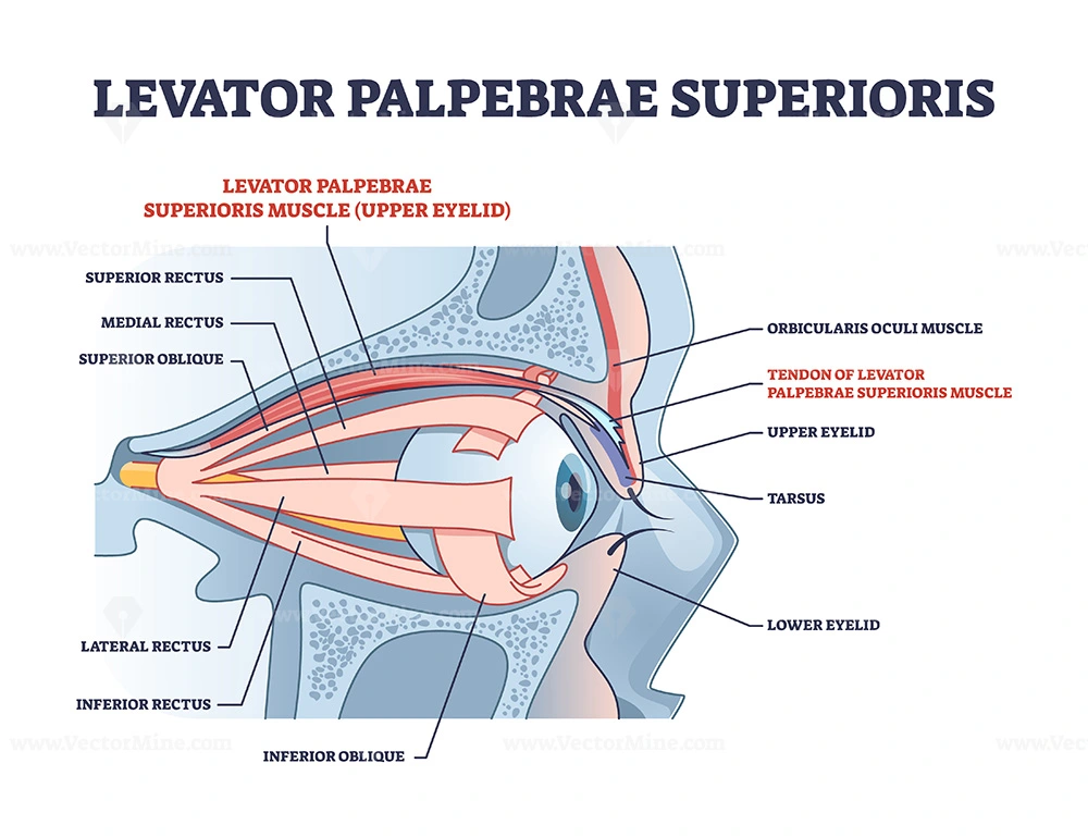
The levator palpebrae superioris originates from an inferior surface of the lesser wing of the sphenoid bone, just above an optic foramen.
It enlarges and decreases in thickness (becomes thinner) and becomes the levator aponeurosis. This portion is inserted on the skin of the upper eyelid, as well as the superior tarsal plate. It is a skeletal muscle. The superior tarsal muscle, a smooth muscle is attached to the levator palpebrae superioris and also inserts on the superior tarsal plate.
The levator palpebrae superioris muscle is a small muscle of a superior orbit that elevates and retracts the upper eyelid. It is not part of the extraocular muscles; it accomplishes not insert on the globe and thus does not produce eye movements. But it is regarded to be one of the facial muscles. It is composed of skeletal striated muscle fibers but on its undersurface, smooth muscle fibers form a superior tarsal muscle (Müller’s muscle), which is under sympathetic control (and sometimes considered a separate muscle).
Origin of levator palpebrae superioris
Levator palpebrae superioris is a triangular muscle that extends along the roof of the orbit, from the apex of the orbit to the superior eyelid. It originates with a short and narrow tendon from the inferior part of the lesser wing of the sphenoid bone, superior and anterior to the common tendinous ring. The muscle belly gradually widens as it runs anteriorly toward the eyelid.
Insertion of levator palpebrae superioris
The muscle fibers infiltrate the upper eyelid, inserting its parts via two aponeurotic fascicles;
Deep fibers connect to the anterior surface of the superior tarsus
Superficial fibers radiate via the eyelid and orbicularis oculi to finally connect to the skin of the superior eyelid.
The most lateral fibers of the muscle’s aponeurosis connect to the orbital tubercle of the zygomatic bone, whereas the most medial fibers attach to the medial palpebral ligament. Some authors acknowledge another element of the levator palpebrae superioris called the superior tarsal muscle. It is a small slip of the smooth muscle that attaches to the underside of the levator palpebrae superioris near its insertion point.
Synonyms: Musculus rectus superior bulbs oculi
Blood supply
The levator palpebrae superioris obtains its blood supply from branches of the ophthalmic artery. specifically, the muscular branches and the supraorbital artery. Blood is drained in the superior ophthalmic vein.
Nerve supply
The levator palpebrae superioris obtains motor innervation from the superior division of the oculomotor nerve. The smooth muscle that originates from its undersurface also called the superior tarsal muscles is innervated by postganglionic sympathetic axons from the superior cervical ganglion.
Function of levator palpebrae superioris
The levator palpebrae superioris elevates the upper eyelids.
The main actions of the levator palpebrae muscle are the retraction and elevation of the superior eyelid and the widening of the palpebral fissure. These actions are limited by many anatomical features of the muscle; Lateral and medial attachments of the muscle’s aponeurosis prevent further elevation of the eyelid once they are stretched maximally. The orbital septum is a fibrous part of the eyelid that when compressed maximally, prevents further retraction of the eyelid.
Antagonistic actions of the orbicularis oculi counteract the levator palpebrae superioris since it pulls the eyelid in the opposite direction.
Levator palpebrae superioris helps the superior rectus and elevates the eyelid when the gaze is directed superiorly. This occurs automatically because the fascial sheaths of the levator palpebrae superioris and superior rectus are fused via examination ligament; this is why when the superior rectus contracts, levator palpebrae superioris follow. Levator palpebrae superioris has a fixed tone that is in equilibrium with the opposing tone of orbicularis oculi, thereby preserving the eyes open and limiting the size of the palpebral fissure.
Being innervated by the sympathetic nervous system, the superior tarsal muscle elevates the eyelid in conditions of a “flight or fight” response. Thus, this muscle causes further widening of the palpebral fissure during moments of excitement, fear, surprise, and others.
Clinical significance
Damage to these muscles and their innervation can cause ptosis, which is the drooping of the eyelid. Lesions in CN III can cause ptosis because, without stimulation from the oculomotor nerve, the levator palpebrae cannot oppose the forces of gravity, and the eyelid droops.
Ptosis can also result from injury to the adjacent superior tarsal muscle or its sympathetic innervation. Such damage to the sympathetic supply originates in Horner’s syndrome and presents as partial ptosis. It is important to differentiate between these two very additional causes of ptosis. This can usually be done clinically without a situation, as each type of ptosis is accompanied by additional specific clinical findings.
The ptosis seen in paralysis of the levator palpebrae superioris is usually additionally noticeable than that seen due to paralysis of the superior tarsal muscle.
Exercise of levator palpebrae superioris
To strengthen levator palpebrae superioris and to relieve annoying eyelid twitching, you should enact targeted eyelid
exercises daily.
First, close your eyelids as tightly as you can and keep that position for ten whole moments. Then open your eyes as broad as possible and maintain them at that extreme for ten seconds. Continue to alternate between squeezing your eyes closed and opening them up wide. Conclude a total of ten sets every day.
Wide eyes
This exercise is extremely easy to perform.
Open the eyes as broadly as possible for about 5 seconds.
Relax the eyes for 5 seconds.
Continue for 30 seconds.
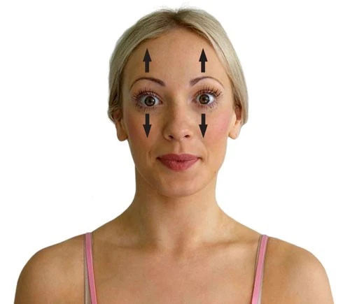
Brow lifter
This exercise targets the frontalis muscles of the forehead, which attach to the levator muscles of the eyelid.
Place the hands on the forehead and press against it with light pressure.
Using the forehead muscles, try to lift the forehead up for 5 seconds.
Repeat 5–10 times.
Rapid lids
A person can execute this exercise by following the steps below.
Use two fingers to press gently against each temple.
Open and close your eyes rapidly.
Continue for about 20 seconds.
Diagonal stretch
This exercise involves a few simple steps.
Lightly place one finger at the end of the individual eyebrow facing forward.
Utilizing the facial muscles, look down as far as possible.
Allow the eyeballs to gaze from one flank to the other like a pendulum.
Continue for about 20 seconds.
Lion’s breath
This exercise is a type of pranayama breathing that people use in yoga to improve energy and eradicate toxins. It may also help
stretch out the muscles closer to the eye and make them more supple.
Open your eyes as wide as possible while gazing upward.
At the same time, attach the tongue out and down as distant as it will go.
Hold for 5–10 seconds while exhaling with a “ha” breath.
Some people may find these exercises worthwhile, while others may notice no difference. Repeating the exercises several times
each day exists likely to produce the most benefit in those who do see a change.
Treatment options
Droopy eyelids do not always need treatment, but some people may fear that they create a tired appearance and wish to repair them.
In some cases, droopy eyelids can hide vision, especially peripheral vision. Also, ptosis in infants can lead to lazy eye (amblyopia), and an ophthalmologist choice usually recommends surgery.
FAQ
What is the origin and insertion of levator palpebrae superioris?
The levator palpebrae superioris originate on the lesser wing of the sphenoid bone just above the optic foramen. It broadens and becomes the levator aponeurosis. This part inserts on the skin of the upper eyelid, as well as the superior tarsal plate. It is a skeletal muscle.
What controls the levator palpebrae superioris muscle?
The striated levator palpebrae superioris (LPS) muscle is innervated by the oculomotor nerve and has a common origin with the superior rectus muscle. Anteriorly, it becomes the levator aponeurosis as it passes anterior to the Whitnall ligament, and inserts into the anterior tarsal surface.
What does the levator muscle do?
The major function of the levator muscle is supporting and raising the pelvic visceral structures. It also assists in proper sexual functioning, defecation, urination, and allowing various structures to pass through it.
How do you test for levator palpebrae superioris?
The examiner is positioned 2–3 feet in front of the patient’s face to permit clear observation of the patient’s eyes. The patient is instructed to tightly squeeze his or her eyelids closed for five to ten seconds. This not just relaxes the levator palpebrae superioris but actively inhibits it.
What nerve controls the levator muscle?
The levator palpebrae superioris muscle receives motor innervation from the superior division of the oculomotor nerve. The smooth muscle that arises from its undersurface, called the superior tarsal muscle is innervated by postganglionic sympathetic axons from the superior cervical ganglion

2 Comments